
mFluor™ Violet Fluorescent Conjugated Antibodies
Expand your flow cytometry possibilities with mFluor™ Violet fluorescent-conjugated antibodies from Bio-Techne.
mFluor Violet dyes are optimally excited by the violet laser (405 nm) for flow cytometry applications. Bio-Techne offers three mFluor Violet dyes conjugated to more than 3,000 targets, allowing for increased flexibility in your flow cytometry panel design.
- Compatible with other conjugated antibodies for multiplex panel design
- Excited by 405 nm laser
- Excellent aqueous solubility
- Discrete emission spectra
- Limited sensitivity to pH changes
- Photostable
- No special disposal requirements
- No special buffers required
- Fixable
What is mFluor Violet?
mFluor Violet Dyes are excited by the violet laser (405 nm) and have discrete emission spectra readily detected by common filter sets of conventional flow cytometers.
Table of Excitation and Emission for mFluor™ Violet Dyes
| Excitation Max (nm) | Emission Max (nm) | Recommended Filter Set | |
| mFluor™ Violet 450 | 406 | 445 | 450/45 |
| mFluor™ Violet 500 | 410 | 501 | 515/20 |
| mFluor™ Violet 610 | 421 | 613 | 605/30 |
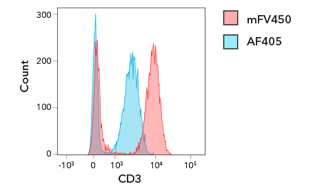
| Marker | Fluorochrome | Staining Index |
| CD3 | mFluor™ Violet 450 | 23.4 |
| Alexa Fluor® 405 | 11.8 |
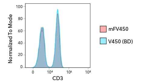
| Marker | Fluorochrome | Staining Index |
| CD3 | mFluor™ Violet 450 | 28.4 |
| V450 | 29 |
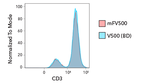
| Marker | Fluorochrome | Staining Index |
| CD3 | mFluor™ Violet 500 | 9 |
| V500 | 9.1 |
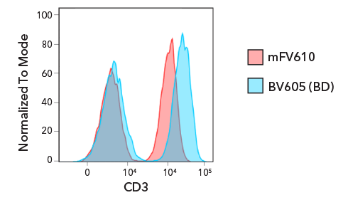
| Marker | Fluorochrome | Staining Index |
| CD3 | mFluor™ Violet 610 | 23.1 |
| Brilliant Violet™ 605 | 40.3 |
Figure 1. mFluor Violet Conjugated Antibodies from Bio-Techne can be detected in common flow cytometry channels. Comparison of mFluor Violet conjugated antibodies to antibodies conjugated to alternative dyes excited by the violet laser.
PBMCs were stained with Anti-Human CD3 conjugated to (A) mFluor Violet 450 and Alexa Fluor® 405, (B) mFluor Violet 405 and V450, (C) mFluor Violet 500 and V500, and (D) mFluor Violet 610 and Brilliant Violet™ 610. Tables show corresponding staining indices for each conjugated antibody. Staining index was calculated as follows: (Median of Positive - Median of Negative) / (SD of Negative * 2).
Why mFluor Violet?
mFluor Violet-conjugated antibodies enable you to do more with Bio-Techne antibodies. mFluor Violet dyes are compatible with other fluorescent dyes and probes to increase the number of targets you can visualize in one flow cytometry panel.
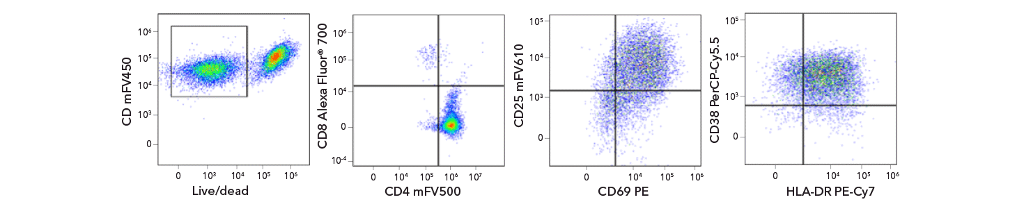
Figure 2. mFluor Violet Conjugated Antibodies are Compatible in Multicolor Flow Cytometry Panels. Human PBMCs were expanded to day 14.
Cells were harvested and stained with anti-human CD3 mFluor Violet 450 (mFV450), CD8 Alexa Fluor 700, CD4 mFluor Violet 500, CD69 PE, CD25 mFluor Violet 610, CD38 PerCP-Cy5.5, and HLA-DR PE-Cy7.

Figure 3. Lag-3 is detectable on expanded T cells using mFluor Violet 610 Conjugated Anti-Human Lag-3. Human PBMCs were expanded for 14 day and analyzed for four exhaustion markers.
Cells were stained with Tim-3 PerCP-Cy5.5, TIGIT PE, PD-1 PE/Cy7 and LAG-3 mFV610.
mFluor™ is used under license from AAT Bioquest, Inc.
Alexa Fluor® is a registered trademark of Molecular Probes, Inc.
Brilliant Violet™ is a trademark of Becton Dickinson.