
CLOCK Antibody - BSA Free
Novus Biologicals | Catalog # NB100-126

Scientific images may be resized, cropped, or adjusted for brightness or contrast for presentation purposes. These updates are limited to presentation and do not affect the underlying scientific interpretation of the data. Where available, additional source data may be provided upon request.
Key Product Details
Species Reactivity
Validated:
Cited:
Applications
Validated:
Cited:
Label
Antibody Source
Format
Product Specifications
Immunogen
Localization
Clonality
Host
Isotype
Scientific Data Images for CLOCK Antibody - BSA Free
Western Blot: CLOCK Antibody [NB100-126]
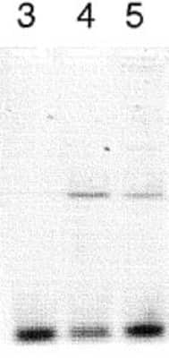
Western Blot: CLOCK Antibody [NB100-126] - Lanes 3-5 contained alpha CLOCK immunoprecipitated mouse DNA (15, 10 and 5 ul of antisera added respectively to the 900 ul of sonicated chromatin sample in the ChIP assay.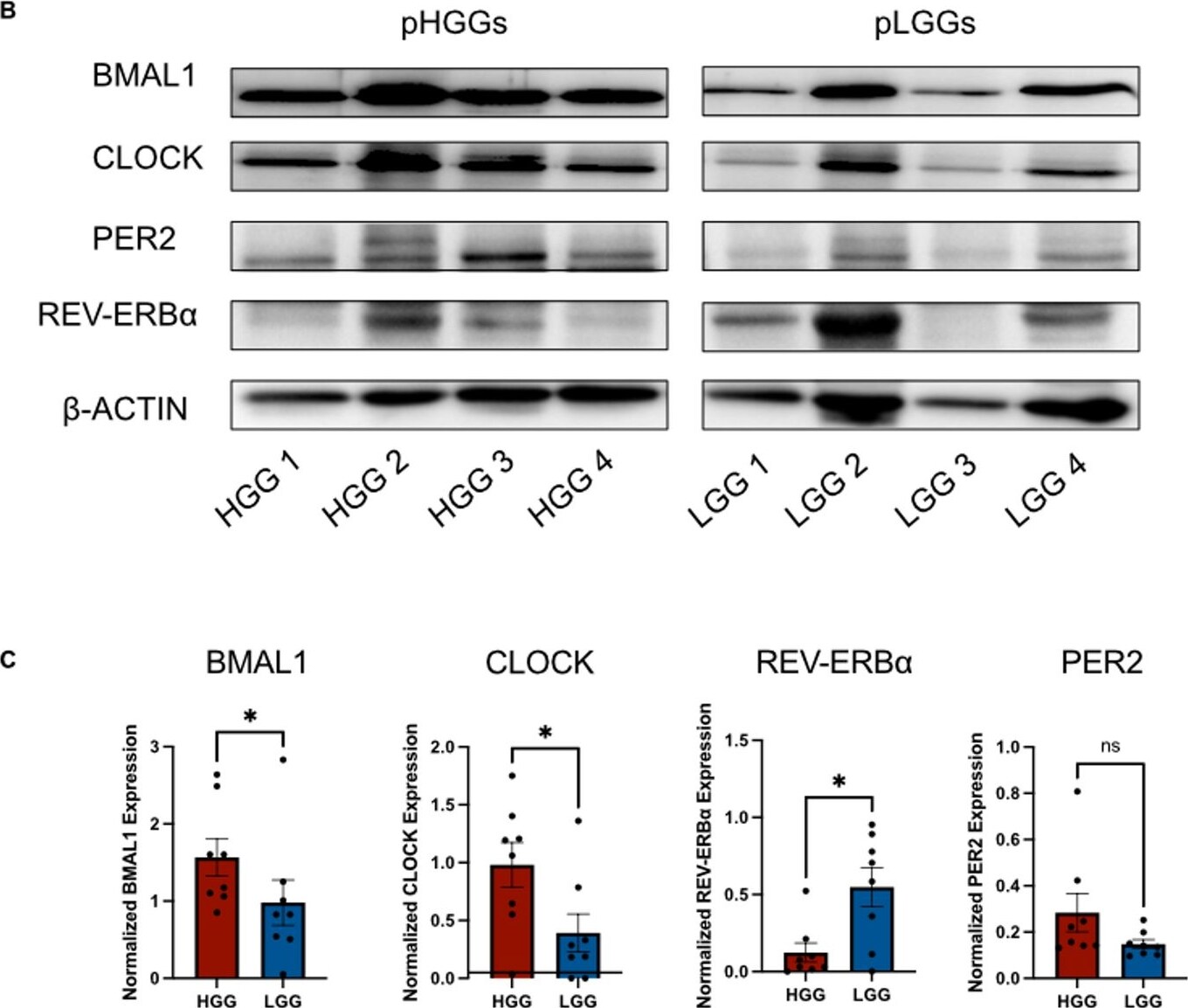
Western Blot: CLOCK Antibody - BSA Free [NB100-126] -
Circadian gene expression varies between pHGGs and pLGGs. (A) Interleaved scatter plots represent bulk RNA sequencing (rsem FPKM) analysis comparing circadian gene expression across pediatric low-grade glioma (pLGG, n = 426, blue) and pediatric high-grade glioma (pHGG, n = 211, red) samples from the Pediatric Brain Tumor Atlas (PBTA, Provisional). Data are expressed as mean +/- SEM, and expression values are log₂-transformed FPKM counts. Statistical analysis was performed using a two-way ANOVA with Benjamini and Hochberg false discovery rate (FDR) to determine differences in gene expression between pHGGs and pLGGs. Each dot represents an individual tumor sample. Asterisks indicate statistically significant differences (*P < 0.05), while “NS" denotes non-significant comparisons. (B) Western blots of four representative pediatric high-grade glioma tissue samples (left) and four pediatric low-grade glioma tissue samples (right) comparing BMAL1, CLOCK, PER2, and REV-ERB alpha protein expression (sections of separate blots displayed; full blots are shown in Supplementary Figure S3). Of note, tissue sample HGG 3 is derived from the same patient as the STN-49 cell line. (C) Normalization to beta -ACTIN control reveals BMAL1 (p < 0.05, 1.57 vs. 0.98) and CLOCK (p < 0.05, 0.98 vs. 0.39) protein expression is higher in high-grade gliomas compared to low-grade gliomas, REV-ERB alpha is lower (p < 0.05, 0.12 vs. 0.55), and there are no significant differences in PER2 protein expression. Data are expressed as mean +/- SEM. Image collected and cropped by CiteAb from the following open publication (https://www.nature.com/articles/s41598-025-17461-9), licensed under a CC-BY license. Not internally tested by Novus Biologicals.Applications for CLOCK Antibody - BSA Free
Chromatin Immunoprecipitation (ChIP)
Western Blot
Formulation, Preparation, and Storage
Purification
Formulation
Format
Preservative
Concentration
Shipping
Stability & Storage
Background: CLOCK
Long Name
Alternate Names
Entrez Gene IDs
Gene Symbol
UniProt
Additional CLOCK Products
Product Documents for CLOCK Antibody - BSA Free
Certificate of Analysis
To download a Certificate of Analysis, please enter a lot or batch number in the search box below.
Product Specific Notices for CLOCK Antibody - BSA Free
This product is for research use only and is not approved for use in humans or in clinical diagnosis. Primary Antibodies are guaranteed for 1 year from date of receipt.
Related Research Areas
Citations for CLOCK Antibody - BSA Free
 Powered by Bioz
Customer Reviews for CLOCK Antibody - BSA Free
There are currently no reviews for this product. Be the first to review CLOCK Antibody - BSA Free and earn rewards!
Have you used CLOCK Antibody - BSA Free?
Submit a review and receive an Amazon gift card!
$25/€18/£15/$25CAN/¥2500 for a review with an image
$10/€7/£6/$10CAN/¥1110 for a review without an image
Submit a review
Protocols
Find general support by application which include: protocols, troubleshooting, illustrated assays, videos and webinars.
- Cellular Response to Hypoxia Protocols
- ChIP Protocol Video
- Chromatin Immunoprecipitation (ChIP) Protocol
- Chromatin Immunoprecipitation Protocol
- R&D Systems Quality Control Western Blot Protocol
- Troubleshooting Guide: Western Blot Figures
- Western Blot Conditions
- Western Blot Protocol
- Western Blot Protocol for Cell Lysates
- Western Blot Troubleshooting
- Western Blot Troubleshooting Guide
- View all Protocols, Troubleshooting, Illustrated assays and Webinars