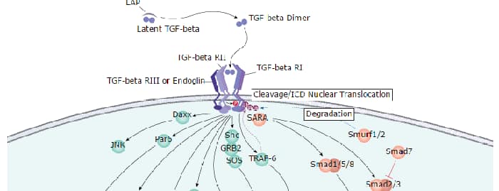
Protein Phosphatase 2A (PP2A) dephosphorylates serine and threonine residues in proteins. This ubiquitously expressed enzyme plays a critical role in modulating cell survival, growth factor responses, and neurotransmission. Phosphorylation near the C-terminus at Y307 of the catalytic subunit decreases the phosphatase activity of PP2A and reduces its ability to bind to proteins such as CD28 and the glutamate receptor GluR5. Phosphorylation of PP2A at Y307 is highest during M-phase and lowest during S-phase of the cell cycle. Transient PP2A phosphorylation at this site is also observed in growth factor-stimulated cells.
phospho-PP2A (Y307), Catalytic Subunit Antibody
R&D Systems | Catalog # AF3989


Scientific images may be resized, cropped, or adjusted for brightness or contrast for presentation purposes. These updates are limited to presentation and do not affect the underlying scientific interpretation of the data. Where available, additional source data may be provided upon request.
Key Product Details
Validated by
Biological Validation
Species Reactivity
Validated:
Human, Mouse, Rat
Cited:
Human, Mouse
Applications
Validated:
Western Blot
Cited:
Western Blot, Neutralization
Label
Unconjugated
Antibody Source
Polyclonal Rabbit IgG
Loading...
Product Specifications
Immunogen
Phosphopeptide containing human PP2A Y307 site
Specificity
Detects human, mouse, and rat PP2A when phosphorylated at Y307. The antigen is identical to the PP4 Y305 phosphorylation site.
Clonality
Polyclonal
Host
Rabbit
Isotype
IgG
Scientific Data Images for phospho-PP2A (Y307), Catalytic Subunit Antibody
Detection of Human Phospho-PP2A (Y307), Catalytic Subunit by Western Blot.
Western blot shows lysates of A549 human lung carcinoma cell line untreated (-) or treated (+) with hydroxyurea. PVDF membrane was probed with 1 µg/mL of Rabbit Anti-Human/Mouse/Rat Phospho-PP2A (Y307), Catalytic Subunit Antigen Affinity-purified Polyclonal Antibody (Catalog # AF3989), followed by HRP-conjugated Anti-Rabbit IgG Secondary Antibody (Catalog # HAF008). A specific band was detected for Phospho-PP2A (Y307), Catalytic Subunit at approximately 36 kDa (as indicated). This experiment was conducted under reducing conditions and using Immunoblot Buffer Group 1.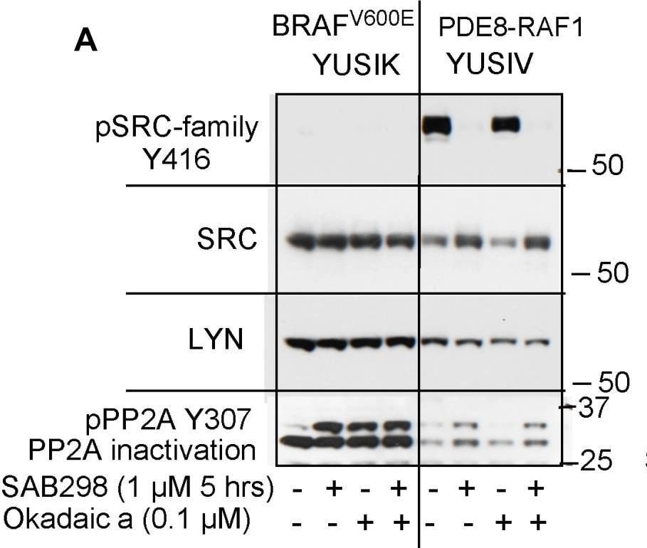
Detection of Phospho-PP2A (Y307), Catalytic Subunit by Western Blot
SAB298 inactivates PP2A, SHP2 via ERBB2 inhibition(A) SAB298 increases the levels of inactivated PP2A (pPP2A Y307) in YUSIK and YUSIV melanoma cells in a fashion similar to okadaic acid (Okadaic a). Active (unphosphorylated) PP2A is not dependent on the presence of pY416 SFK. Panels B and C show SHP2 inactivation by SAB298 in YUSIK, YUROB and YUSOC, but not YUGASP melanoma cells (pSHP2 Y542). Suppression of SHP2 is associated with SAB298 induced inactivation of ERBB2 (pERBB2 Y1196) (C, D). Image collected and cropped by CiteAb from the following open publication (https://pubmed.ncbi.nlm.nih.gov/31040916), licensed under a CC-BY license. Not internally tested by R&D Systems.Applications for phospho-PP2A (Y307), Catalytic Subunit Antibody
Application
Recommended Usage
Western Blot
1 µg/mL
Sample: Hydroxyurea-treated A549 human lung carcinoma cell line
Sample: Hydroxyurea-treated A549 human lung carcinoma cell line
Reviewed Applications
Read 6 reviews rated 4 using AF3989 in the following applications:
Formulation, Preparation, and Storage
Purification
Antigen Affinity-purified
Reconstitution
Reconstitute at 0.2 mg/mL in sterile PBS. For liquid material, refer to CoA for concentration.
Loading...
Formulation
Lyophilized from a 0.2 μm filtered solution in PBS with Trehalose. *Small pack size (SP) is supplied either lyophilized or as a 0.2 µm filtered solution in PBS.
Shipping
Lyophilized product is shipped at ambient temperature. Liquid small pack size (-SP) is shipped with polar packs. Upon receipt, store immediately at the temperature recommended below.
Stability & Storage
Use a manual defrost freezer and avoid repeated freeze-thaw cycles.
- 12 months from date of receipt, -20 to -70 °C as supplied.
- 1 month, 2 to 8 °C under sterile conditions after reconstitution.
- 6 months, -20 to -70 °C under sterile conditions after reconstitution.
Calculators
Background: PP2A
Additional PP2A Products
Product Documents for phospho-PP2A (Y307), Catalytic Subunit Antibody
Certificate of Analysis
To download a Certificate of Analysis, please enter a lot or batch number in the search box below.
Note: Certificate of Analysis not available for kit components.
Product Specific Notices for phospho-PP2A (Y307), Catalytic Subunit Antibody
For research use only
Related Research Areas
Citations for phospho-PP2A (Y307), Catalytic Subunit Antibody
 Powered by Bioz
Customer Reviews for phospho-PP2A (Y307), Catalytic Subunit Antibody (6)
4 out of 5
6 Customer Ratings
Have you used phospho-PP2A (Y307), Catalytic Subunit Antibody?
Submit a review and receive an Amazon gift card!
$25/€18/£15/$25CAN/¥2500 for a review with an image
$10/€7/£6/$10CAN/¥1110 for a review without an image
Submit a review
Customer Images

Showing
1
-
5 of
6 reviews
Showing All
Filter By:
-
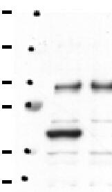
Application: Western BlotSample Tested: Adult brainSpecies: MouseVerified Customer | Posted 02/06/2021The extracted proteins were separated by SDS gel electrophoresis and transferred to nitrocellulose membranes. After incubation with the primary antibody overnight, the blots were washed in TBST and incubated at room temperature with anti-rabbit secondary antibody (1:10000 dilution).

-
Application: Western BlotSample Tested: Liver cellsSpecies: HumanVerified Customer | Posted 11/06/2020
-
Application: MicroarraysSample Tested: EDTA PlasmaSpecies: HumanVerified Customer | Posted 06/10/2020
-
Application: ELISASample Tested: PlasmaSpecies: HumanVerified Customer | Posted 11/10/2018
-
Application: MicroarraysSample Tested: EDTA PlasmaSpecies: HumanVerified Customer | Posted 11/07/2018
-
Application: MicroarraySample Tested: EDTA PlasmaSpecies: HumanVerified Customer | Posted 11/02/2018
There are no reviews that match your criteria.
Protocols
Find general support by application which include: protocols, troubleshooting, illustrated assays, videos and webinars.
- Cellular Response to Hypoxia Protocols
- R&D Systems Quality Control Western Blot Protocol
- Troubleshooting Guide: Western Blot Figures
- Western Blot Conditions
- Western Blot Protocol
- Western Blot Protocol for Cell Lysates
- Western Blot Troubleshooting
- Western Blot Troubleshooting Guide
- View all Protocols, Troubleshooting, Illustrated assays and Webinars
Loading...
Associated Pathways