
Loading...
Key Product Details
Species Reactivity
Validated:
Human
Predicted:
Mouse (96%), Rat (96%). Backed by our 100% Guarantee.
Applications
Immunohistochemistry, Immunohistochemistry-Paraffin
Label
Unconjugated
Antibody Source
Polyclonal Rabbit IgG
Format
BSA Free
Loading...
Product Specifications
Immunogen
This antibody was developed against Recombinant Protein corresponding to amino acids: RLAHLDLSYNNFSHVPADMFQEAHGLVHIDLSHNPWLRRVHPQAFQGLMQLRDLDLSYGGLAFLSLEALEGLPGLVTLQIGGNPWVCGCTMEPLLKWLRNRIQRCTADSQ
Clonality
Polyclonal
Host
Rabbit
Isotype
IgG
Applications for LRRC55 Antibody - BSA Free
Application
Recommended Usage
Immunohistochemistry
1:50 - 1:200
Immunohistochemistry-Paraffin
1:50 - 1:200
Application Notes
For IHC-Paraffin, HIER pH 6 retrieval is recommended.
Reviewed Applications
Read 1 review rated 1 using NBP1-81890 in the following applications:
Formulation, Preparation, and Storage
Purification
Affinity purified
Formulation
PBS (pH 7.2) and 40% Glycerol
Format
BSA Free
Preservative
0.02% Sodium Azide
Concentration
Concentrations vary lot to lot. See vial label for concentration. If unlisted please contact technical services.
Shipping
The product is shipped with polar packs. Upon receipt, store it immediately at the temperature recommended below.
Stability & Storage
Store at 4C short term. Aliquot and store at -20C long term. Avoid freeze-thaw cycles.
Background: LRRC55
Long Name
Leucine-rich Repeat Containing 55
Alternate Names
FLJ45686
Gene Symbol
LRRC55
Additional LRRC55 Products
Product Documents for LRRC55 Antibody - BSA Free
Certificate of Analysis
To download a Certificate of Analysis, please enter a lot or batch number in the search box below.
Product Specific Notices for LRRC55 Antibody - BSA Free
This product is for research use only and is not approved for use in humans or in clinical diagnosis. Primary Antibodies are guaranteed for 1 year from date of receipt.
Related Research Areas
Customer Reviews for LRRC55 Antibody - BSA Free (1)
1 out of 5
1 Customer Rating
Have you used LRRC55 Antibody - BSA Free?
Submit a review and receive an Amazon gift card!
$25/€18/£15/$25CAN/¥2500 Yen for a review with an image
$10/€7/£6/$10CAN/¥1110 Yen for a review without an image
Submit a review
Customer Images
Showing
1
-
1 of
1 review
Showing All
Filter By:
-
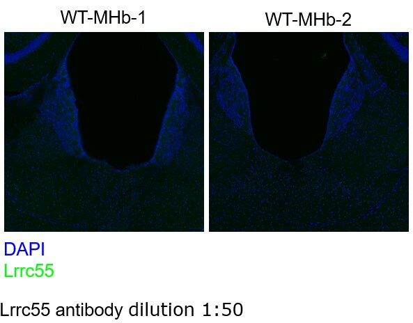
Application: Immunohistochemistry-FrozenSample Tested: Adult mouse brain tissueSpecies: MouseVerified Customer | Posted 06/17/2021Lrrc55 is known highly expressed in the medial habenula (MHb) of mouse brain. However, the NBP1-81890-25ul Rabbit Polyclonal LRRC55 Antibody did not have any signal in the MHb.1. Wash in PBS 10min x 3 2. Incubation in 5% goat serum and 0.3% Triton X-100 for 2h at RT 3. 5% goat serum and 0.3% Triton X-100 4. Rabbit Polyclonal LRRC55 Antibody (1:50) 5. Goat anti-rabbit 488 6. PBS 10min x 3 7. DAPI 5min at RT 8. PBS 10min x 3 Mice were transcardially perfused with phosphate-buffered saline (PBS) followed by 4% paraformaldehyde. The brain was removed and post-fixed in 4% paraformaldehyde overnight at 4°C and dehydrated in 30% sucrose for 48 h. Coronal section (50 μm) containing the medial habenula were collected using a cryostat. The sections were rinsed 3 times with PBS for 10 min each and blocked with 5% goat serum and 0.3% Triton X-100 for 2 hours at room temperature and incubated for overnight at 4°C with following primary antibodies: anti-Lrrc55 (1:50, NBP1-81890-25UL). After 3 rinses with PBS for 10 min, secondary antibodies (1:1000, conjugated with Alexa 594) were incubated for 2 hours at room temperature. Then the sections were washed 3 times with PBS for 10 min each and stained with DAPI (1:10000 of 5 mg/mL). Images were acquired using a Zeiss 780 inverted confocal microscope.
 Bio-Techne ResponseThis review was submitted through the legacy Novus Innovators Program, reflecting a new species or application tested on a primary antibody.
There are no reviews that match your criteria.
Protocols
Find general support by application which include: protocols, troubleshooting, illustrated assays, videos and webinars.
- Antigen Retrieval Protocol (PIER)
- Antigen Retrieval for Frozen Sections Protocol
- Appropriate Fixation of IHC/ICC Samples
- Cellular Response to Hypoxia Protocols
- Chromogenic IHC Staining of Formalin-Fixed Paraffin-Embedded (FFPE) Tissue Protocol
- Chromogenic Immunohistochemistry Staining of Frozen Tissue
- ClariTSA™ Fluorophore Kits
- Detection & Visualization of Antibody Binding
- Fluorescent IHC Staining of Frozen Tissue Protocol
- Graphic Protocol for Heat-induced Epitope Retrieval
- Graphic Protocol for the Preparation and Fluorescent IHC Staining of Frozen Tissue Sections
- Graphic Protocol for the Preparation and Fluorescent IHC Staining of Paraffin-embedded Tissue Sections
- Graphic Protocol for the Preparation of Gelatin-coated Slides for Histological Tissue Sections
- IHC Sample Preparation (Frozen sections vs Paraffin)
- Immunofluorescent IHC Staining of Formalin-Fixed Paraffin-Embedded (FFPE) Tissue Protocol
- Immunohistochemistry (IHC) and Immunocytochemistry (ICC) Protocols
- Immunohistochemistry Frozen Troubleshooting
- Immunohistochemistry Paraffin Troubleshooting
- Preparing Samples for IHC/ICC Experiments
- Preventing Non-Specific Staining (Non-Specific Binding)
- Primary Antibody Selection & Optimization
- Protocol for Heat-Induced Epitope Retrieval (HIER)
- Protocol for Making a 4% Formaldehyde Solution in PBS
- Protocol for VisUCyte™ HRP Polymer Detection Reagent
- Protocol for the Preparation & Fixation of Cells on Coverslips
- Protocol for the Preparation and Chromogenic IHC Staining of Frozen Tissue Sections
- Protocol for the Preparation and Chromogenic IHC Staining of Frozen Tissue Sections - Graphic
- Protocol for the Preparation and Chromogenic IHC Staining of Paraffin-embedded Tissue Sections
- Protocol for the Preparation and Chromogenic IHC Staining of Paraffin-embedded Tissue Sections - Graphic
- Protocol for the Preparation and Fluorescent IHC Staining of Frozen Tissue Sections
- Protocol for the Preparation and Fluorescent IHC Staining of Paraffin-embedded Tissue Sections
- Protocol for the Preparation of Gelatin-coated Slides for Histological Tissue Sections
- TUNEL and Active Caspase-3 Detection by IHC/ICC Protocol
- The Importance of IHC/ICC Controls
- Troubleshooting Guide: Immunohistochemistry
- View all Protocols, Troubleshooting, Illustrated assays and Webinars
Loading...