PLVAP Antibody - BSA Free
Novus Biologicals | Catalog # NBP1-83911
![Immunohistochemistry-Paraffin: PLVAP Antibody [NBP1-83911]](https://resources.rndsystems.com/images/products/PLVAP-Antibody-Immunohistochemistry-Paraffin-NBP1-83911-img0006.jpg "Immunohistochemistry-Paraffin: PLVAP Antibody [NBP1-83911]")
Loading...
Key Product Details
Species Reactivity
Validated:
Human
Cited:
Human
Applications
Validated:
Immunohistochemistry, Immunohistochemistry-Paraffin
Cited:
Immunohistochemistry-Paraffin, Western Blot, Electron Microscopy
Label
Unconjugated
Antibody Source
Polyclonal Rabbit IgG
Format
BSA Free
Loading...
Product Specifications
Immunogen
This antibody was developed against Recombinant Protein corresponding to amino acids: KEQLQKVQALCLPLDKDKFEMDLRNLWRDSIIPRSLDNLGYNLYHPLGSELASIRRACDHMPSLMSSKVEELARSLRADIERVARENSDLQRQKLEAQQGLRASQEAKQKVEKEAQAREAKLQAECSR
Clonality
Polyclonal
Host
Rabbit
Isotype
IgG
Scientific Data Images for PLVAP Antibody - BSA Free
Immunohistochemistry-Paraffin: PLVAP Antibody [NBP1-83911]
Immunohistochemistry-Paraffin: PLVAP Antibody [NBP1-83911] - Staining of human duodenum shows strong membranous positivity in endothelial cells.![Immunohistochemistry-Paraffin: PLVAP Antibody [NBP1-83911]](https://resources.rndsystems.com/images/products/PLVAP-Antibody-Immunohistochemistry-Paraffin-NBP1-83911-img0007.jpg "Immunohistochemistry-Paraffin: PLVAP Antibody [NBP1-83911]")
Immunohistochemistry-Paraffin: PLVAP Antibody [NBP1-83911]
Immunohistochemistry-Paraffin: PLVAP Antibody [NBP1-83911] - Staining of human fallopian tube shows strong membranous positivity in endothelial cells.![Immunohistochemistry-Paraffin: PLVAP Antibody [NBP1-83911]](https://resources.rndsystems.com/images/products/PLVAP-Antibody-Immunohistochemistry-Paraffin-NBP1-83911-img0008.jpg "Immunohistochemistry-Paraffin: PLVAP Antibody [NBP1-83911]")
Immunohistochemistry-Paraffin: PLVAP Antibody [NBP1-83911]
Immunohistochemistry-Paraffin: PLVAP Antibody [NBP1-83911] - Staining of human testis shows no positivity in cells in seminiferous ducts as expected.![Immunohistochemistry-Paraffin: PLVAP Antibody [NBP1-83911]](https://resources.rndsystems.com/images/products/PLVAP-Antibody-Immunohistochemistry-Paraffin-NBP1-83911-img0009.jpg "Immunohistochemistry-Paraffin: PLVAP Antibody [NBP1-83911]")
Immunohistochemistry-Paraffin: PLVAP Antibody [NBP1-83911]
Immunohistochemistry-Paraffin: PLVAP Antibody [NBP1-83911] - Staining of human tonsil shows strong membranous positivity in endothelial cells.Applications for PLVAP Antibody - BSA Free
Application
Recommended Usage
Immunohistochemistry
1:500 - 1:1000
Immunohistochemistry-Paraffin
1:500 - 1:1000
Application Notes
For IHC-Paraffin, HIER pH 6 retrieval is recommended.
Reviewed Applications
Read 3 reviews rated 4.3 using NBP1-83911 in the following applications:
Formulation, Preparation, and Storage
Purification
Affinity purified
Formulation
PBS (pH 7.2) and 40% Glycerol
Format
BSA Free
Preservative
0.02% Sodium Azide
Concentration
Concentrations vary lot to lot. See vial label for concentration. If unlisted please contact technical services.
Shipping
The product is shipped with polar packs. Upon receipt, store it immediately at the temperature recommended below.
Stability & Storage
Store at 4C short term. Aliquot and store at -20C long term. Avoid freeze-thaw cycles.
Background: PLVAP
Long Name
Plasmalemma Vesicle Associated Protein
Alternate Names
FELS, gp68, PV-1, PV1
Gene Symbol
PLVAP
Additional PLVAP Products
Product Documents for PLVAP Antibody - BSA Free
Certificate of Analysis
To download a Certificate of Analysis, please enter a lot or batch number in the search box below.
Product Specific Notices for PLVAP Antibody - BSA Free
This product is for research use only and is not approved for use in humans or in clinical diagnosis. Primary Antibodies are guaranteed for 1 year from date of receipt.
Related Research Areas
Citations for PLVAP Antibody - BSA Free
 Powered by Bioz
Customer Reviews for PLVAP Antibody - BSA Free (3)
4.3 out of 5
3 Customer Ratings
Have you used PLVAP Antibody - BSA Free?
Submit a review and receive an Amazon gift card!
$25/€18/£15/$25CAN/¥2500 Yen for a review with an image
$10/€7/£6/$10CAN/¥1110 Yen for a review without an image
Submit a review
Customer Images
Showing
1
-
3 of
3 reviews
Showing All
Filter By:
-
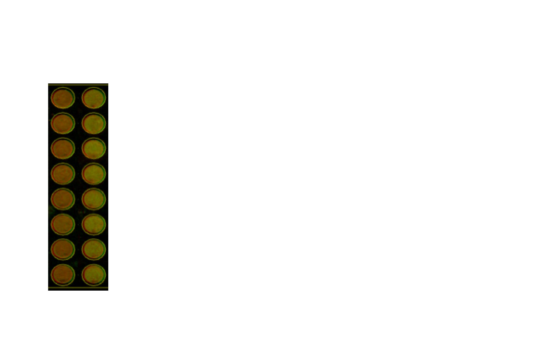
Application: In-Cell WesternSample Tested: Human retinal endothelial cells and U937Species: HumanVerified Customer | Posted 03/14/2019PLVAP antibody, NBP1-83911, [1:140 ] followed by Goat anti-rabbit IRDYE 800 secondary antibody. CellTag 700 used for normalization. Left = untreated cells and right = 24 hour VEGFA treatment.

-
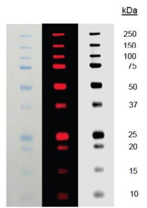
Application: Western BlotSample Tested: Human retinal endothelial cellsSpecies: HumanVerified Customer | Posted 02/26/2019Immunoblot with NBP1-83911 (PLVAP antibody) at a dilution of (1:1000) and over night incubation at 4 C. Left = untreated cells and right = 24 hr VEGFA treatment.Green band = PLVAP & Red band = ActinCell lysate from Human Retinal Microvascular Endothelial Cells (HRMECs) was prepared and loaded onto Mini-protean TGX gel followed by western blot with NBP1-83911 (PLVAP antibody) at a dilution of (1:1000) and incubated over night at 4 C. Left = untreated cells and right = 24 hr VEGFA treatment. Green band = PLVAP and Red band = Actin
-
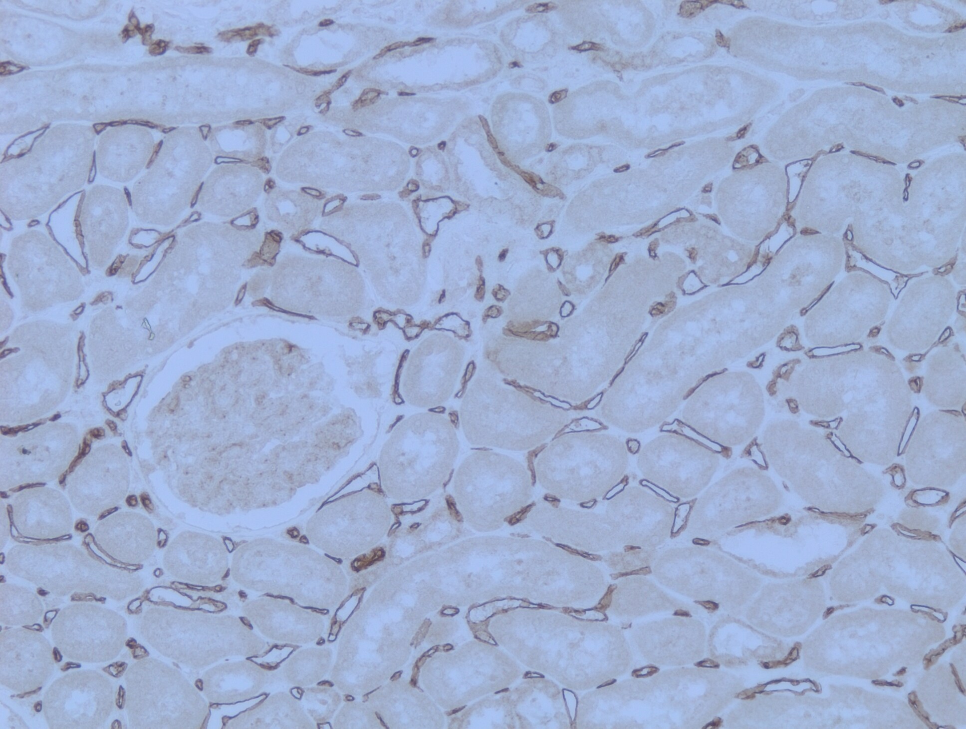
Application: Immunohistochemistry-ParaffinSample Tested: Human kidneySpecies: HumanVerified Customer | Posted 05/22/2018Heat mediated antigen retrieval was performed by heating in citrate buffer (pH6) at 95C for 20 minutes
There are no reviews that match your criteria.
Protocols
Find general support by application which include: protocols, troubleshooting, illustrated assays, videos and webinars.
- Antigen Retrieval Protocol (PIER)
- Antigen Retrieval for Frozen Sections Protocol
- Appropriate Fixation of IHC/ICC Samples
- Cellular Response to Hypoxia Protocols
- Chromogenic IHC Staining of Formalin-Fixed Paraffin-Embedded (FFPE) Tissue Protocol
- Chromogenic Immunohistochemistry Staining of Frozen Tissue
- ClariTSA™ Fluorophore Kits
- Detection & Visualization of Antibody Binding
- Fluorescent IHC Staining of Frozen Tissue Protocol
- Graphic Protocol for Heat-induced Epitope Retrieval
- Graphic Protocol for the Preparation and Fluorescent IHC Staining of Frozen Tissue Sections
- Graphic Protocol for the Preparation and Fluorescent IHC Staining of Paraffin-embedded Tissue Sections
- Graphic Protocol for the Preparation of Gelatin-coated Slides for Histological Tissue Sections
- IHC Sample Preparation (Frozen sections vs Paraffin)
- Immunofluorescent IHC Staining of Formalin-Fixed Paraffin-Embedded (FFPE) Tissue Protocol
- Immunohistochemistry (IHC) and Immunocytochemistry (ICC) Protocols
- Immunohistochemistry Frozen Troubleshooting
- Immunohistochemistry Paraffin Troubleshooting
- Preparing Samples for IHC/ICC Experiments
- Preventing Non-Specific Staining (Non-Specific Binding)
- Primary Antibody Selection & Optimization
- Protocol for Heat-Induced Epitope Retrieval (HIER)
- Protocol for Making a 4% Formaldehyde Solution in PBS
- Protocol for VisUCyte™ HRP Polymer Detection Reagent
- Protocol for the Preparation & Fixation of Cells on Coverslips
- Protocol for the Preparation and Chromogenic IHC Staining of Frozen Tissue Sections
- Protocol for the Preparation and Chromogenic IHC Staining of Frozen Tissue Sections - Graphic
- Protocol for the Preparation and Chromogenic IHC Staining of Paraffin-embedded Tissue Sections
- Protocol for the Preparation and Chromogenic IHC Staining of Paraffin-embedded Tissue Sections - Graphic
- Protocol for the Preparation and Fluorescent IHC Staining of Frozen Tissue Sections
- Protocol for the Preparation and Fluorescent IHC Staining of Paraffin-embedded Tissue Sections
- Protocol for the Preparation of Gelatin-coated Slides for Histological Tissue Sections
- TUNEL and Active Caspase-3 Detection by IHC/ICC Protocol
- The Importance of IHC/ICC Controls
- Troubleshooting Guide: Immunohistochemistry
- View all Protocols, Troubleshooting, Illustrated assays and Webinars
Loading...