
3-methylcytosine (3-mC) Antibody (PSH03-22)
Novus Biologicals | Catalog # NBP3-31985
Recombinant Monoclonal Antibody

Loading...
Key Product Details
Species Reactivity
Non-species specific
Applications
ELISA, Immunocytochemistry/ Immunofluorescence, Dot Blot
Label
Unconjugated
Antibody Source
Recombinant Monoclonal Rabbit IgG Clone # PSH03-22 expressed in HEK293
Loading...
Product Specifications
Immunogen
3-mc-OVA
Clonality
Monoclonal
Host
Rabbit
Isotype
IgG
Scientific Data Images for 3-methylcytosine (3-mC) Antibody (PSH03-22)
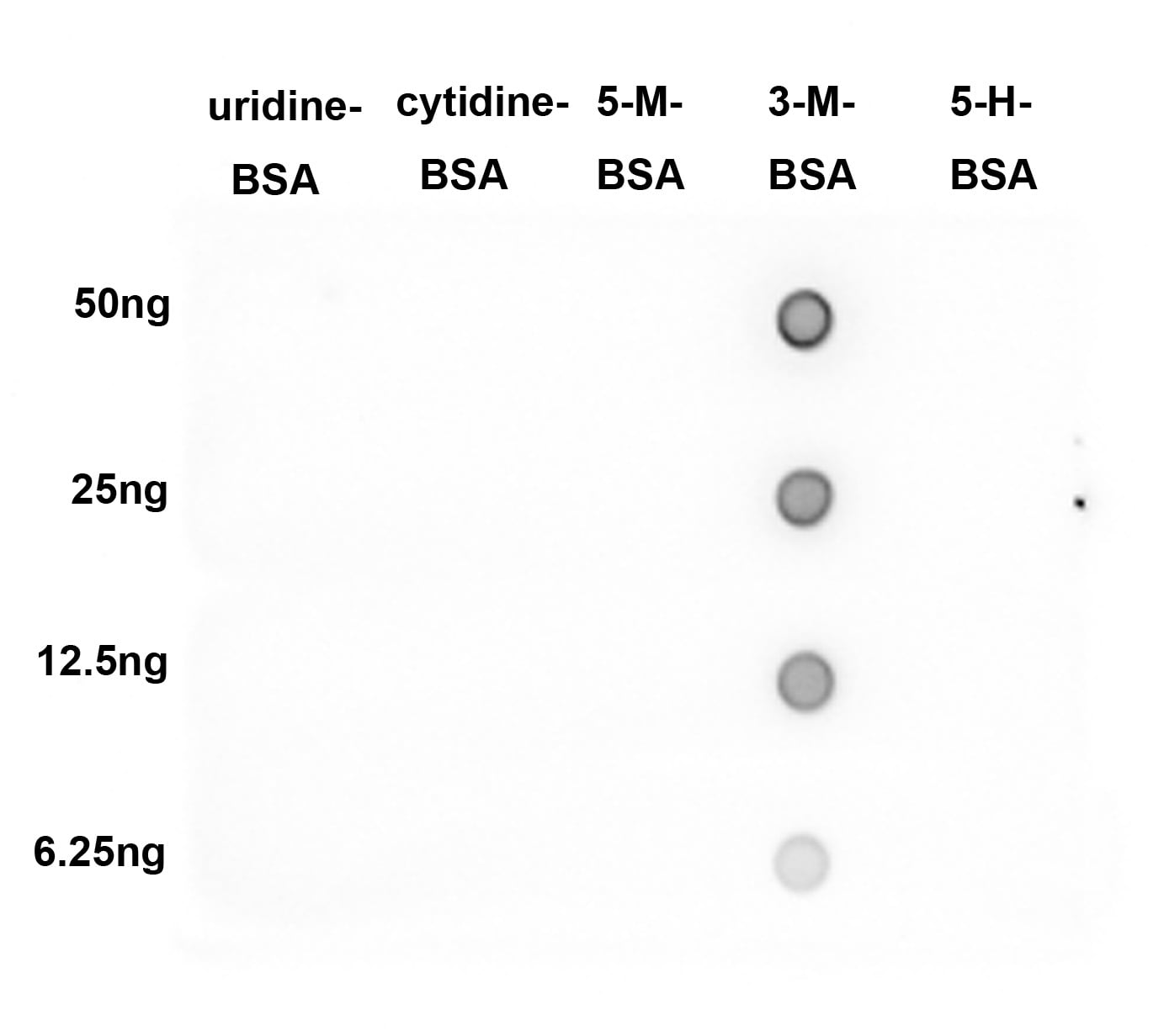
Dot Blot: 3-methylcytosine (3-mC) Antibody (PSH03-22) [NBP3-31985]
Dot Blot: 3-methylcytosine (3-mC) Antibody (PSH03-22) [NBP3-31985] - Dot blot analysis of 3-methylcytosine (3-mC) on different proteins with Rabbit anti-3mC antibody (NBP3-31985) at 1/2,000 dilution. Goat Anti-Rabbit IgG - HRP Secondary Antibody at 1/50,000 dilution for 1 hour at room temperature.Lane 1: Uridine-BSA (negative)
Lane 2: Cytidine-BSA (negative)
Lane 3: 5-Methylcytosine-BSA (negative)
Lane 4: 3-Methylcytosine-BSA (positive)
Lane 5: 5-Hydroxymethylcytidine-BSA (negative)
Proteins loading: 50ng, 25ng, 12.5ng, 6.25ng;
Blocking and dilution buffer: 5% NFDM/TBST;
Exposure time: 1 minute.
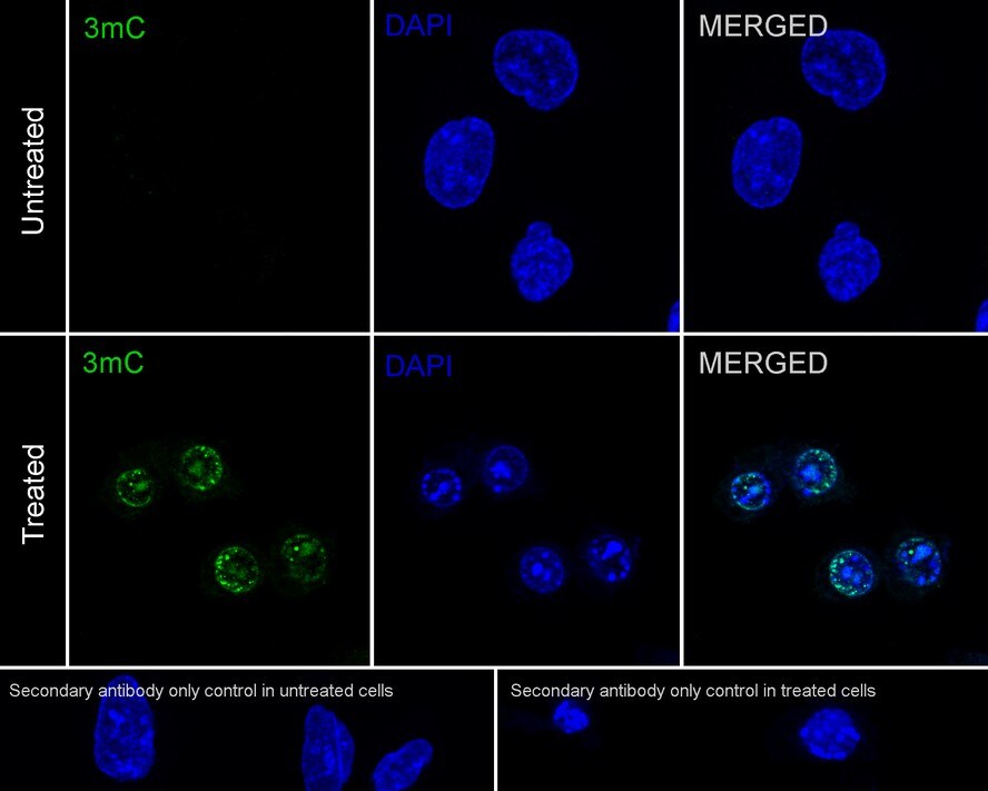
Immunocytochemistry/ Immunofluorescence: 3-methylcytosine (3-mC) Antibody (PSH03-22) [NBP3-31985]
Immunocytochemistry/ Immunofluorescence: 3-methylcytosine (3-mC) Antibody (PSH03-22) [NBP3-31985] - Immunocytochemistry analysis of U-2 OS cells treated with 4mM Methyl Methanesulfonate for 1 hour labeling 3-methylcytosine (3-mC) with Rabbit anti-3-methylcytosine (3-mC) antibody (NBP3-31985) at 1/100 dilution.Cells were fixed in 4% paraformaldehyde for 20 minutes at room temperature, permeabilized with 0.1% Triton X-100 in PBS for 5 minutes at room temperature, then blocked with 1% BSA in 10% negative goat serum for 1 hour at room temperature. Cells were then incubated with Rabbit anti-3-methylcytosine (3-mC) antibody (NBP3-31985) at 1/100 dilution in 1% BSA in PBST overnight at 4 ℃. Goat Anti-Rabbit IgG H&L (iFluor™ 488) was used as the secondary antibody at 1/1,000 dilution. PBS instead of the primary antibody was used as the secondary antibody only control. Nuclear DNA was labelled in blue with DAPI.
Applications for 3-methylcytosine (3-mC) Antibody (PSH03-22)
Application
Recommended Usage
Dot Blot
1:2000
Immunocytochemistry/ Immunofluorescence
1:100
Formulation, Preparation, and Storage
Purification
Protein A purified
Formulation
PBS (pH7.4), 0.1% BSA and 40% Glycerol
Preservative
0.05% Sodium Azide
Concentration
1 mg/ml
Shipping
The product is shipped with polar packs. Upon receipt, store it immediately at the temperature recommended below.
Stability & Storage
Store at 4C short term. Aliquot and store at -20C long term. Avoid freeze-thaw cycles.
Background: 3-methylcytosine (3-mC)
Alternate Names
3-mC, 3-Methylcytosine, 6-amino-1-methylpyrimidin-2-one
Additional 3-methylcytosine (3-mC) Products
Product Documents for 3-methylcytosine (3-mC) Antibody (PSH03-22)
Certificate of Analysis
To download a Certificate of Analysis, please enter a lot or batch number in the search box below.
Product Specific Notices for 3-methylcytosine (3-mC) Antibody (PSH03-22)
This product is for research use only and is not approved for use in humans or in clinical diagnosis. Primary Antibodies are guaranteed for 1 year from date of receipt.
Customer Reviews for 3-methylcytosine (3-mC) Antibody (PSH03-22)
There are currently no reviews for this product. Be the first to review 3-methylcytosine (3-mC) Antibody (PSH03-22) and earn rewards!
Have you used 3-methylcytosine (3-mC) Antibody (PSH03-22)?
Submit a review and receive an Amazon gift card!
$25/€18/£15/$25CAN/¥2500 Yen for a review with an image
$10/€7/£6/$10CAN/¥1110 Yen for a review without an image
Submit a review
Protocols
Find general support by application which include: protocols, troubleshooting, illustrated assays, videos and webinars.
- Appropriate Fixation of IHC/ICC Samples
- Cellular Response to Hypoxia Protocols
- ClariTSA™ Fluorophore Kits
- Detection & Visualization of Antibody Binding
- ELISA Sample Preparation & Collection Guide
- ELISA Troubleshooting Guide
- How to Run an R&D Systems DuoSet ELISA
- How to Run an R&D Systems Quantikine ELISA
- How to Run an R&D Systems Quantikine™ QuicKit™ ELISA
- ICC Cell Smear Protocol for Suspension Cells
- ICC Immunocytochemistry Protocol Videos
- ICC for Adherent Cells
- Immunocytochemistry (ICC) Protocol
- Immunocytochemistry Troubleshooting
- Immunofluorescence of Organoids Embedded in Cultrex Basement Membrane Extract
- Immunohistochemistry (IHC) and Immunocytochemistry (ICC) Protocols
- Preparing Samples for IHC/ICC Experiments
- Preventing Non-Specific Staining (Non-Specific Binding)
- Primary Antibody Selection & Optimization
- Protocol for VisUCyte™ HRP Polymer Detection Reagent
- Protocol for the Fluorescent ICC Staining of Cell Smears - Graphic
- Protocol for the Fluorescent ICC Staining of Cultured Cells on Coverslips - Graphic
- Protocol for the Preparation and Fluorescent ICC Staining of Cells on Coverslips
- Protocol for the Preparation and Fluorescent ICC Staining of Non-adherent Cells
- Protocol for the Preparation and Fluorescent ICC Staining of Stem Cells on Coverslips
- Protocol for the Preparation of a Cell Smear for Non-adherent Cell ICC - Graphic
- Quantikine HS ELISA Kit Assay Principle, Alkaline Phosphatase
- Quantikine HS ELISA Kit Principle, Streptavidin-HRP Polymer
- Sandwich ELISA (Colorimetric) – Biotin/Streptavidin Detection Protocol
- Sandwich ELISA (Colorimetric) – Direct Detection Protocol
- TUNEL and Active Caspase-3 Detection by IHC/ICC Protocol
- The Importance of IHC/ICC Controls
- Troubleshooting Guide: ELISA
- View all Protocols, Troubleshooting, Illustrated assays and Webinars
Loading...