ARNT/HIF-1 beta Antibody (H1beta234) - BSA Free
Novus Biologicals | Catalog # NB100-124
Key Product Details
Validated by
Biological Validation
Species Reactivity
Validated:
Human, Mouse, Rat, Bovine, Ferret, Primate, Sheep
Cited:
Human, Mouse, Rat
Applications
Validated:
Immunohistochemistry, Immunohistochemistry-Paraffin, Western Blot, Immunoblotting, Immunocytochemistry/ Immunofluorescence, Immunoprecipitation, Chromatin Immunoprecipitation (ChIP), Chromatin Immunoprecipitation Sequencing, Gel Super Shift Assays
Cited:
Immunohistochemistry, Immunohistochemistry-Paraffin, Western Blot, Immunoblotting, Immunocytochemistry/ Immunofluorescence, Immunoprecipitation, Chemotaxis, Chip Cytometry, Gel Supershift Assay, IF/IHC
Label
Unconjugated
Antibody Source
Monoclonal Mouse IgG1 kappa Clone # H1beta234
Format
BSA Free
Loading...
Product Specifications
Immunogen
ARNT/HIF-1 beta Antibody (H1beta234) was developed against a fusion protein containing amino acids 496-789 of human HIF-1 beta. [Uniprot: P27540]
Clonality
Monoclonal
Host
Mouse
Isotype
IgG1 kappa
Theoretical MW
86.6 kDa.
Disclaimer note: The observed molecular weight of the protein may vary from the listed predicted molecular weight due to post translational modifications, post translation cleavages, relative charges, and other experimental factors.
Disclaimer note: The observed molecular weight of the protein may vary from the listed predicted molecular weight due to post translational modifications, post translation cleavages, relative charges, and other experimental factors.
Scientific Data Images for ARNT/HIF-1 beta Antibody (H1beta234) - BSA Free
![Western Blot: ARNT/HIF-1 beta Antibody (H1beta234)BSA Free [NB100-124]](https://resources.rndsystems.com/images/products/ARNT-HIF-1-beta-Antibody-H1beta234-Western-Blot-NB100-124-img0009.jpg "Western Blot: ARNT/HIF-1 beta Antibody (H1beta234)BSA Free [NB100-124]")
![Western Blot: ARNT/HIF-1 beta Antibody (H1beta234)BSA Free [NB100-124]](https://resources.rndsystems.com/images/products/ARNT-HIF-1-beta-Antibody-H1beta234-Western-Blot-NB100-124-img0010.jpg "Western Blot: ARNT/HIF-1 beta Antibody (H1beta234)BSA Free [NB100-124]")
Western Blot: ARNT/HIF-1 beta Antibody (H1beta234)BSA Free [NB100-124]
ARNT-HIF-1-beta-Antibody-H1beta234-Western-Blot-NB100-124-img0010.jpg![Immunohistochemistry: ARNT/HIF-1 beta Antibody (H1beta234) - BSA Free [NB100-124]](https://resources.rndsystems.com/images/products/ARNT-HIF-1-beta-Antibody-H1beta234-Immunohistochemistry-NB100-124-img0005.jpg "Immunohistochemistry: ARNT/HIF-1 beta Antibody (H1beta234) - BSA Free [NB100-124]")
Immunohistochemistry: ARNT/HIF-1 beta Antibody (H1beta234) - BSA Free [NB100-124]
Immunohistochemistry: ARNT/HIF-1 beta Antibody (H1beta234) [NB100-124] - Staining of human glioblastoma multi-forme utilizing ARNT/HIF-1 beta antibody (H1beta234) [NB100-124].![Immunocytochemistry/ Immunofluorescence: ARNT/HIF-1 beta Antibody (H1beta234) - BSA Free [NB100-124]](https://resources.rndsystems.com/images/products/ARNT-HIF-1-beta-Antibody-H1beta234-Immunocytochemistry-Immunofluorescence-NB100-124-img0011.jpg "Immunocytochemistry/ Immunofluorescence: ARNT/HIF-1 beta Antibody (H1beta234) - BSA Free [NB100-124]")
Immunocytochemistry/ Immunofluorescence: ARNT/HIF-1 beta Antibody (H1beta234) - BSA Free [NB100-124]
ARNT-HIF-1-beta-Antibody-H1beta234-Immunocytochemistry-Immunofluorescence-NB100-124-img0011.jpg![Western Blot: ARNT/HIF-1 beta Antibody (H1beta234)BSA Free [NB100-124]](https://resources.rndsystems.com/images/products/ARNT-HIF-1-beta-Antibody-H1beta234-Western-Blot-NB100-124-img0006.jpg "Western Blot: ARNT/HIF-1 beta Antibody (H1beta234)BSA Free [NB100-124]")
Western Blot: ARNT/HIF-1 beta Antibody (H1beta234)BSA Free [NB100-124]
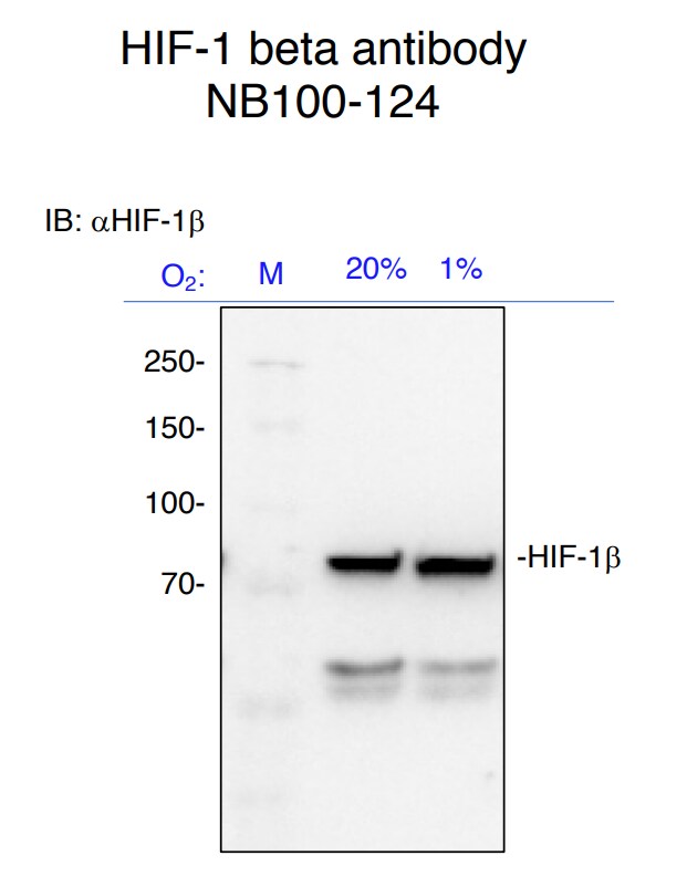
Western Blot: ARNT/HIF-1 beta Antibody (H1beta234) [NB100-124] - Analysis of HIF-1 beta in HeLa nuclear extract using ARNT/HIF-1 beta antibody (H1beta234) [NB100-124]. Theoretical molecular weight 86.6 kDa. Observed molecular weight ~85 kDa.![Immunocytochemistry/ Immunofluorescence: ARNT/HIF-1 beta Antibody (H1beta234) - BSA Free [NB100-124]](https://resources.rndsystems.com/images/products/ARNT-HIF-1-beta-Antibody-H1beta234-Immunocytochemistry-Immunofluorescence-NB100-124-img0007.jpg "Immunocytochemistry/ Immunofluorescence: ARNT/HIF-1 beta Antibody (H1beta234) - BSA Free [NB100-124]")
Immunocytochemistry/ Immunofluorescence: ARNT/HIF-1 beta Antibody (H1beta234) - BSA Free [NB100-124]
Immunocytochemistry/Immunofluorescence: ARNT/HIF-1 beta Antibody (H1beta234) [NB100-124] - HeLa cells were fixed for 10 minutes using 10% formalin and then permeabilized for 5 minutes using 1X TBS + 0.5% Triton X-100. The cells were incubated with ARNT/HIF-1 beta antibody (H1beta234) [NB100-124] at 5 ug/mL overnight at 4C and detected with an anti-mouse DyLight 488 (Green) at a 1:500 dilution. Actin was detected with Phalloidin 568 (Red) at a 1:200 dilution. Nuclei were counterstained with DAPI (Blue). Cells were imaged using a 40X objective. - BSA Free [NB100-124] -")
Western Blot: ARNT/HIF-1 beta Antibody (H1beta234) - BSA Free [NB100-124] -
Western Blot: ARNT/HIF-1 beta Antibody (H1beta234) - BSA Free [NB100-124] - PGE1 induces HIF-1 alpha protein accumulation in vascular-derived cells.Human aortic smooth muscle cells (HASMCs) (A & B), human umbilical vein endothelial cells (HUVECs) (C & D), & HEK293 cells (G) were exposed to 1 or 10 µM PGE1 under 20% O2 or 1% O2 conditions for 4 h. After treatment, cells were harvested & whole-cell lysates were subjected to immunoblot assay for HIF-1 alpha, HIF-1 beta, & beta -actin protein expression. Experiments were repeated thrice (A, C & G). Representative immunoblots are shown (A & C). Band intensities were analyzed densitometrically (B & D). Fold induction relative to lane 1 was plotted as mean ± S.D. ∗P < 0.05 compared with the control. HASMCs (E) & HUVECs (F) were exposed to 1 µM PGE1 for the indicated times under 20% O2 & were harvested for immunoblot assay for HIF-1 alpha protein. Experiments were repeated twice. Representative immunoblots are shown. (H) HASMCs & HUVECs were exposed to 10 µM PGE1 for 4 h under 20% O2 & were harvested for immunoblot assay for HIF-2 alpha protein. Experiments were repeated twice. Representative immunoblots are shown. I. HASMCs were exposed to 1 µM PGE1, lipo-PGE1 & PGE1-alfadex under 20% O2 conditions for 4 h. After treatment, cells were harvested & whole-cell lysates were subjected to immunoblot assay for HIF-1 alpha. Image collected & cropped by CiteAb from the following publication (https://pubmed.ncbi.nlm.nih.gov/24349900), licensed under a CC-BY license. Not internally tested by Novus Biologicals. - BSA Free [NB100-124] -")
Western Blot: ARNT/HIF-1 beta Antibody (H1beta234) - BSA Free [NB100-124] -
Western Blot: ARNT/HIF-1 beta Antibody (H1beta234) - BSA Free [NB100-124] - Effect of PGE1 on stability & synthesis of HIF-1 alpha.(A) Human aortic smooth muscle cells (HASMCs) & human umbilical vein endothelial cells (HUVECs) were exposed to 1 µM PGE1 or 100 µM DFX or incubated for 4 h, & CHX was added to a final concentration of 100 µM. The cells were incubated for 0 to 30 min, & whole-cell lysates were subjected to immunoblot assay using anti-HIF-1 alpha or -beta antibodies. (B) Serum-starved HASMCs were pretreated with no drug & 1 µM PGE1 for 30 min in Met-free medium. [35S]Met-Cys was added, & the cells were incubated for 60 min prior to preparation of cell lysates. Aliquots of 1 mg of the lysates were subjected to immunoprecipitation with anti-HIF-1 alpha antibody, separated by SDS-PAGE & exposed. Aliquots of 50 µg of the same lysate were separately subjected to immunoblotting analysis with anti-beta -actin antibody. (C) HASMCs & HUVECs were exposed to vehicle or 1 µM PGE1 for 4 h in the presence of 10 µM LY294002 (LY), 50 µM PD98059 (PD), or 5 µM GF109203X (GF). The cells were harvested & the whole-cell lysates were subjected to immunoblot assay for HIF-1 alpha & beta -actin protein expression. Experiments were repeated at least twice. Representative immunoblots are shown. (D) HASMCs were exposed to vehicle or 1 µM PGE1 for 12 h in the presence of 10 µM LY294002 (LY), 50 µM PD98059 (PD), or 5 µM GF109203X (GF). Cells were harvested & subjected to semi-quantitative RT-PCR for VEGF & GLUT1. Experiments were repeated three times. Fold induction relative to that under 20% O2 without PGE1 treatment was plotted. ∗P < 0.05 compared with the control (20%, PGE1 treatment without any kinase inhibitors). Image collected & cropped by CiteAb from the following publication (https://pubmed.ncbi.nlm.nih.gov/24349900), licensed under a CC-BY license. Not internally tested by Novus Biologicals. - BSA Free [NB100-124] -")
Western Blot: ARNT/HIF-1 beta Antibody (H1beta234) - BSA Free [NB100-124] -
Western Blot: ARNT/HIF-1 beta Antibody (H1beta234) - BSA Free [NB100-124] - PGE1 induces HIF-1 alpha protein accumulation in vascular-derived cells.Human aortic smooth muscle cells (HASMCs) (A & B), human umbilical vein endothelial cells (HUVECs) (C & D), & HEK293 cells (G) were exposed to 1 or 10 µM PGE1 under 20% O2 or 1% O2 conditions for 4 h. After treatment, cells were harvested & whole-cell lysates were subjected to immunoblot assay for HIF-1 alpha, HIF-1 beta, & beta -actin protein expression. Experiments were repeated thrice (A, C & G). Representative immunoblots are shown (A & C). Band intensities were analyzed densitometrically (B & D). Fold induction relative to lane 1 was plotted as mean ± S.D. ∗P < 0.05 compared with the control. HASMCs (E) & HUVECs (F) were exposed to 1 µM PGE1 for the indicated times under 20% O2 & were harvested for immunoblot assay for HIF-1 alpha protein. Experiments were repeated twice. Representative immunoblots are shown. (H) HASMCs & HUVECs were exposed to 10 µM PGE1 for 4 h under 20% O2 & were harvested for immunoblot assay for HIF-2 alpha protein. Experiments were repeated twice. Representative immunoblots are shown. I. HASMCs were exposed to 1 µM PGE1, lipo-PGE1 & PGE1-alfadex under 20% O2 conditions for 4 h. After treatment, cells were harvested & whole-cell lysates were subjected to immunoblot assay for HIF-1 alpha. Image collected & cropped by CiteAb from the following publication (https://pubmed.ncbi.nlm.nih.gov/24349900), licensed under a CC-BY license. Not internally tested by Novus Biologicals. - BSA Free [NB100-124] -")
Western Blot: ARNT/HIF-1 beta Antibody (H1beta234) - BSA Free [NB100-124] -
Western Blot: ARNT/HIF-1 beta Antibody (H1beta234) - BSA Free [NB100-124] - Differential involvement of EP receptors in PGE1-induced HIF-1 alpha protein accumulation.(A) Expression of EP1, EP2, EP3, & EP4 receptors in human aortic smooth muscle cells (HASMCs), human umbilical vein endothelial cells (HUVECs), & cells of the neuroblastoma cell line SH-SY5Y. HASMCs, HUVECs, & SH-SY5Y cells were cultured under 20% O2 & harvested for semi-quantitative RT-PCR for EP1-4 receptors. Experiments were repeated at least three times in triplicate. Fold induction relative to expression in SH-SY5Y cells was plotted as mean ± S.D. HASMCs & HUVECs were exposed to 1 µM of EP-receptor-specific agonists (ONO-DI-004 for EP1, ONO-AE1-259-01 for EP2, ONO-AE-248 for EP3, & ONO-AE1-329 for EP4) for 4 h (B). HASMCs & HUVECs were exposed to 1 µM PGE1 with or without 1 µM EP-receptor-specific antagonists (ONO-8713 against EP1, ONO-AE3-240 against EP3, & ONO-AE2-227 against EP4) for 4 h (C). The cells were harvested & the whole-cell lysates were subjected to immunoblot assay for HIF-1 alpha & beta -actin protein expression. Experiments were repeated twice. Representative immunoblots are shown. Image collected & cropped by CiteAb from the following publication (https://pubmed.ncbi.nlm.nih.gov/24349900), licensed under a CC-BY license. Not internally tested by Novus Biologicals. - BSA Free [NB100-124] -")
Western Blot: ARNT/HIF-1 beta Antibody (H1beta234) - BSA Free [NB100-124] -
Western Blot: ARNT/HIF-1 beta Antibody (H1beta234) - BSA Free [NB100-124] - HIF was not the only factor stabilizing activated EGFR in VHL-deficient ccRCC cells.A. Western blot analysis of 786-VHL & 786-mock cells stably expressing shRNA constructs. For HIF2 alpha & HIF1 beta analysis, nuclear extracts were generated & analyzed. Anti-HA blot detected HA-VHL. B. 786-VHL cells expressing SCR (control), HIF2a-566 & HIF2a-1631 (shRNA constructs against HIF2 alpha ) were treated with EGF & analyzed as described in Fig. 1A. C. 786-mock cells expressing SCR (control sequence), HIF2a-566 & HIF2a-1631 (shRNA constructs against HIF2 alpha ) were treated with EGF & analyzed as described in Fig. 2B. D. The EGFR signals in Fig. 2B & 2C were normalized over Vinculin via densitometry & plotted over time. Means & SDs of three separate experiments were shown. Image collected & cropped by CiteAb from the following publication (https://dx.plos.org/10.1371/journal.pone.0023936), licensed under a CC-BY license. Not internally tested by Novus Biologicals. - BSA Free [NB100-124] -")
Immunocytochemistry/ Immunofluorescence: ARNT/HIF-1 beta Antibody (H1beta234) - BSA Free [NB100-124] -
Immunocytochemistry/ Immunofluorescence: ARNT/HIF-1 beta Antibody (H1beta234) - BSA Free [NB100-124] - Co-localization of HIF-2 alpha with HIF-1 beta & RNAPII. (a) Immunofluorescence detecting indicated proteins using antibodies labelled with Alexa Fluor 555 (red, pseudocolour) & Alexa Fluor 488 (green, pseudocolour). ‘Merge’ is the red image superimposed onto the green image of the co-stained nuclear proteins. ‘Coloc’ is the co-localization channel calculated using ImageJ plugin Co-localization Threshold. White indicates pixels where both red & green signal is found (i.e. co-localization). ‘Overlay’ is the co-localization image superimposed onto the merged image. Inlay is the magnified region (white square). White arrows highlight regions of co-localization. Scale bar, 5 μm. Abbreviations: RNAPII, RNA Polymerase II phospho-serine 5. (b) Immunofluorescence images were analysed using ImageJ plugin Co-localization Threshold with use of the Costes et al. [31] method to automatically create a threshold prior to calculating the Mander's coefficient for both proteins. The results are given as, for example, the percentage of protein A (HIF-2 alpha ) that co-localized with protein B (HIF-1 beta or RNAPII) & vice versa. Image collected & cropped by CiteAb from the following publication (https://pubmed.ncbi.nlm.nih.gov/27655733), licensed under a CC-BY license. Not internally tested by Novus Biologicals. - BSA Free [NB100-124] -")
Western Blot: ARNT/HIF-1 beta Antibody (H1beta234) - BSA Free [NB100-124] -
Western Blot: ARNT/HIF-1 beta Antibody (H1beta234) - BSA Free [NB100-124] - Differential involvement of EP receptors in PGE1-induced HIF-1 alpha protein accumulation.(A) Expression of EP1, EP2, EP3, & EP4 receptors in human aortic smooth muscle cells (HASMCs), human umbilical vein endothelial cells (HUVECs), & cells of the neuroblastoma cell line SH-SY5Y. HASMCs, HUVECs, & SH-SY5Y cells were cultured under 20% O2 & harvested for semi-quantitative RT-PCR for EP1-4 receptors. Experiments were repeated at least three times in triplicate. Fold induction relative to expression in SH-SY5Y cells was plotted as mean ± S.D. HASMCs & HUVECs were exposed to 1 µM of EP-receptor-specific agonists (ONO-DI-004 for EP1, ONO-AE1-259-01 for EP2, ONO-AE-248 for EP3, & ONO-AE1-329 for EP4) for 4 h (B). HASMCs & HUVECs were exposed to 1 µM PGE1 with or without 1 µM EP-receptor-specific antagonists (ONO-8713 against EP1, ONO-AE3-240 against EP3, & ONO-AE2-227 against EP4) for 4 h (C). The cells were harvested & the whole-cell lysates were subjected to immunoblot assay for HIF-1 alpha & beta -actin protein expression. Experiments were repeated twice. Representative immunoblots are shown. Image collected & cropped by CiteAb from the following publication (https://pubmed.ncbi.nlm.nih.gov/24349900), licensed under a CC-BY license. Not internally tested by Novus Biologicals. - BSA Free [NB100-124] -")
Immunocytochemistry/ Immunofluorescence: ARNT/HIF-1 beta Antibody (H1beta234) - BSA Free [NB100-124] -
Immunocytochemistry/ Immunofluorescence: ARNT/HIF-1 beta Antibody (H1beta234) - BSA Free [NB100-124] - Sub-nuclear localization of HIF-1 alpha & HIF-2 alpha. (a) HeLa cells ectopically expressing HIF-1 alpha & HIF-2 alpha EGFP fusions compared with endogenous HIF-1 alpha & HIF-2 alpha labelled using immunostaining. Images of HIF-1 alpha were taken following DMOG treatment (6 h; 0.5 mM). Scale bar, 5 μm. (b) HeLa cells transiently transfected with plasmids encoding (i) clover-HIF-2 alpha (green, pseudocolour), (ii) dsRED-HIF-2 alpha (red, pseudocolour), (iii) HIF-2 alpha -venus (yellow pseudocolour) & (iv) Halotag-HIF-2 alpha (green, pseudocolour). The cells expressing Halotag-HIF-2 alpha were labelled with the fluorescent Oregon Green Halotag ligand (HL-OregonGreen; Promega, WI, USA) to visualize the fusion protein. (c) Confocal images of C2C12 (mouse myoblast; top) & HEK293T (Human embryonic kidney cells; bottom) cells ectopically expressing EGFP-HIF-2 alpha. Scale bar, 5 μm. (d) HeLa cells transiently transfected with EGFP-HIF-2 alpha were imaged with a CCD camera. One thousand frames were acquired per cell in normoxia, hypoxia (1% v/v O2, 16 h) or following treatment with DMOG (0.5 mM, 6 h). The average (±s.d.) number of speckles per nucleus in each condition was 64 ± 49 (n = 25), 44 ± 24 (n = 24) & 96 ± 33 (n = 22), respectively. Mean of the sample data represented by the red dashed line. (e) Using the images from (d) the average speckle area per nucleus over the 1000 frames. The mean values (±s.d.) for each condition were 0.24 ± 0.09 µm (n = 25), 0.21 ± 0.07 µm (n = 24) & 0.27 ± 0.09 µm (n = 22), respectively. The mean values for hypoxia & DMOG were compared with the normoxic values (independent t-test, significance value set at 5%). Mean of the sample data represented by the red dashed line. Image collected & cropped by CiteAb from the following publication (https://pubmed.ncbi.nlm.nih.gov/27655733), licensed under a CC-BY license. Not internally tested by Novus Biologicals. - BSA Free [NB100-124] -")
Western Blot: ARNT/HIF-1 beta Antibody (H1beta234) - BSA Free [NB100-124] -
Western Blot: ARNT/HIF-1 beta Antibody (H1beta234) - BSA Free [NB100-124] - PGE1 induces HIF-1 alpha protein accumulation in vascular-derived cells.Human aortic smooth muscle cells (HASMCs) (A & B), human umbilical vein endothelial cells (HUVECs) (C & D), & HEK293 cells (G) were exposed to 1 or 10 µM PGE1 under 20% O2 or 1% O2 conditions for 4 h. After treatment, cells were harvested & whole-cell lysates were subjected to immunoblot assay for HIF-1 alpha, HIF-1 beta, & beta -actin protein expression. Experiments were repeated thrice (A, C & G). Representative immunoblots are shown (A & C). Band intensities were analyzed densitometrically (B & D). Fold induction relative to lane 1 was plotted as mean ± S.D. ∗P < 0.05 compared with the control. HASMCs (E) & HUVECs (F) were exposed to 1 µM PGE1 for the indicated times under 20% O2 & were harvested for immunoblot assay for HIF-1 alpha protein. Experiments were repeated twice. Representative immunoblots are shown. (H) HASMCs & HUVECs were exposed to 10 µM PGE1 for 4 h under 20% O2 & were harvested for immunoblot assay for HIF-2 alpha protein. Experiments were repeated twice. Representative immunoblots are shown. I. HASMCs were exposed to 1 µM PGE1, lipo-PGE1 & PGE1-alfadex under 20% O2 conditions for 4 h. After treatment, cells were harvested & whole-cell lysates were subjected to immunoblot assay for HIF-1 alpha. Image collected & cropped by CiteAb from the following publication (https://pubmed.ncbi.nlm.nih.gov/24349900), licensed under a CC-BY license. Not internally tested by Novus Biologicals. - BSA Free [NB100-124] -")
Western Blot: ARNT/HIF-1 beta Antibody (H1beta234) - BSA Free [NB100-124] -
Western Blot: ARNT/HIF-1 beta Antibody (H1beta234) - BSA Free [NB100-124] - PGE1 induces HIF-1 alpha protein accumulation in vascular-derived cells.Human aortic smooth muscle cells (HASMCs) (A & B), human umbilical vein endothelial cells (HUVECs) (C & D), & HEK293 cells (G) were exposed to 1 or 10 µM PGE1 under 20% O2 or 1% O2 conditions for 4 h. After treatment, cells were harvested & whole-cell lysates were subjected to immunoblot assay for HIF-1 alpha, HIF-1 beta, & beta -actin protein expression. Experiments were repeated thrice (A, C & G). Representative immunoblots are shown (A & C). Band intensities were analyzed densitometrically (B & D). Fold induction relative to lane 1 was plotted as mean ± S.D. ∗P < 0.05 compared with the control. HASMCs (E) & HUVECs (F) were exposed to 1 µM PGE1 for the indicated times under 20% O2 & were harvested for immunoblot assay for HIF-1 alpha protein. Experiments were repeated twice. Representative immunoblots are shown. (H) HASMCs & HUVECs were exposed to 10 µM PGE1 for 4 h under 20% O2 & were harvested for immunoblot assay for HIF-2 alpha protein. Experiments were repeated twice. Representative immunoblots are shown. I. HASMCs were exposed to 1 µM PGE1, lipo-PGE1 & PGE1-alfadex under 20% O2 conditions for 4 h. After treatment, cells were harvested & whole-cell lysates were subjected to immunoblot assay for HIF-1 alpha. Image collected & cropped by CiteAb from the following publication (https://pubmed.ncbi.nlm.nih.gov/24349900), licensed under a CC-BY license. Not internally tested by Novus Biologicals.Applications for ARNT/HIF-1 beta Antibody (H1beta234) - BSA Free
Application
Recommended Usage
Chromatin Immunoprecipitation (ChIP)
1:10-1:500
Gel Super Shift Assays
reported in scientific literature (PMID 11325839)
Immunoblotting
1:100 - 1:2000
Immunocytochemistry/ Immunofluorescence
reported in scientific literature (PMID 25343232)
Immunohistochemistry
1:100
Immunohistochemistry-Paraffin
1:100
Western Blot
1:500
Application Notes
In Western blot, a band at approximately 92 kDa is seen.
Reviewed Applications
Read 4 reviews rated 4.8 using NB100-124 in the following applications:
Formulation, Preparation, and Storage
Purification
Protein G purified
Formulation
PBS
Format
BSA Free
Preservative
0.05% Sodium Azide
Concentration
1 mg/ml
Shipping
The product is shipped with polar packs. Upon receipt, store it immediately at the temperature recommended below.
Stability & Storage
Aliquot and store at -20C or -80C. Avoid freeze-thaw cycles.
Background: ARNT/HIF-1 beta
ARNT has an important role in two specific signaling pathways - the aryl hydrocarbon receptor (AhR) and the hypoxia inducible factor (HIF) pathway (1). In the AhR pathway, AhR in the cytosol is typically inactive and bound to heat shock protein 90 (hsp90) (3). Upon activation and ligand binding by environmental pollutants such as dioxins, AhR is translocated to the nucleus, dissociates from hsp90, and dimerizes with ARNT, leading to binding to response elements and expression of target genes including monooxygenases (1, 3). In the HIF pathway, under hypoxia (low oxygen) conditions prolylhydroxylase domain (PHD) enzymes and factor inhibiting HIF (FIH) are inhibited. HIF-1 alpha (or HIF-2 alpha) accumulates and is transported to the nucleus where it heterodimerizes with ARNT, allowing for binding to target gene's hypoxia response element (HRE), recruitment of coactivators, and transcription (1, 3). HIF-induced gene transcription plays a large role in tumor progression by promoting invasion, metastasis, de-differentiation and altered metabolism, and angiogenesis (1). While HIF-1 alpha's stability is dependent upon oxygen conditions, HIF-1 beta is stable in both normoxia and hypoxia (1-3).
The bHLH-PAS family and ARNT have been linked with a variety of pathologies and diseases including cancer, metabolic diseases, autoimmune diseases, and psychiatric disorders (2). ARNT/AHR is expressed in the skin and its pathway activation enhances skin barrier function and epidermal terminal differentiation, thus AHR agonists are currently being used as therapeutics for atopic dermatitis and psoriasis (4). Accordingly, studies of Arnt-deficient mice show profound abnormalities in skin barrier function and keratinization (4). Additionally, studies suggest that ARNT plays an important role in diabetes and beta-cell function (5). Islets from patients with type 2 diabetes have a significantly decreased ARNT expression compared to glucose-tolerant control donors (5). Modulation and stimulation of the HIF pathway may be a potential therapeutic strategy for treating type 2 diabetes and metabolic syndrome (5).
Alternate names for ARNT/HIF-1 beta include aryl hydrocarbon receptor nuclear translocator, BHLHE2, class E basic helix-loop-helix protein 2, Dixon receptor nuclear translocator, Hypoxia-inducible factor 1-beta, nuclear translocator, and TANGO.
References
1. Mandl, M., & Depping, R. (2014). Hypoxia-inducible aryl hydrocarbon receptor nuclear translocator (ARNT) (HIF-1beta): is it a rare exception?. Molecular medicine (Cambridge, Mass.). https://doi.org/10.2119/molmed.2014.00032
2. Wu, D., & Rastinejad, F. (2017). Structural characterization of mammalian bHLH-PAS transcription factors. Current opinion in structural biology. https://doi.org/10.1016/j.sbi.2016.09.011
3. Esser, C., & Rannug, A. (2015). The aryl hydrocarbon receptor in barrier organ physiology, immunology, and toxicology. Pharmacological reviews.https://doi.org/10.1124/pr.114.009001
4. Furue, M., Hashimoto-Hachiya, A., & Tsuji, G. (2019). Aryl Hydrocarbon Receptor in Atopic Dermatitis and Psoriasis. International journal of molecular sciences. https://doi.org/10.3390/ijms20215424
5. Girgis, C. M., Cheng, K., Scott, C. H., & Gunton, J. E. (2012). Novel links between HIFs, type 2 diabetes, and metabolic syndrome. Trends in endocrinology and metabolism: TEM, https://doi.org/10.1016/j.tem.2012.05.003
Long Name
Aryl Hydrocarbon Receptor Nuclear Translocator
Alternate Names
HIF-1 beta, HIF1 beta, TANGO
Gene Symbol
ARNT
Additional ARNT/HIF-1 beta Products
Product Documents for ARNT/HIF-1 beta Antibody (H1beta234) - BSA Free
Certificate of Analysis
To download a Certificate of Analysis, please enter a lot or batch number in the search box below.
Product Specific Notices for ARNT/HIF-1 beta Antibody (H1beta234) - BSA Free
This product is for research use only and is not approved for use in humans or in clinical diagnosis. Primary Antibodies are guaranteed for 1 year from date of receipt.
Citations for ARNT/HIF-1 beta Antibody (H1beta234) - BSA Free
 Powered by Bioz
Customer Reviews for ARNT/HIF-1 beta Antibody (H1beta234) - BSA Free (4)
4.8 out of 5
4 Customer Ratings
Have you used ARNT/HIF-1 beta Antibody (H1beta234) - BSA Free?
Submit a review and receive an Amazon gift card!
$25/€18/£15/$25CAN/¥2500 Yen for a review with an image
$10/€7/£6/$10CAN/¥1110 Yen for a review without an image
Submit a review
Customer Images
Showing
1
-
4 的
4 reviews
Showing All
Filter By:
-
Application: Western BlotSample Tested: Hep3B human hepatocellular carcinoma cell lineSpecies: HumanVerified Customer | Posted 03/15/2021HIF-1beta expression in Hep3B cells exposed to 24 hours of hypoxia

-
Application: Western BlotSample Tested: HUMAN CANCER CELL LINES, WHOLE CELL LYSATESSpecies: HumanVerified Customer | Posted 07/05/2019
-
Application: Western BlotSample Tested: SH-SY5Y whole cell lysateSpecies: OtherVerified Customer | Posted 05/28/2014
-
Application: Western BlotSample Tested: T47D cell nuclear extractSpecies: HumanVerified Customer | Posted 01/04/2011
There are no reviews that match your criteria.
Protocols
View specific protocols for ARNT/HIF-1 beta Antibody (H1beta234) - BSA Free (NB100-124):
Western Blot Procedure
1. Resolve aliquots (15 mg) of induced nuclear protein extracts on a SDS/6% polyacrylamide gel.
2. Transfer to nitrocellulose membranes in 20 mM Tris-HCl (pH 8.0)/150 mM glycine/20% (vol/vol) methanol.
3. Block membranes for 1.5 hours with 1X western wash buffer containing 5% non-fat dry milk (NFDM).
4. Incubate membranes for 1.5 hours at room temperature (RT) in NB 100-124 diluted 1:1,500 in 1X western wash/5% NFDM.
5. Wash with 1X western wash for 35 minutes at RT (1 X 15 minutes, 2 X 10 minutes).
6. Incubate membranes with HRP conjugated anti-Mouse IgG for 1 hour (RT) in 1X western wash/5% NFDM.
7. Wash with 1X western wash for 35 minutes at RT (1 X 15 minutes, 2 X 10 minutes).
8. Drain membrane and place on saran wrap.
9. Using Amersham ECL Kit, mix equal volumes of two reagents. Pour over membrane (protein side facing up). Let solution sit on membrane for 15-20 seconds.
10. Drain membrane and place on new saran wrap
11. Wrap up membrane and expose to film.
12. Develop accordingly.
10X Western wash
24.2g Tris
80g NaCl Tween-20 to 1% pH 7.6 and QS to 4L
Stripping buffer
100 mM BME
2% SDS
62.5 mM Tris (pH 6.7)
Incubate membrane for 30 minutes at 56 degrees C. Wash membrane 15 minutes with several changes of 1X western wash.
Notes: If hypoxia treatment is not hypoxic enough (less than 2% oxygen to get an induction), signal will be absent. Also, if the harvest time is too slow or there are not enough protease inhibitors, etc., the induced protein will be rapidly lost as HIF-1beta has a very short half-life. Whole cell extracts or nuclear extracts of hypoxia induced cell lines (293, Hep3B, COS7, Hepa) are useful as a positive control. Nuclear Extract
Preparation Reference: Wang and Semenza. "Purification and Characterization of Hypoxia-Inducible Factor 1". Journal of Biological Chemistry. 270(3): 1230-1237, 1995.
Find general support by application which include: protocols, troubleshooting, illustrated assays, videos and webinars.
- Antigen Retrieval Protocol (PIER)
- Antigen Retrieval for Frozen Sections Protocol
- Appropriate Fixation of IHC/ICC Samples
- Cellular Response to Hypoxia Protocols
- ChIP Protocol Video
- Chromatin Immunoprecipitation (ChIP) Protocol
- Chromatin Immunoprecipitation Protocol
- Chromogenic IHC Staining of Formalin-Fixed Paraffin-Embedded (FFPE) Tissue Protocol
- Chromogenic Immunohistochemistry Staining of Frozen Tissue
- ClariTSA™ Fluorophore Kits
- Detection & Visualization of Antibody Binding
- Fluorescent IHC Staining of Frozen Tissue Protocol
- Graphic Protocol for Heat-induced Epitope Retrieval
- Graphic Protocol for the Preparation and Fluorescent IHC Staining of Frozen Tissue Sections
- Graphic Protocol for the Preparation and Fluorescent IHC Staining of Paraffin-embedded Tissue Sections
- Graphic Protocol for the Preparation of Gelatin-coated Slides for Histological Tissue Sections
- ICC Cell Smear Protocol for Suspension Cells
- ICC Immunocytochemistry Protocol Videos
- ICC for Adherent Cells
- IHC Sample Preparation (Frozen sections vs Paraffin)
- Immunocytochemistry (ICC) Protocol
- Immunocytochemistry Troubleshooting
- Immunofluorescence of Organoids Embedded in Cultrex Basement Membrane Extract
- Immunofluorescent IHC Staining of Formalin-Fixed Paraffin-Embedded (FFPE) Tissue Protocol
- Immunohistochemistry (IHC) and Immunocytochemistry (ICC) Protocols
- Immunohistochemistry Frozen Troubleshooting
- Immunohistochemistry Paraffin Troubleshooting
- Immunoprecipitation Protocol
- Preparing Samples for IHC/ICC Experiments
- Preventing Non-Specific Staining (Non-Specific Binding)
- Primary Antibody Selection & Optimization
- Protocol for Heat-Induced Epitope Retrieval (HIER)
- Protocol for Making a 4% Formaldehyde Solution in PBS
- Protocol for VisUCyte™ HRP Polymer Detection Reagent
- Protocol for the Fluorescent ICC Staining of Cell Smears - Graphic
- Protocol for the Fluorescent ICC Staining of Cultured Cells on Coverslips - Graphic
- Protocol for the Preparation & Fixation of Cells on Coverslips
- Protocol for the Preparation and Chromogenic IHC Staining of Frozen Tissue Sections
- Protocol for the Preparation and Chromogenic IHC Staining of Frozen Tissue Sections - Graphic
- Protocol for the Preparation and Chromogenic IHC Staining of Paraffin-embedded Tissue Sections
- Protocol for the Preparation and Chromogenic IHC Staining of Paraffin-embedded Tissue Sections - Graphic
- Protocol for the Preparation and Fluorescent ICC Staining of Cells on Coverslips
- Protocol for the Preparation and Fluorescent ICC Staining of Non-adherent Cells
- Protocol for the Preparation and Fluorescent ICC Staining of Stem Cells on Coverslips
- Protocol for the Preparation and Fluorescent IHC Staining of Frozen Tissue Sections
- Protocol for the Preparation and Fluorescent IHC Staining of Paraffin-embedded Tissue Sections
- Protocol for the Preparation of Gelatin-coated Slides for Histological Tissue Sections
- Protocol for the Preparation of a Cell Smear for Non-adherent Cell ICC - Graphic
- R&D Systems Quality Control Western Blot Protocol
- TUNEL and Active Caspase-3 Detection by IHC/ICC Protocol
- The Importance of IHC/ICC Controls
- Troubleshooting Guide: Immunohistochemistry
- Troubleshooting Guide: Western Blot Figures
- Western Blot Conditions
- Western Blot Protocol
- Western Blot Protocol for Cell Lysates
- Western Blot Troubleshooting
- Western Blot Troubleshooting Guide
- View all Protocols, Troubleshooting, Illustrated assays and Webinars
FAQs for ARNT/HIF-1 beta Antibody (H1beta234) - BSA Free
Showing
1
-
5 的
7 FAQs
Showing All
-
Q: Can you please tell me the country of origin for antibody NB100-124 HIF-1 Beta [H1beta234] (ARNT) antibody?
A: It was produced in the USA.
-
Q: Do you provide any images of Human, WB citing the use of NB100-124?
A: Unfortunately, we do not have any images other than those listed on the website although at least 10 of the publications listed using this product mention using it for western blot.
-
Q: Does your 100% guarantee apply to Western Blot for NB100-124?
A: Yes; use in Western Blot has been validated for NB100-124 through at least 50 publications found in the "Reviews & Publications" tab. If this product fails to work as stated, our technical service team will work with you to troubleshoot the issue. If this process is not successful, Novus will provide you with a free replacement or a 100% refund for the product. Guaranteed. For untested species and applications we offer our Innovator's program.
-
Q: For use in Western Blot with HIF-1 beta antibodies, what molecular weight of the band should I expect to see?
A: The theoretical molecular weight determined by our technical team for ARNT/HIF-1 beta antibodies is 86.6 kDa.
-
Q: If this product is used in an application or species as a part of a customer review, will that validate this product in the application/species?
A: If any of our primary antibodes are used in an untested application or species and it is shown to work through images from customer reviews or through publications, this validates the application/species for this product, allowing the tested application/species to fall under our 100% guarantee. Please check out our Innovator's Reward Program if you decide to test a primary antibody with a species or application that is not currently listed. Please note that the Innovator's Reward Program only applies to our primary antibodies.
-
Q: Is NBP2-80582 the same as NB100-124?
A: NBP2-80582 is the Azide and BSA Free version of NB100-124. These products are the same except NBP2-80582 contains no preservative and NB100-124 contains 0.05% Sodium Azide as a preservative.
-
Q: Is this target appropriate for use in Chromatin Research?
A: Yes; here is a list of research areas that we have deemed appropriate for the target "ARNT/HIF-1 beta": Angiogenesis, Autophagy, Cancer, Cellular Markers, Chromatin Research, HIF Target Genes, Hypoxia, Transcription Factors and Regulators, Cardiovascular Biology, Lipid and Metabolism.
-
Q: Can you please tell me the country of origin for antibody NB100-124 HIF-1 Beta [H1beta234] (ARNT) antibody?
A: It was produced in the USA.
-
Q: Do you provide any images of Human, WB citing the use of NB100-124?
A: Unfortunately, we do not have any images other than those listed on the website although at least 10 of the publications listed using this product mention using it for western blot.
-
Q: Does your 100% guarantee apply to Western Blot for NB100-124?
A: Yes; use in Western Blot has been validated for NB100-124 through at least 50 publications found in the "Reviews & Publications" tab. If this product fails to work as stated, our technical service team will work with you to troubleshoot the issue. If this process is not successful, Novus will provide you with a free replacement or a 100% refund for the product. Guaranteed. For untested species and applications we offer our Innovator's program.
-
Q: For use in Western Blot with HIF-1 beta antibodies, what molecular weight of the band should I expect to see?
A: The theoretical molecular weight determined by our technical team for ARNT/HIF-1 beta antibodies is 86.6 kDa.
-
Q: If this product is used in an application or species as a part of a customer review, will that validate this product in the application/species?
A: If any of our primary antibodes are used in an untested application or species and it is shown to work through images from customer reviews or through publications, this validates the application/species for this product, allowing the tested application/species to fall under our 100% guarantee. Please check out our Innovator's Reward Program if you decide to test a primary antibody with a species or application that is not currently listed. Please note that the Innovator's Reward Program only applies to our primary antibodies.
-
Q: Is NBP2-80582 the same as NB100-124?
A: NBP2-80582 is the Azide and BSA Free version of NB100-124. These products are the same except NBP2-80582 contains no preservative and NB100-124 contains 0.05% Sodium Azide as a preservative.
-
Q: Is this target appropriate for use in Chromatin Research?
A: Yes; here is a list of research areas that we have deemed appropriate for the target "ARNT/HIF-1 beta": Angiogenesis, Autophagy, Cancer, Cellular Markers, Chromatin Research, HIF Target Genes, Hypoxia, Transcription Factors and Regulators, Cardiovascular Biology, Lipid and Metabolism.
Loading...
Associated Pathways

