![Western Blot: GCC185 Antibody [NBP2-04024]](https://resources.rndsystems.com/images/products/GCC185-Antibody-Western-Blot-NBP2-04024-img0003.jpg "Western Blot: GCC185 Antibody [NBP2-04024]")
Key Product Details
Validated by
Independent Antibodies
Species Reactivity
Human
Applications
Western Blot, Immunocytochemistry/ Immunofluorescence, Immunoprecipitation
Label
Unconjugated
Antibody Source
Polyclonal Rabbit IgG
Format
BSA Free
Loading...
Product Specifications
Immunogen
The immunogen this antibody was made to, maps to a region between residue 1 and 50 of human 185 kDa Golgi coiled-coil protein using the numbering given in entry NP_852118.1 (GeneID 9648).
Clonality
Polyclonal
Host
Rabbit
Isotype
IgG
Scientific Data Images for GCC185 Antibody - BSA Free
Western Blot: GCC185 Antibody [NBP2-04024]
Western Blot: GCC185 Antibody [NBP2-04024] - Detection of human GCC185 by western blot and immunoprecipitation. Samples: Whole cell lysate from HeLa (15 and 50 ug for WB; 1 mg for IP, 20% of IP loaded), HEK293T (T; 50 ug) and Jurkat (J; 50 ug) cells. Antibodies: Affinity purified rabbit anti-GCC185 antibody NBP2-04024 used for WB at 0.1 ug/ml (A) and 1 ug/ml (B) and used for IP at 6 ug/mg lysate. GCC185 was also immunoprecipitated by another rabbit anti-GCC185 antibody, which recognizes a downstream epitope. Detection: Chemiluminescence with exposure times of 30 seconds (A) and 10 seconds (B).![Immunocytochemistry/ Immunofluorescence: GCC185 Antibody [NBP2-04024]](https://resources.rndsystems.com/images/products/GCC185-Antibody-Immunocytochemistry-Immunofluorescence-NBP2-04024-img0004.jpg "Immunocytochemistry/ Immunofluorescence: GCC185 Antibody [NBP2-04024]")
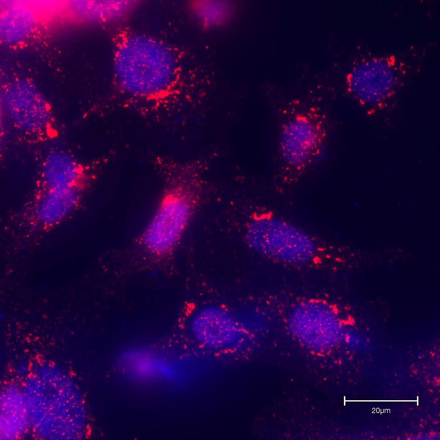
Immunocytochemistry/ Immunofluorescence: GCC185 Antibody [NBP2-04024]
Immunocytochemistry/Immunofluorescence: GCC185 Antibody [NBP2-04024] - PC-3 cells (human) stained with GCC185 antibody (red). Nucleus stained with DAPI (blue). EVOS(TM) M5000 Imaging System - 63x oil objective. ICC/IF image submitted by a verified customer review.Applications for GCC185 Antibody - BSA Free
Application
Recommended Usage
Immunoprecipitation
2 - 10 ug/mg
Western Blot
1:2000 - 1:10000
Application Notes
This GCC185 antibody is validated for ICC/IF from a verified customer review. Western blot of lysates performed using standard western blot reagents and 4-8% SDS-PAGE.
Reviewed Applications
Read 1 review rated 5 using NBP2-04024 in the following applications:
Formulation, Preparation, and Storage
Purification
Immunogen affinity purified
Formulation
Tris-Citrate/Phosphate (pH 7.0 - 8.0)
Format
BSA Free
Preservative
0.09% Sodium Azide
Concentration
1.0 mg/ml
Shipping
The product is shipped with polar packs. Upon receipt, store it immediately at the temperature recommended below.
Stability & Storage
Store at 4C. Do not freeze.
Background: GCC185
Alternate Names
185 kDa Golgi coiled-coil protein, CLL-associated antigen KW-11, CTCL tumor antigen se1-1, GCC protein, 185-kD, GCC185GRIP and coiled-coil domain-containing 2, Golgi coiled-coil protein GCC185, GRIP and coiled-coil domain containing 2, GRIP and coiled-coil domain-containing protein 2, KIAA0336REN53, Ran-binding protein 2-like 4, RANBP2L4, Renal carcinoma antigen NY-REN-53
Gene Symbol
GCC2
UniProt
Additional GCC185 Products
Product Documents for GCC185 Antibody - BSA Free
Certificate of Analysis
To download a Certificate of Analysis, please enter a lot or batch number in the search box below.
Product Specific Notices for GCC185 Antibody - BSA Free
This product is for research use only and is not approved for use in humans or in clinical diagnosis. Primary Antibodies are guaranteed for 1 year from date of receipt.
Customer Reviews for GCC185 Antibody - BSA Free (1)
5 out of 5
1 Customer Rating
Have you used GCC185 Antibody - BSA Free?
Submit a review and receive an Amazon gift card!
$25/€18/£15/$25CAN/¥2500 Yen for a review with an image
$10/€7/£6/$10CAN/¥1110 Yen for a review without an image
Submit a review
Customer Images
Showing
1
-
1 的
1 review
Showing All
Filter By:
-
Application: ImmunofluorescenceSample Tested: PC-3 human prostate cancer cell lineSpecies: HumanVerified Customer | Posted 08/28/2020EVOS™ M5000 Imaging System - 63x oil objectivePC-3 cells stained with GCC185 Ab - red, blue - nucleus, DAPI.

There are no reviews that match your criteria.
Protocols
Find general support by application which include: protocols, troubleshooting, illustrated assays, videos and webinars.
- Appropriate Fixation of IHC/ICC Samples
- Cellular Response to Hypoxia Protocols
- ClariTSA™ Fluorophore Kits
- Detection & Visualization of Antibody Binding
- ICC Cell Smear Protocol for Suspension Cells
- ICC Immunocytochemistry Protocol Videos
- ICC for Adherent Cells
- Immunocytochemistry (ICC) Protocol
- Immunocytochemistry Troubleshooting
- Immunofluorescence of Organoids Embedded in Cultrex Basement Membrane Extract
- Immunohistochemistry (IHC) and Immunocytochemistry (ICC) Protocols
- Immunoprecipitation Protocol
- Preparing Samples for IHC/ICC Experiments
- Preventing Non-Specific Staining (Non-Specific Binding)
- Primary Antibody Selection & Optimization
- Protocol for VisUCyte™ HRP Polymer Detection Reagent
- Protocol for the Fluorescent ICC Staining of Cell Smears - Graphic
- Protocol for the Fluorescent ICC Staining of Cultured Cells on Coverslips - Graphic
- Protocol for the Preparation and Fluorescent ICC Staining of Cells on Coverslips
- Protocol for the Preparation and Fluorescent ICC Staining of Non-adherent Cells
- Protocol for the Preparation and Fluorescent ICC Staining of Stem Cells on Coverslips
- Protocol for the Preparation of a Cell Smear for Non-adherent Cell ICC - Graphic
- R&D Systems Quality Control Western Blot Protocol
- TUNEL and Active Caspase-3 Detection by IHC/ICC Protocol
- The Importance of IHC/ICC Controls
- Troubleshooting Guide: Western Blot Figures
- Western Blot Conditions
- Western Blot Protocol
- Western Blot Protocol for Cell Lysates
- Western Blot Troubleshooting
- Western Blot Troubleshooting Guide
- View all Protocols, Troubleshooting, Illustrated assays and Webinars
Loading...