Loading...
Key Product Details
Applications
Flow Cytometry
Loading...
Product Specifications
Description
The GFP Compensation Beads serve as an easy-to-use single color compensation control in multicolor flow cytometry. The surface of the particle is labeled with rrGFP (Renilla reniformis Green Fluorescence Protein).
Concentration: 10^7 particles / mL
Particle size: 3.0 -3.4 micron
Particle material: polystyrene
Concentration: 10^7 particles / mL
Particle size: 3.0 -3.4 micron
Particle material: polystyrene
Application Notes
Resuspend by vortexing before use. To achieve optimum particle suspension, sonicate the reagent after vortex mixing.
Spectra Viewer
Plan Your Experiments
Use our spectra viewer to interactively plan your experiments, assessing multiplexing options. View the excitation and emission spectra for our fluorescent dye range and other commonly used dyes.
Spectra Viewer
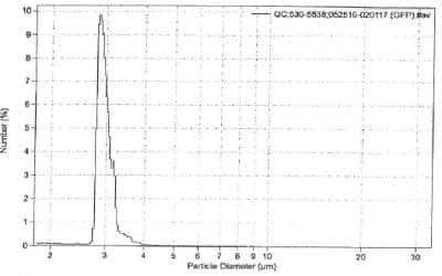
Scientific Data Images for GFP Comp-Bead Particles
GFP Comp-Bead Particles [NBP3-00503] - Coulter M3 analysis: Mean particle size 3.0 micron.
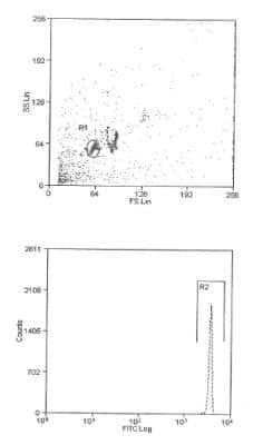
GFP Comp-Bead Particles [NBP3-00503] - Dot plot shows front scatter population. Histogram shows positive fluorescence in the FITC channel.
Formulation, Preparation, and Storage
NBP3-00503
| Formulation | 0.016 M PBS (pH 7.4), 0.2% BSA |
| Preservative | 0.02% Sodium Azide |
| Concentration | Please see the protocols for proper use of this product. If no protocol is available, contact technical services for assistance. |
| Shipping | The product is shipped with polar packs. Upon receipt, store it immediately at the temperature recommended below. |
| Stability & Storage | Store at 4C in the dark. Do not freeze. |
Background: GFP Comp-Bead Particles
Compensation beads are a highly desirable alternative to traditional cell-based single-color compensation. Positive beads can come pre-loaded with anti-IgG for conjugated antibody capture of mouse, rat, or hamster origin. Uncoated (negative) beads ensure standardization of the autofluorescence in each channel. Beads are preferable to cell-based compensation for several reasons:
1) No precious sample is wasted, and more sample can be used for experimental acquisition.
2) Since 100% of positive beads are able to capture the antibody, the exact experimental fluorochrome can be used regardless of a marker's cellular expression. Dimly expressed markers may not be bright enough for compensation using cells.
3) Reduced autofluorescence of beads allows for more precise calculation of spectral overlap.
Alternate Names
Comp Beads, Compensation Beads, Flow Cytometry Compensation, GFP Compensation Beads
Additional GFP Comp-Bead Particles Products
Product Documents for GFP Comp-Bead Particles
Certificate of Analysis
To download a Certificate of Analysis, please enter a lot or batch number in the search box below.
Product Specific Notices for GFP Comp-Bead Particles
This product is for research use only and is not approved for use in humans or in clinical diagnosis. Support products are guaranteed for 6 months from date of receipt.
Customer Reviews for GFP Comp-Bead Particles
There are currently no reviews for this product. Be the first to review GFP Comp-Bead Particles and earn rewards!
Have you used GFP Comp-Bead Particles?
Submit a review and receive an Amazon gift card!
$25/€18/£15/$25CAN/¥2500 Yen for a review with an image
$10/€7/£6/$10CAN/¥1110 Yen for a review without an image
Submit a review
Protocols
Find general support by application which include: protocols, troubleshooting, illustrated assays, videos and webinars.
- 7-Amino Actinomycin D (7-AAD) Cell Viability Flow Cytometry Protocol
- Extracellular Membrane Flow Cytometry Protocol
- Flow Cytometry Protocol for Cell Surface Markers
- Flow Cytometry Protocol for Staining Membrane Associated Proteins
- Flow Cytometry Staining Protocols
- Flow Cytometry Troubleshooting Guide
- Intracellular Flow Cytometry Protocol Using Alcohol (Methanol)
- Intracellular Flow Cytometry Protocol Using Detergents
- Intracellular Nuclear Staining Flow Cytometry Protocol Using Detergents
- Intracellular Staining Flow Cytometry Protocol Using Alcohol Permeabilization
- Intracellular Staining Flow Cytometry Protocol Using Detergents to Permeabilize Cells
- Propidium Iodide Cell Viability Flow Cytometry Protocol
- Protocol for Liperfluo
- Protocol for the Characterization of Human Th22 Cells
- Protocol for the Characterization of Human Th9 Cells
- Protocol: Annexin V and PI Staining by Flow Cytometry
- Protocol: Annexin V and PI Staining for Apoptosis by Flow Cytometry
- Troubleshooting Guide: Fluorokine Flow Cytometry Kits
- View all Protocols, Troubleshooting, Illustrated assays and Webinars
Loading...