

Scientific images may be resized, cropped, or adjusted for brightness or contrast for presentation purposes. These updates are limited to presentation and do not affect the underlying scientific interpretation of the data. Where available, additional source data may be provided upon request.
Key Product Details
Validated by
Biological Validation
Species Reactivity
Validated:
Human
Cited:
Human
Applications
Validated:
Immunoprecipitation
Cited:
Flow Cytometry
Label
Unconjugated
Antibody Source
Monoclonal Mouse IgG1 Clone # 632525
Loading...
Product Specifications
Immunogen
Chinese hamster ovary cell line CHO-derived recombinant human ADAM12
Arg29-Ser513
Accession # O43184
Arg29-Ser513
Accession # O43184
Specificity
Detects human ADAM12 in direct ELISAs.
In direct ELISAs, no cross-reactivity
with recombinant human ADAM8, 9, 10, 15, 17, 19, 22, 23, 32, or 33 is observed.
Clonality
Monoclonal
Host
Mouse
Isotype
IgG1
Scientific Data Images for Human ADAM12 Antibody (632525)
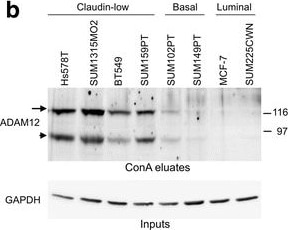
Detection of Human ADAM12 by Western Blot
ADAM12 expression is upregulated in claudin-low breast cancer cells and in subpopulations enriched for CSCs. a. Cell surface expression of ADAM12 protein in breast cancer cell lines was evaluated by flow cytometry. Red, anti-ADAM12 antibody staining; gray, isotype control antibody staining. b Total cellular expression of ADAM12 was analyzed by Western blotting after partial purification of ADAM12 on concanavalin A (conA) agarose, as described [42]. GAPDH in the input fractions shows comparable conA agarose loading for all cells. Arrow, the nascent full length ADAM12; arrowhead, the processed form lacking the N-terminal pro-domain. c CSC signature score versus ADAM12 mRNA expression in 51 breast cancer cell lines. The CSC signature scores were calculated based on ref. [22] and microarray expression data retrieved from GEO:GSE69017, as described in Methods. Cell lines analyzed in panels a and b are shown in red. d ADAM12 protein levels in total lysates of SUM159PT cells grown as attached monolayers or as mammospheres, analyzed by Western blotting. Arrow, the nascent full length ADAM12; arrowhead, the processed form lacking the N-terminal pro-domain; *, a non-specific band. Positive control represents ADAM12 after partial purification on conA agarose. e Cell surface expression of ADAM12 in SUM159PT cells treated for 6 days with DMSO (control) or with 10 nM paclitaxel (PTX), and then allowed to recover for 6 days without PTX, was examined by flow cytometry. The population of cells with the highest expression of ADAM12 is shown in red. FSC, forward scatter Image collected and cropped by CiteAb from the following publication (https://pubmed.ncbi.nlm.nih.gov/28148288), licensed under a CC-BY license. Not internally tested by R&D Systems.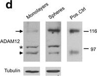
Detection of Human ADAM12 by Western Blot
ADAM12 expression is upregulated in claudin-low breast cancer cells and in subpopulations enriched for CSCs. a. Cell surface expression of ADAM12 protein in breast cancer cell lines was evaluated by flow cytometry. Red, anti-ADAM12 antibody staining; gray, isotype control antibody staining. b Total cellular expression of ADAM12 was analyzed by Western blotting after partial purification of ADAM12 on concanavalin A (conA) agarose, as described [42]. GAPDH in the input fractions shows comparable conA agarose loading for all cells. Arrow, the nascent full length ADAM12; arrowhead, the processed form lacking the N-terminal pro-domain. c CSC signature score versus ADAM12 mRNA expression in 51 breast cancer cell lines. The CSC signature scores were calculated based on ref. [22] and microarray expression data retrieved from GEO:GSE69017, as described in Methods. Cell lines analyzed in panels a and b are shown in red. d ADAM12 protein levels in total lysates of SUM159PT cells grown as attached monolayers or as mammospheres, analyzed by Western blotting. Arrow, the nascent full length ADAM12; arrowhead, the processed form lacking the N-terminal pro-domain; *, a non-specific band. Positive control represents ADAM12 after partial purification on conA agarose. e Cell surface expression of ADAM12 in SUM159PT cells treated for 6 days with DMSO (control) or with 10 nM paclitaxel (PTX), and then allowed to recover for 6 days without PTX, was examined by flow cytometry. The population of cells with the highest expression of ADAM12 is shown in red. FSC, forward scatter Image collected and cropped by CiteAb from the following publication (https://pubmed.ncbi.nlm.nih.gov/28148288), licensed under a CC-BY license. Not internally tested by R&D Systems.Applications for Human ADAM12 Antibody (632525)
Application
Recommended Usage
Immunoprecipitation
25 µg/mL
Sample: Conditioned cell culture medium spiked with Recombinant Human ADAM12 (Catalog # 4416-AD), see our available Western blot detection antibodies
Sample: Conditioned cell culture medium spiked with Recombinant Human ADAM12 (Catalog # 4416-AD), see our available Western blot detection antibodies
Reviewed Applications
Read 1 review rated 5 using MAB44161 in the following applications:
Formulation, Preparation, and Storage
Purification
Protein A or G purified from hybridoma culture supernatant
Reconstitution
Sterile PBS to a final concentration of 0.5 mg/mL. For liquid material, refer to CoA for concentration.
Loading...
Formulation
Lyophilized from a 0.2 μm filtered solution in PBS with Trehalose. *Small pack size (SP) is supplied either lyophilized or as a 0.2 µm filtered solution in PBS.
Shipping
Lyophilized product is shipped at ambient temperature. Liquid small pack size (-SP) is shipped with polar packs. Upon receipt, store immediately at the temperature recommended below.
Stability & Storage
Use a manual defrost freezer and avoid repeated freeze-thaw cycles.
- 12 months from date of receipt, -20 to -70 °C as supplied.
- 1 month, 2 to 8 °C under sterile conditions after reconstitution.
- 6 months, -20 to -70 °C under sterile conditions after reconstitution.
Calculators
Background: ADAM12
Long Name
A Disintegrin and Metalloprotease-like Domain 12
Alternate Names
Meltrin alpha
Entrez Gene IDs
8038 (Human)
Gene Symbol
ADAM12
UniProt
Additional ADAM12 Products
Product Documents for Human ADAM12 Antibody (632525)
Certificate of Analysis
To download a Certificate of Analysis, please enter a lot or batch number in the search box below.
Note: Certificate of Analysis not available for kit components.
Product Specific Notices for Human ADAM12 Antibody (632525)
For research use only
Related Research Areas
Citations for Human ADAM12 Antibody (632525)
 Powered by Bioz
Customer Reviews for Human ADAM12 Antibody (632525) (1)
5 out of 5
1 Customer Rating
Have you used Human ADAM12 Antibody (632525)?
Submit a review and receive an Amazon gift card!
$25/€18/£15/$25CAN/¥2500 for a review with an image
$10/€7/£6/$10CAN/¥1110 for a review without an image
Submit a review
Customer Images
Showing
1
-
1 的
1 review
Showing All
Filter By:
-
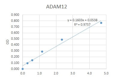
Application: ELISASample Tested: Serum and PlasmaSpecies: HumanVerified Customer | Posted 07/06/2021we used this antibody for an ELISA to quantify human ADAM12 in serum, which works very well.

There are no reviews that match your criteria.
Protocols
Find general support by application which include: protocols, troubleshooting, illustrated assays, videos and webinars.
Loading...