
Fc Receptor-Like 5 (FCRL5), also known as FcRH5, IRTA2, and CD307, is a 120 kDa protein with sequence homology to classical Fc receptors. The type 1 transmembrane FCRL proteins contain from three to nine immunoglobulin-like domains. They are differentially expressed within the B cell lineage and can either promote or inhibit B cell proliferation and activation (1, 2). According to R&D Systems testing, FCRL5 binds to purified human IgG with high affinity. Mature human FCRL5 consists of a 836 amino acid (aa) extracellular domain (ECD) with nine Ig-like domains, a 21 aa transmembrane segment, and a 105 aa cytoplasmic domain with one immunotyrosine activation motif (ITAM) and two immunotyrosine inhibitory motifs (ITIMs) (1, 3). Mouse FCRL5 contains only five Ig-like domains in its ECD. It shares 49% aa sequence identity with human FCRL5 within common regions. Alternate splicing of human FCRL5 generates isoforms that consist of approximately the first one, six, or eight Ig-like domains (3, 4). FCRL5 expression is restricted to mature B lineage cells in lymphoid tissues and blood (3, 5‑7). Its ligation inhibits signaling through the B cell antigen receptor (8). Epstein-Barr virus transformation of B cells induces the up‑regulation of surface FCRL5 by a direct effect of its EBNA2 protein on FCRL5 gene transcription (9). The FCRL5 gene maps to the 1q21 chromosomal locus, a common site of rearrangements in B cell malignancies, and the FCRL5 protein is preferentially expressed in cell lines with 1q21 abnormalities (3). FCRL5 is up‑regulated on tumor cells in some types of B cell malignancies (6, 10‑12). In addition, soluble FCRL5 is elevated in the serum of many B cell leukemia patients (11, 13).

Scientific images may be resized, cropped, or adjusted for brightness or contrast for presentation purposes. These updates are limited to presentation and do not affect the underlying scientific interpretation of the data. Where available, additional source data may be provided upon request.
Key Product Details
Species Reactivity
Applications
Label
Antibody Source
Product Specifications
Immunogen
Gln16-Arg844
Accession # AAK93971
Specificity
Clonality
Host
Isotype
Scientific Data Images for Human FCRL5 Antibody
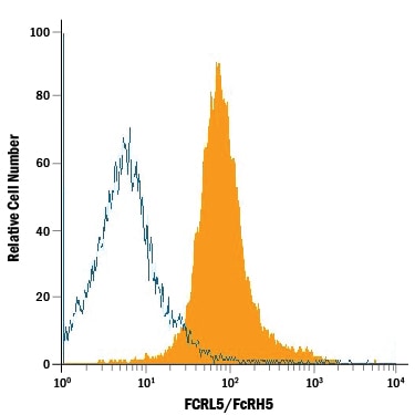
Detection of FCRL5 in CD19+Human PBMC Lymphocytes by Flow Cytometry.
CD19+human peripheral blood mononuclear cell (PBMC) lymphocytes were stained with Goat Anti-Human FCRL5 Antigen Affinity-purified Polyclonal Antibody (Catalog # AF2087, filled histogram) or isotype control antibody (Catalog # AB-108-C, open histogram), followed by Phycoerythrin-conjugated Anti-Goat IgG Secondary Antibody (Catalog # F0107).Applications for Human FCRL5 Antibody
CyTOF-ready
Flow Cytometry
Sample: CD19+ human peripheral blood mononuclear cell (PBMC) lymphocytes
Western Blot
Sample: Recombinant Human FCRL5/FcRH5 (Catalog # 2078-FC)
Reviewed Applications
Read 1 review rated 4 using AF2087 in the following applications:
Flow Cytometry Panel Builder
Bio-Techne Knows Flow Cytometry
Save time and reduce costly mistakes by quickly finding compatible reagents using the Panel Builder Tool.
Advanced Features
- Spectra Viewer - Custom analysis of spectra from multiple fluorochromes
- Spillover Popups - Visualize the spectra of individual fluorochromes
- Antigen Density Selector - Match fluorochrome brightness with antigen density

Formulation, Preparation, and Storage
Purification
Reconstitution
Reconstitute at 0.2 mg/mL in sterile PBS. For liquid material, refer to CoA for concentration.
Formulation
Shipping
Stability & Storage
- 12 months from date of receipt, -20 to -70 °C as supplied.
- 1 month, 2 to 8 °C under sterile conditions after reconstitution.
- 6 months, -20 to -70 °C under sterile conditions after reconstitution.
Calculators
Background: FCRL5
References
- Davis, R.S. (2007) Annu. Rev. Immunol. 25:525.
- Maltais, L.J. et al. (2006) Nat. Immunol. 7:431.
- Hatzivassiliou, G. et al. (2001) Immunity 14:277.
- SwissProt # Q96RD9.
- Miller, I. et al. (2002) Blood 99:2662.
- Polson, A.G. et al. (2006) Int. Immunol. 18:1363.
- Vidal-Laliena, M. et al. (2005) Cell. Immunol. 236:6.
- Haga, C.L. et al. (2007) Proc. Natl. Acad. Sci. 104:9770.
- Mohan, J. et al. (2006) Blood 107:4433.
- Ise, T. et al. (2005) Clin. Cancer Res. 11:87.
- Ise, T. et al. (2007) Leukemia 21:169.
- Kazemi, T. et al. (2009) Cancer Immunol. Immunother. 58:989.
- Ise, T. et al. (2006) Clin. Chem. Lab. Med. 44:594.
Long Name
Alternate Names
Gene Symbol
UniProt
Additional FCRL5 Products
Product Documents for Human FCRL5 Antibody
Certificate of Analysis
To download a Certificate of Analysis, please enter a lot or batch number in the search box below.
Note: Certificate of Analysis not available for kit components.
Product Specific Notices for Human FCRL5 Antibody
For research use only
Related Research Areas
Customer Reviews for Human FCRL5 Antibody (1)
Have you used Human FCRL5 Antibody?
Submit a review and receive an Amazon gift card!
$25/€18/£15/$25CAN/¥2500 for a review with an image
$10/€7/£6/$10CAN/¥1110 for a review without an image
Submit a review
Customer Images
-
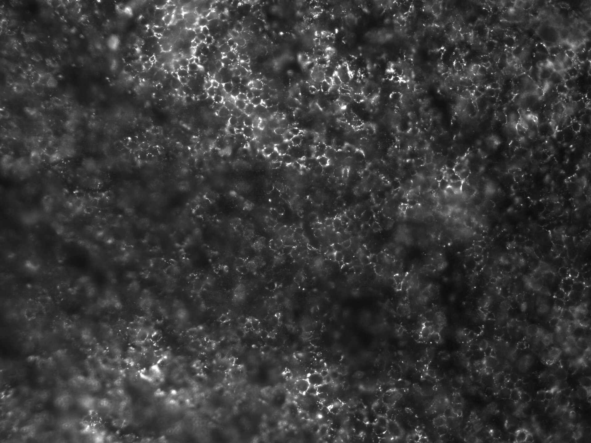
Application: Immunofluorescence - fresh frozenSample Tested: human tonsilSpecies: HumanVerified Customer | Posted 11/18/2018FcRH5-Alexa647 on fresh-frozen human tonsil

There are no reviews that match your criteria.
Protocols
Find general support by application which include: protocols, troubleshooting, illustrated assays, videos and webinars.
- 7-Amino Actinomycin D (7-AAD) Cell Viability Flow Cytometry Protocol
- Cellular Response to Hypoxia Protocols
- Extracellular Membrane Flow Cytometry Protocol
- Flow Cytometry Protocol for Cell Surface Markers
- Flow Cytometry Protocol for Staining Membrane Associated Proteins
- Flow Cytometry Staining Protocols
- Flow Cytometry Troubleshooting Guide
- Intracellular Flow Cytometry Protocol Using Alcohol (Methanol)
- Intracellular Flow Cytometry Protocol Using Detergents
- Intracellular Nuclear Staining Flow Cytometry Protocol Using Detergents
- Intracellular Staining Flow Cytometry Protocol Using Alcohol Permeabilization
- Intracellular Staining Flow Cytometry Protocol Using Detergents to Permeabilize Cells
- Propidium Iodide Cell Viability Flow Cytometry Protocol
- Protocol for Liperfluo
- Protocol for the Characterization of Human Th22 Cells
- Protocol for the Characterization of Human Th9 Cells
- Protocol: Annexin V and PI Staining by Flow Cytometry
- Protocol: Annexin V and PI Staining for Apoptosis by Flow Cytometry
- R&D Systems Quality Control Western Blot Protocol
- Troubleshooting Guide: Fluorokine Flow Cytometry Kits
- Troubleshooting Guide: Western Blot Figures
- Western Blot Conditions
- Western Blot Protocol
- Western Blot Protocol for Cell Lysates
- Western Blot Troubleshooting
- Western Blot Troubleshooting Guide
- View all Protocols, Troubleshooting, Illustrated assays and Webinars