
TGF-beta 1 (transforming growth factor beta 1) and the closely related TGF-beta 2 and -beta 3 are members of the large TGF-beta superfamily. TGF‑ beta proteins are highly pleiotropic cytokines that regulate processes such as immune function, proliferation and epithelial-mesenchymal transition (1‑3). Human TGF-beta 1 cDNA encodes a 390 amino acid (aa) precursor that contains a 29 aa signal peptide and a 361 aa proprotein (4). A furin-like convertase processes the proprotein within the trans-Golgi to generate an N‑terminal 249 aa (aa 30-278) latency-associated peptide (LAP) and a C-terminal 112 aa mature TGF-beta 1 (aa 279-390) (4‑6). Disulfide-linked homodimers of LAP and TGF-beta 1 remain non‑covalently associated after secretion, forming the small latent TGF-beta 1 complex (4‑8). Purified LAP is also capable of associating with active TGF‑ beta with high affinity, and can neutralize TGF-beta activity (9). Covalent linkage of LAP to one of three latent TGF-beta binding proteins (LTBPs) creates a large latent complex that may interact with the extracellular matrix (5-7). TGF-beta activation from latency is controlled both spatially and temporally, by multiple pathways that include actions of proteases such as plasmin and MMP9, and/or by thrombospondin 1 or selected integrins (5, 8). The LAP portion of human TGF-beta 1 shares 91%, 92%, 85%, 86% and 88% aa identity with porcine, canine, mouse, rat and equine TGF-beta 1 LAP, respectively, while mature human TGF-beta 1 portion shares 100% aa identity with porcine, canine and bovine TGF-beta 1, and 99% aa identity with mouse, rat and equine TGF-beta 1. Although different isoforms of TGF-beta are naturally associated with their own distinct LAPs, the TGF-beta 1 LAP is capable of complexing with, and inactivating, all other human TGF-beta isoforms and those of most other species (9). Mutations within the LAP are associated with Camurati-Engelmann disease, a rare sclerosing bone dysplasia characterized by inappropriate presence of active TGF‑ beta 1 (10).
Human LAP (TGF-beta 1) Antibody
R&D Systems | Catalog # AF-246-NA


Scientific images may be resized, cropped, or adjusted for brightness or contrast for presentation purposes. These updates are limited to presentation and do not affect the underlying scientific interpretation of the data. Where available, additional source data may be provided upon request.
Key Product Details
Species Reactivity
Validated:
Cited:
Applications
Validated:
Cited:
Label
Antibody Source
Product Specifications
Immunogen
Leu30-Ser390
Accession # P01137
Specificity
Clonality
Host
Isotype
Endotoxin Level
Scientific Data Images for Human LAP (TGF-beta 1) Antibody
Detection of Human LAP (TGF-beta 1) by Western Blot.
Western blot shows lysates of Saos-2 human osteosarcoma cell line. PVDF membrane was probed with 2 µg/mL of Goat Anti-Human LAP (TGF-beta 1) Antigen Affinity-purified Polyclonal Antibody (Catalog # AF-246-NA) followed by HRP-conjugated Anti-Goat IgG Secondary Antibody (Catalog # HAF017). A specific band was detected for LAP (TGF-beta 1) at approximately 48 kDa (as indicated). This experiment was conducted under reducing conditions and using Immunoblot Buffer Group 1.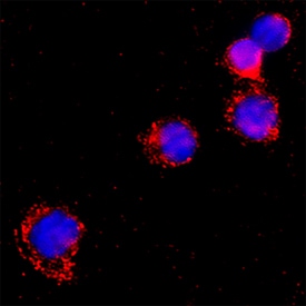
LAP (TGF-beta 1) in Human PBMCs.
LAP (TGF-beta 1) was detected in immersion fixed Human peripheral blood mononuclear cells using Goat Anti-Human LAP (TGF-beta 1) Antigen Affinity-purified Polyclonal Antibody (Catalog # AF-246-NA) at 15 µg/mL for 3 hours at room temperature. Cells were stained using the NorthernLights™ 557-conjugated Anti-Goat IgG Secondary Antibody (red; NL001) and counterstained with DAPI (blue). Specific staining was localized to cytoplasm. Staining was performed using our protocol for Fluorescent ICC Staining of Non-adherent Cells.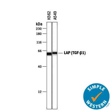
Detection of Human LAP (TGF-beta 1) by Simple WesternTM.
Simple Western lane view shows lysates of K562 human chronic myelogenous leukemia cell line and A549 human lung carcinoma cell line, loaded at 0.2 mg/mL. A specific band was detected for LAP (TGF-beta 1) at approximately 61 kDa (as indicated) using 20 µg/mL of Goat Anti-Human LAP (TGF-beta 1) Antigen Affinity-purified Polyclonal Antibody (Catalog # AF-246-NA) followed by 1:50 dilution of HRP-conjugated Anti-Goat IgG Secondary Antibody (Catalog # HAF109). This experiment was conducted under reducing conditions and using the 12-230 kDa separation system.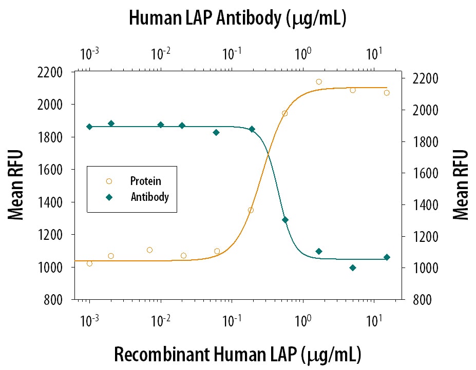
LAP TGF‑ beta 1 Inhibition of TGF‑ beta 1 Activity and Neutralization by Human LAP TGF‑ beta 1 Antibody.
Recombinant Human LAP TGF-beta 1 (Catalog # 246-LP) inhibits Recom-binant Human TGF-beta 1 (Catalog # 240-B) growth inhibition activity in the HT-2 mouse T cell line in a dose-dependent manner (orange line). Inhibition of Recom-binant Human TGF-beta 1 (1 ng/mL) activity elicited by Recombin-ant Human LAP TGF-beta 1 (500 ng/mL) is neutralized (green line) by increasing concentrations of Goat Anti-Human LAP TGF-beta 1 Antigen Affinity-purified Polyclonal Antibody (Catalog # AF-246-NA). The ND50 is typically 0.4-2 µg/mL.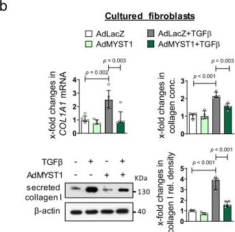
Detection of LAP (TGF-beta 1) by Western Blot
Re-expression of MYST1 inhibits TGF beta -dependent fibroblast activation and prevents experimental fibrosis.a, b Overexpression of MYST1 in cultured human dermal fibroblasts: a Expression of alpha SMA (n = 4 biological replicates per group) and formation of stress fibers (n = 5 biological replicates per group). Representative images and quantification. Horizontal scale bar, 100 µm. b mRNA levels of COL1A1 analyzed by qRT-PCR (n = 5 biological replicates per group), total collagen as analyzed by hydroxyproline assays (n = 4 biological replicates per group) and type I collagen protein as analyzed by western blot (n = 5 biological replicates per group). c–e Adenoviral overexpression of MYST1 in murine models. c T beta RIact-induced skin fibrosis: Representative trichrome stainings for, dermal thickening, myofibroblast counts and hydroxyproline content (n = 4 biological replicates per group). d Bleomycin-induced pulmonary fibrosis: Representative Trichrome stainings, fibrotic area, myofibroblast counts, Ashcroft scores and hydroxyproline content (n = 4 biological replicates for the group AdMyst1 treated with TGF beta and n = 3 for other groups). e Bleomycin-induced dermal fibrosis: Representative trichrome stainings shown, dermal thickening, myofibroblast counts and hydroxyproline content (n = 4 biological replicates for bleomycin treated groups and n = 3 for saline treated groups). Horizontal scale bars, 100 µm. All data are presented as median ± IQR. p-values were determined by ANOVA one-way with Tukey’s multiple comparison post hoc test and are indicated in the figure. See source data for more detailed information. rel.: relative, Ad: adenovirus, conc.: concentration, int.: intensity, T beta RIact: constitutively active TGF beta receptor type I, Bleo: bleomycin. Image collected and cropped by CiteAb from the following open publication (https://pubmed.ncbi.nlm.nih.gov/34285225), licensed under a CC-BY license. Not internally tested by R&D Systems.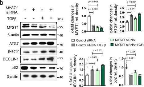
Detection of LAP (TGF-beta 1) by Western Blot
Downregulation of MYST1 mediates the stimulatory effects of TGF beta on autophagy.a, b Effects of siRNA-mediated knockdown of MYST1 on autophagy in healthy human dermal fibroblasts as assessed by quantification of the mRNA levels of ATG7 (n = 4 biological replicates per group and n = 3 for MYST1 siRNA + TGF beta ), BECLIN1 (n = 4 biological replicates per group) and respective protein quantifications (n = 4 biological replicates per group). Confirmation of reduced mRNA (n = 4 biological replicates per group) (a) and protein (b; n = 4 biological replicates per group) levels of MYST1 by qRT-PCR and western blot. c EGFP/RFP-LC3 reporter activity upon knockdown of MYST1; representative images and quantification (n = 5 biological replicates per group). Horizontal scale bar, 5 µm. d, e mRNA expression of ATG7, BECLIN1, and MYST1 (n = 6 biological replicates per group) (d) and respective protein quantification (e) (n = 4 biological replicates per group) levels after adenoviral overexpression of MYST1 in healthy human dermal fibroblasts (e). All data are presented as mean ± IQR. p-values were determined by ANOVA one-way with Tukey’s multiple comparison post hoc test. See source data for more detailed information. rel.: relative, Ad: adenovirus. The results in panel c are part of a large set of experiments and share controls with Supplementary Fig. 8a. Data are presented separately just for the purpose of this figure. Image collected and cropped by CiteAb from the following open publication (https://pubmed.ncbi.nlm.nih.gov/34285225), licensed under a CC-BY license. Not internally tested by R&D Systems.Applications for Human LAP (TGF-beta 1) Antibody
Immunocytochemistry
Sample: Immersion fixed Human peripheral blood mononuclear cells
Simple Western
Sample: K562 human chronic myelogenous leukemia cell line and A549 human lung carcinoma cell line
Western Blot
Sample: Saos‑2 human osteosarcoma cell line
Neutralization
Reviewed Applications
Read 4 reviews rated 4 using AF-246-NA in the following applications:
Formulation, Preparation, and Storage
Purification
Reconstitution
Reconstitute at 0.2 mg/mL in sterile PBS. For liquid material, refer to CoA for concentration.
Formulation
Shipping
Stability & Storage
- 12 months from date of receipt, -20 to -70 °C as supplied.
- 1 month, 2 to 8 °C under sterile conditions after reconstitution.
- 6 months, -20 to -70 °C under sterile conditions after reconstitution.
Calculators
Background: LAP (TGF-beta 1)
References
- Dunker, N. & K. Krieglstein (2000) Eur. J. Biochem. 267:6982.
- Wahl, S.M. (2006) Immunol. Rev. 213:213.
- Chang, H. et al. (2002) Endocr. Rev. 23:787.
- Derynck, R. et al. (1985) Nature 316:701.
- Dabovic, B. and D.B. Rifkin (2008) “TGF-beta Bioavailability” in The TGF-beta Family. Derynck, R. and K. Miyazono (eds): Cold Spring Harbor Laboratory Press, p. 179.
- Brunner, A.M. et al. (1989) J. Biol. Chem. 264:13660.
- Miyazono, K. et al. (1991) EMBO J. 10:1091.
- Oklu, R. and R. Hesketh (2000) Biochem. J. 352:601.
- Miller, D.M. et al. (1992) Mol. Endocrinol. 6:694.
- Janssens, K. et al. (2003) J. Biol. Chem. 278:7718.
Long Name
Alternate Names
Entrez Gene IDs
Gene Symbol
UniProt
Additional LAP (TGF-beta 1) Products
Product Documents for Human LAP (TGF-beta 1) Antibody
Certificate of Analysis
To download a Certificate of Analysis, please enter a lot or batch number in the search box below.
Note: Certificate of Analysis not available for kit components.
Product Specific Notices for Human LAP (TGF-beta 1) Antibody
For research use only
Citations for Human LAP (TGF-beta 1) Antibody
 Powered by Bioz
Customer Reviews for Human LAP (TGF-beta 1) Antibody (4)
Have you used Human LAP (TGF-beta 1) Antibody?
Submit a review and receive an Amazon gift card!
$25/€18/£15/$25CAN/¥2500 for a review with an image
$10/€7/£6/$10CAN/¥1110 for a review without an image
Submit a review
Customer Images
-
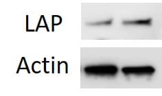
Application: Western BlotSample Tested: Pancreatic cancer cellsSpecies: HumanVerified Customer | Posted 01/27/2021

-
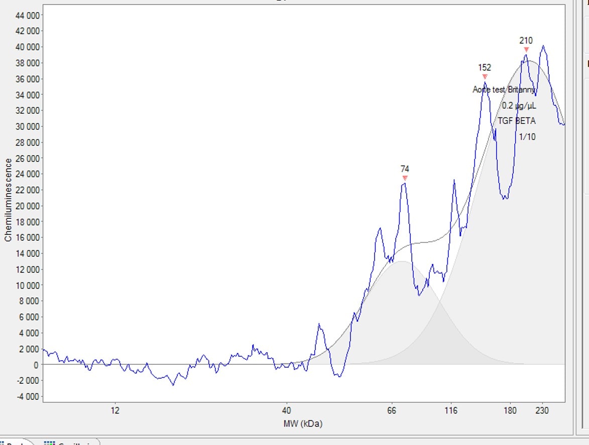
Application: Western BlotSample Tested: Aorta tissueSpecies: RatVerified Customer | Posted 02/03/2020
-
Application: Western BlotSample Tested: See PMID 21998117Species: OtherVerified Customer | Posted 01/07/2015
-
Application: Western BlotSample Tested: See PMID 22835852Species: OtherVerified Customer | Posted 01/07/2015
There are no reviews that match your criteria.
Protocols
Find general support by application which include: protocols, troubleshooting, illustrated assays, videos and webinars.
- Appropriate Fixation of IHC/ICC Samples
- Cellular Response to Hypoxia Protocols
- ClariTSA™ Fluorophore Kits
- Detection & Visualization of Antibody Binding
- ICC Cell Smear Protocol for Suspension Cells
- ICC Immunocytochemistry Protocol Videos
- ICC for Adherent Cells
- Immunocytochemistry (ICC) Protocol
- Immunocytochemistry Troubleshooting
- Immunofluorescence of Organoids Embedded in Cultrex Basement Membrane Extract
- Immunohistochemistry (IHC) and Immunocytochemistry (ICC) Protocols
- Preparing Samples for IHC/ICC Experiments
- Preventing Non-Specific Staining (Non-Specific Binding)
- Primary Antibody Selection & Optimization
- Protocol for VisUCyte™ HRP Polymer Detection Reagent
- Protocol for the Fluorescent ICC Staining of Cell Smears - Graphic
- Protocol for the Fluorescent ICC Staining of Cultured Cells on Coverslips - Graphic
- Protocol for the Preparation and Fluorescent ICC Staining of Cells on Coverslips
- Protocol for the Preparation and Fluorescent ICC Staining of Non-adherent Cells
- Protocol for the Preparation and Fluorescent ICC Staining of Stem Cells on Coverslips
- Protocol for the Preparation of a Cell Smear for Non-adherent Cell ICC - Graphic
- R&D Systems Quality Control Western Blot Protocol
- TUNEL and Active Caspase-3 Detection by IHC/ICC Protocol
- The Importance of IHC/ICC Controls
- Troubleshooting Guide: Western Blot Figures
- Western Blot Conditions
- Western Blot Protocol
- Western Blot Protocol for Cell Lysates
- Western Blot Troubleshooting
- Western Blot Troubleshooting Guide
- View all Protocols, Troubleshooting, Illustrated assays and Webinars