Cell Division Cycle 25B (Cdc25B) phosphatase removes inorganic phosphate groups covalently attached to tyrosine, serine and threonine residues in proteins (1). Breast cancer patients bearing tumors containing high levels of Cdc25B have been found to have a greater incidence of aggressive, high-grade tumors than those with low Cdc25B levels (2). In cells, the levels of Cdc25B activity are highest during the G2/M transition of the cell cycle, where it is suspected to be involved in “checkpoint” control of cell cycle progression (3). Overexpression of Cdc25B reduces the G2/M cell cycle block caused by ionizing radiation (4). Although activated by phosphorylation, Ser323 phosphorylation causes the enzyme to bind the protein 14-3-3, preventing substrate access to the catalytic site (5). One of the major substrates of Cdc25B is Cdc2, a kinase that is activated by dephosphorylation (6). The recombinant protein is truncated to remove the N‑terminal regulatory domains and is fully active.

Key Product Details
Species Reactivity
Validated:
Human, Mouse, Rat
Cited:
Human
Applications
Validated:
Western Blot, Immunocytochemistry
Cited:
Western Blot
Label
Unconjugated
Antibody Source
Polyclonal Goat IgG
Loading...
Product Specifications
Immunogen
E. coli-derived recombinant human CDC25B
Glu391-Gln580
Accession # P30305
Glu391-Gln580
Accession # P30305
Specificity
Detects human, mouse, and rat CDC25B. Four splice variants of CDC25B, with molecular weights of 61, 63, 65, and 67 kDa, are known. The immunogen selected for this antibody is common to all variants. Immunoreactivity consistent with at least 3 variants has been detected in Western blots. In Western blots, this antibody does not cross-react with recombinant human (rh) CDC25A or rhCDC25C.
Clonality
Polyclonal
Host
Goat
Isotype
IgG
Scientific Data Images for CDC25B Antibody
Detection of Human/Mouse/Rat CDC25B by Western Blot.
Western blot shows lysates of HeLa human cervical epithelial carcinoma cell line, DA3 mouse myeloma cell line, and Nb2-11 rat lymphoma cell line. PVDF membrane was probed with 0.3 µg/mL of Goat Anti-Human/Mouse/Rat CDC25B Antigen Affinity-purified Polyclonal Antibody (Catalog # AF1649) followed by HRP-conjugated Anti-Goat IgG Secondary Antibody (Catalog # HAF017). A specific band was detected for CDC25B at approximately 61 - 67 kDa (as indicated). This experiment was conducted under reducing conditions and using Immunoblot Buffer Group 1.
CDC25B in HL‑60 Human Cell Line.
CDC25B was detected in immersion fixed HL-60 human acute promyelocytic leukemia cell line using Goat Anti-Human/Mouse/Rat CDC25B Antigen Affinity-purified Polyclonal Antibody (Catalog # AF1649) at 15 µg/mL for 3 hours at room temperature. Cells were stained using the NorthernLights™ 557-conjugated Anti-Goat IgG Secondary Antibody (red; Catalog # NL001) and counterstained with DAPI (blue). Specific staining was localized to cytoplasm. View our protocol for Fluorescent ICC Staining of Non-adherent Cells.Applications for CDC25B Antibody
Application
Recommended Usage
Immunocytochemistry
10-25 µg/mL
Sample: Immersion fixed human peripheral blood mononuclear cells and HL‑60 human acute promyelocytic leukemia cell line
Sample: Immersion fixed human peripheral blood mononuclear cells and HL‑60 human acute promyelocytic leukemia cell line
Western Blot
0.3 µg/mL
Sample: HeLa human cervical epithelial carcinoma cell line, DA3 mouse myeloma cell line, and Nb2-11 rat lymphoma cell line
Sample: HeLa human cervical epithelial carcinoma cell line, DA3 mouse myeloma cell line, and Nb2-11 rat lymphoma cell line
Reviewed Applications
Read 1 review rated 5 using AF1649 in the following applications:
Formulation, Preparation, and Storage
Purification
Antigen Affinity-purified
Reconstitution
Reconstitute at 0.2 mg/mL in sterile PBS. For liquid material, refer to CoA for concentration.
Loading...
Formulation
Lyophilized from a 0.2 μm filtered solution in PBS with Trehalose. *Small pack size (SP) is supplied either lyophilized or as a 0.2 µm filtered solution in PBS.
Shipping
Lyophilized product is shipped at ambient temperature. Liquid small pack size (-SP) is shipped with polar packs. Upon receipt, store immediately at the temperature recommended below.
Stability & Storage
Use a manual defrost freezer and avoid repeated freeze-thaw cycles.
- 12 months from date of receipt, -20 to -70 °C as supplied.
- 1 month, 2 to 8 °C under sterile conditions after reconstitution.
- 6 months, -20 to -70 °C under sterile conditions after reconstitution.
Calculators
Background: CDC25B
References
- Draetta, G. and J. Eckstein (1997) Biochim. Biophys. Acta 1332:M53.
- Galaktionov, K. et al. (1995) Science 269:1575.
- Lammer, C. et al. (1998) J. Cell Sci. 111:2445.
- Miyata, H. et al. (2001) Cancer Res. 61:3188.
- Forrest, A. and B. Gabrielli (2001) Oncogene 20:4393.
- Gautier, J. et al. (1991) Cell 67:197.
Long Name
Cell Division Cycle 25B
Alternate Names
CDC25HU2, cell division cycle 25 homolog B (S. cerevisiae), cell division cycle 25 homolog B (S. pombe), cell division cycle 25B, Dual specificity phosphatase Cdc25B, EC 3.1.3.48, M-phase inducer phosphatase 2
Entrez Gene IDs
994 (Human)
Gene Symbol
CDC25B
UniProt
Additional CDC25B Products
Product Documents for CDC25B Antibody
Certificate of Analysis
To download a Certificate of Analysis, please enter a lot or batch number in the search box below.
Note: Certificate of Analysis not available for kit components.
Product Specific Notices for CDC25B Antibody
For research use only
Related Research Areas
Citations for CDC25B Antibody
 Powered by Bioz
Customer Reviews for CDC25B Antibody (1)
5 out of 5
1 Customer Rating
Have you used CDC25B Antibody?
Submit a review and receive an Amazon gift card!
$25/€18/£15/$25CAN/¥2500 Yen for a review with an image
$10/€7/£6/$10CAN/¥1110 Yen for a review without an image
Submit a review
Customer Images
Showing
1
-
1 的
1 review
Showing All
Filter By:
-
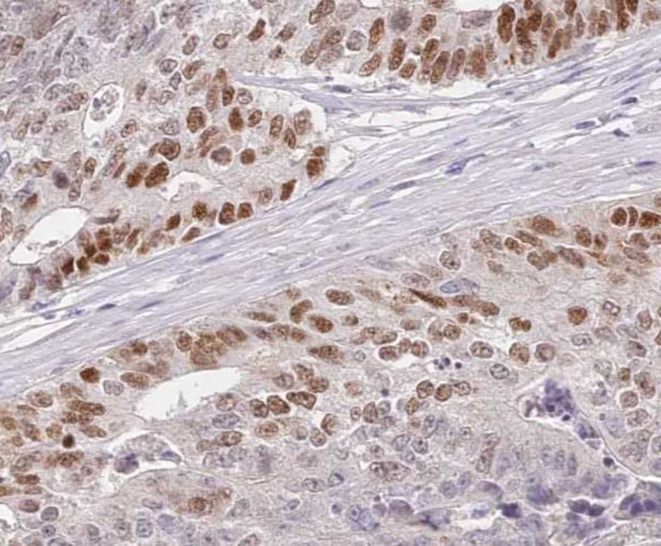
Application: ImmunohistochemistrySample Tested: Colon cancer tissueSpecies: HumanVerified Customer | Posted 09/22/2021

There are no reviews that match your criteria.
Protocols
Find general support by application which include: protocols, troubleshooting, illustrated assays, videos and webinars.
- Appropriate Fixation of IHC/ICC Samples
- Cellular Response to Hypoxia Protocols
- ClariTSA™ Fluorophore Kits
- Detection & Visualization of Antibody Binding
- ICC Cell Smear Protocol for Suspension Cells
- ICC Immunocytochemistry Protocol Videos
- ICC for Adherent Cells
- Immunocytochemistry (ICC) Protocol
- Immunocytochemistry Troubleshooting
- Immunofluorescence of Organoids Embedded in Cultrex Basement Membrane Extract
- Immunohistochemistry (IHC) and Immunocytochemistry (ICC) Protocols
- Preparing Samples for IHC/ICC Experiments
- Preventing Non-Specific Staining (Non-Specific Binding)
- Primary Antibody Selection & Optimization
- Protocol for VisUCyte™ HRP Polymer Detection Reagent
- Protocol for the Fluorescent ICC Staining of Cell Smears - Graphic
- Protocol for the Fluorescent ICC Staining of Cultured Cells on Coverslips - Graphic
- Protocol for the Preparation and Fluorescent ICC Staining of Cells on Coverslips
- Protocol for the Preparation and Fluorescent ICC Staining of Non-adherent Cells
- Protocol for the Preparation and Fluorescent ICC Staining of Stem Cells on Coverslips
- Protocol for the Preparation of a Cell Smear for Non-adherent Cell ICC - Graphic
- R&D Systems Quality Control Western Blot Protocol
- TUNEL and Active Caspase-3 Detection by IHC/ICC Protocol
- The Importance of IHC/ICC Controls
- Troubleshooting Guide: Western Blot Figures
- Western Blot Conditions
- Western Blot Protocol
- Western Blot Protocol for Cell Lysates
- Western Blot Troubleshooting
- Western Blot Troubleshooting Guide
- View all Protocols, Troubleshooting, Illustrated assays and Webinars
Loading...