
The p53 tumor suppressor protein is a multi-functional transcription factor that regulates cellular decisions regarding proliferation, cell cycle checkpoints, and apoptosis. The importance of p53 is underscored by its mutation in over 50% of human cancers. Mice that lack one or both copies of p53 also showed an increased incidence of tumors, which makes the p53 deficient mouse a model system for studying cancer generation and progression.

Scientific images may be resized, cropped, or adjusted for brightness or contrast for presentation purposes. These updates are limited to presentation and do not affect the underlying scientific interpretation of the data. Where available, additional source data may be provided upon request.
Key Product Details
Validated by
Biological Validation
Species Reactivity
Validated:
Human, Mouse, Rat
Cited:
Human
Applications
Validated:
Western Blot
Cited:
Western Blot
Label
HRP
Antibody Source
Polyclonal Goat IgG
Loading...
Product Specifications
Immunogen
E. coli-derived recombinant human p53
Asp7-Asp393
Accession # P04637
Asp7-Asp393
Accession # P04637
Specificity
Detects human, mouse, and rat p53 in Western blots.
Clonality
Polyclonal
Host
Goat
Isotype
IgG
Scientific Data Images for p53 Antibody [HRP]
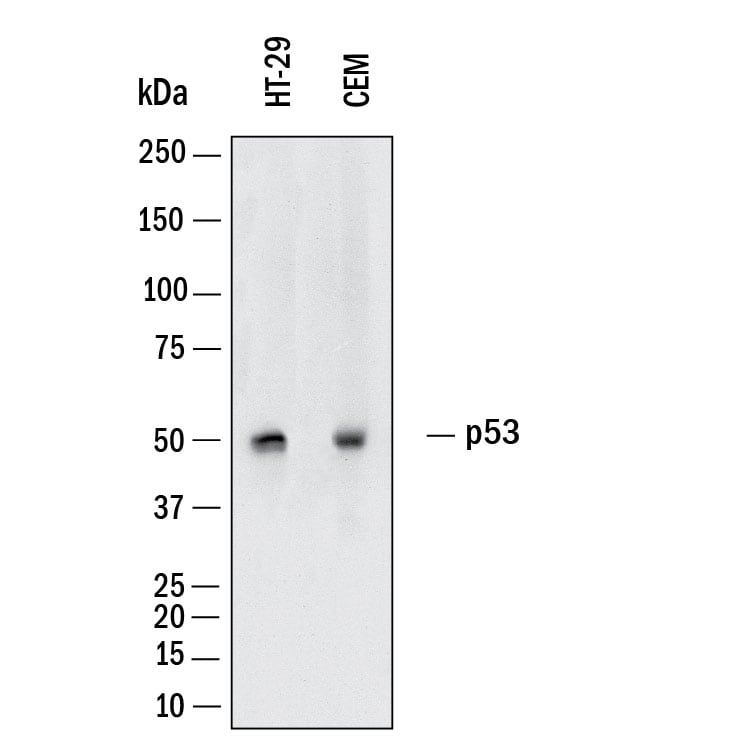
Detection of Human p53 by Western Blot.
Western Blot shows lysates of HT‑29 human colon adenocarcinoma cell line and CEM human T-lymphoblastoid cell line. PVDF membrane was probed with 1:5000 dilution of Goat Anti-Human/Mouse/Rat p53 HRP-conjugated Antigen Affinity-purified Polyclonal Antibody (Catalog # HAF1355). A specific band was detected for p53 at approximately 53 kDa (as indicated). This experiment was conducted under reducing conditions and using Western Blot Buffer Group 1.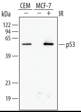
Detection of Human p53 by Western Blot.
Western blot shows lysates of CEM human T‑lymphoblastoid cell line and MCF‑7 human breast cancer cell line untreated (-) or treated (+) with 10 Gy ionizing radiation (IR) for 1 hour. PVDF membrane was probed with 1:5000 dilution of Human/Mouse/Rat p53 HRP-conjugated Antigen Affinity-purified Polyclonal Antibody (Catalog # HAF1355). A specific band was detected for p53 at approximately 53 kDa (as indicated). This experiment was conducted under reducing conditions and using Immunoblot Buffer Group 1.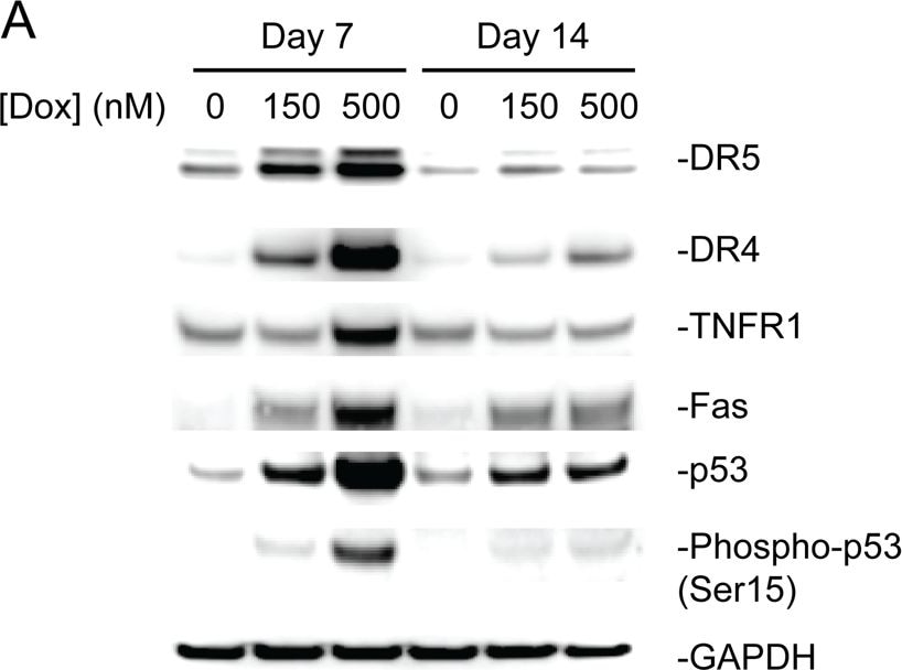
Detection of Human p53 by Western Blot
Western blotting confirmed doxorubicin-induced increase in expression of p53 and DRs.a Immunoblot and b densitometric quantification demonstrate after exposing cardiomyocytes to 150 nM or 500 nM of doxorubicin for 48 h the phosphorylation of p53 and total expression of p53, TNFR1, DR4, DR5, and Fas increase. TNFR1 protein expression increased only after exposure to 500 nM of doxorubicin. The protein expression of p53 targets, DR4 and DR5, decrease after a 7-day washout period. Fas remains slightly upregulated after 7-day washout Image collected and cropped by CiteAb from the following publication (https://pubmed.ncbi.nlm.nih.gov/31231550), licensed under a CC-BY license. Not internally tested by R&D Systems.Applications for p53 Antibody [HRP]
Application
Recommended Usage
Western Blot
1:5000 dilution
Sample: HT-29 human colon adenocarcinoma cell line, CEM human T‑lymphoblastoid cell line and ionizing radiation-treated MCF‑7 human breast cancer cell line
Sample: HT-29 human colon adenocarcinoma cell line, CEM human T‑lymphoblastoid cell line and ionizing radiation-treated MCF‑7 human breast cancer cell line
Reviewed Applications
Read 1 review rated 5 using HAF1355 in the following applications:
Formulation, Preparation, and Storage
Purification
Antigen Affinity-purified
Formulation
A 0.2 μm filtered solution in Tris with stabilizers and preservatives.
Shipping
The product is shipped with polar packs. Upon receipt, store it immediately at the temperature recommended below.
Stability & Storage
Do not freeze.
- 6 months from date of receipt, 2 to 8 °C as supplied.
Background: p53
Alternate Names
BCC7, LFS1, TP53, TRP53
Gene Symbol
TP53
UniProt
Additional p53 Products
Product Documents for p53 Antibody [HRP]
Certificate of Analysis
To download a Certificate of Analysis, please enter a lot or batch number in the search box below.
Note: Certificate of Analysis not available for kit components.
Product Specific Notices for p53 Antibody [HRP]
For research use only
Citations for p53 Antibody [HRP]
 Powered by Bioz
Customer Reviews for p53 Antibody [HRP] (1)
5 out of 5
1 Customer Rating
Have you used p53 Antibody [HRP]?
Submit a review and receive an Amazon gift card!
$25/€18/£15/$25CAN/¥2500 for a review with an image
$10/€7/£6/$10CAN/¥1110 for a review without an image
Submit a review
Customer Images
![p53 Antibody [HRP] HAF1355](https://resources.rndsystems.com/images/reviews/review_haf1355_27821_0.png)
Showing
1
-
1 的
1 review
Showing All
Filter By:
-
Application: Western BlotSample Tested: Cancer cell lysatesSpecies: HumanVerified Customer | Posted 07/11/2018Total cell lysates of MDA-MB-231 (breast), OVCAR-3 (ovarian), and A549 (lung) were subjected to western blot. PVDF membrane were probed with 1mm/ml Human/Mouse/Rat p53 HRP-conjugated Antibody (HAF1355). A specific band was detected for P53 at approximately 53kDa. This experiment was conducted under reducing conditions
![p53 Antibody [HRP] HAF1355](data:image/png;base64,R0lGODlhAQABAAD/ACwAAAAAAQABAAACADs=)
There are no reviews that match your criteria.
Protocols
Find general support by application which include: protocols, troubleshooting, illustrated assays, videos and webinars.
- Cellular Response to Hypoxia Protocols
- R&D Systems Quality Control Western Blot Protocol
- Troubleshooting Guide: Western Blot Figures
- Western Blot Conditions
- Western Blot Protocol
- Western Blot Protocol for Cell Lysates
- Western Blot Troubleshooting
- Western Blot Troubleshooting Guide
- View all Protocols, Troubleshooting, Illustrated assays and Webinars
Loading...
Associated Pathways


