
Serum Albumin Antibody
R&D Systems | Catalog # AF3329


Scientific images may be resized, cropped, or adjusted for brightness or contrast for presentation purposes. These updates are limited to presentation and do not affect the underlying scientific interpretation of the data. Where available, additional source data may be provided upon request.
Key Product Details
Species Reactivity
Validated:
Human, Mouse
Cited:
Human, Mouse, Transgenic Mouse
Applications
Validated:
Western Blot, Immunocytochemistry, Simple Western
Cited:
Immunohistochemistry, Western Blot, Immunocytochemistry, Functional Assay
Label
Unconjugated
Antibody Source
Polyclonal Goat IgG
Loading...
Product Specifications
Immunogen
Mouse Serum Albumin
Specificity
Detects human and mouse Serum Albumin in direct ELISAs and Western blots.
Clonality
Polyclonal
Host
Goat
Isotype
IgG
Scientific Data Images for Serum Albumin Antibody
Detection of Human and Mouse Serum Albumin by Western Blot.
Western blot shows lysates of human liver tissue and mouse liver tissue. PVDF membrane was probed with 0.1 µg/mL of Goat Anti-Human/Mouse Serum Albumin Antigen Affinity-purified Polyclonal Antibody (Catalog # AF3329) followed by HRP-conjugated Anti-Goat IgG Secondary Antibody (Catalog # HAF017). A specific band was detected for Serum Albumin at approximately 65-70 kDa (as indicated). This experiment was conducted under reducing conditions and using Immunoblot Buffer Group 1.
Serum Albumin in HepG2 Human Cell Line.
Serum Albumin was detected in immersion fixed HepG2 human hepatocellular carcinoma cell line using Goat Anti-Human/Mouse Serum Albumin Antigen Affinity-purified Polyclonal Antibody (Catalog # AF3329) at 10 µg/mL for 3 hours at room temperature. Cells were stained using the NorthernLights™ 557-conjugated Anti-Goat IgG Secondary Antibody (red; Catalog # NL001) and counterstained with DAPI (blue). Specific staining was localized to cytoplasm. View our protocol for Fluorescent ICC Staining of Cells on Coverslips.
Detection of Human and Mouse Albumin by Simple WesternTM.
Simple Western lane view shows lysates of human liver tissue and mouse liver tissue, loaded at 0.2 mg/mL. A specific band was detected for Albumin at approximately 64 kDa (as indicated) using 10 µg/mL of Goat Anti-Human/Mouse Serum Albumin Antigen Affinity-purified Polyclonal Antibody (Catalog # AF3329) followed by 1:50 dilution of HRP-conjugated Anti-Goat IgG Secondary Antibody (Catalog # HAF109). This experiment was conducted under reducing conditions and using the 12-230 kDa separation system.
Detection of Human Albumin by Simple WesternTM.
Simple Western lane view shows lysates of iBJ6 human induced pluripotent stem cell line undifferentiated (0) and hepatocyte differentiated for 14 and 20 days, loaded at 0.2 mg/mL. A specific band was detected for Albumin at approximately 64 kDa (as indicated) using 10 µg/mL of Goat Anti-Human/Mouse Serum Albumin Antigen Affinity-purified Polyclonal Antibody (Catalog # AF3329) followed by 1:50 dilution of HRP-conjugated Anti-Goat IgG Secondary Antibody (Catalog # HAF109). This experiment was conducted under reducing conditions and using the 12-230 kDa separation system.
Detection of Human Albumin by Simple WesternTM.
Simple Western lane view shows lysates of human serum, loaded at 1:25000. A specific band was detected for Albumin at approximately 64 kDa (as indicated) using 5 µg/mL of Goat Anti-Human/Mouse Serum Albumin Antigen Affinity-purified Polyclonal Antibody (Catalog # AF3329) followed by 1:50 dilution of HRP-conjugated Anti-Goat IgG Secondary Antibody (Catalog # HAF109). This experiment was conducted under reducing conditions and using the 12-230 kDa separation system.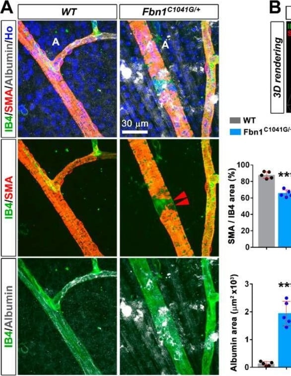
Detection of Serum Albumin by Immunohistochemistry
The fibrillin-1 C1041G mutation impairs retinal arteriole integrity and functionality in adult mice. (A) Retinal arteriole from 4 month old WT (left panels) and Fbn1C1041G/+ (right panels) mice stained for the VSMC marker alpha SMA (red), IB4 (green), albumin (white) and the nucleus marker Hoechst (blue). Note the focal loss of VSMCs around the retinal arteriole (middle panels, red arrowheads) and extravasated albumin (bottom panels) in 4 month old Fbn1C1041G/+ mice. Quantifications of the alpha SMA positive area over the IB4 positive area and albumin area per field are shown, (n = 5 WT and 5 Fbn1C1041G/+ mice). (B) 3D volume reconstruction of the same image of the Fbn1C1041G/+ arteriole displayed in (A) showing focal loss of VSMCs (left panel, red arrowheads) and extravasated albumin (right panel). (C) Retina whole-mounts from 4 month old WT (left panels) and Fbn1C1041G/+ mice (right panels) stained for IB4 (green), the adherens junction marker VE-cadherin (white) and the nucleus marker Hoechst (Ho, blue) (top panel). The single VE-cadherin channel is shown (bottom panels). The red line delineates the contours of a single endothelial cell. Quantifications of the VE-cadherin positive area over the IB4 positive area and the mean EC surface area are shown (n = 30 ECs from 5 WT and 39 ECs from 6 Fbn1C1041G/+ mice). A—arteriole. *** p < 0.001 vs. WT mice (Student’s t tests). Image collected and cropped by CiteAb from the following open publication (https://pubmed.ncbi.nlm.nih.gov/36291539), licensed under a CC-BY license. Not internally tested by R&D Systems.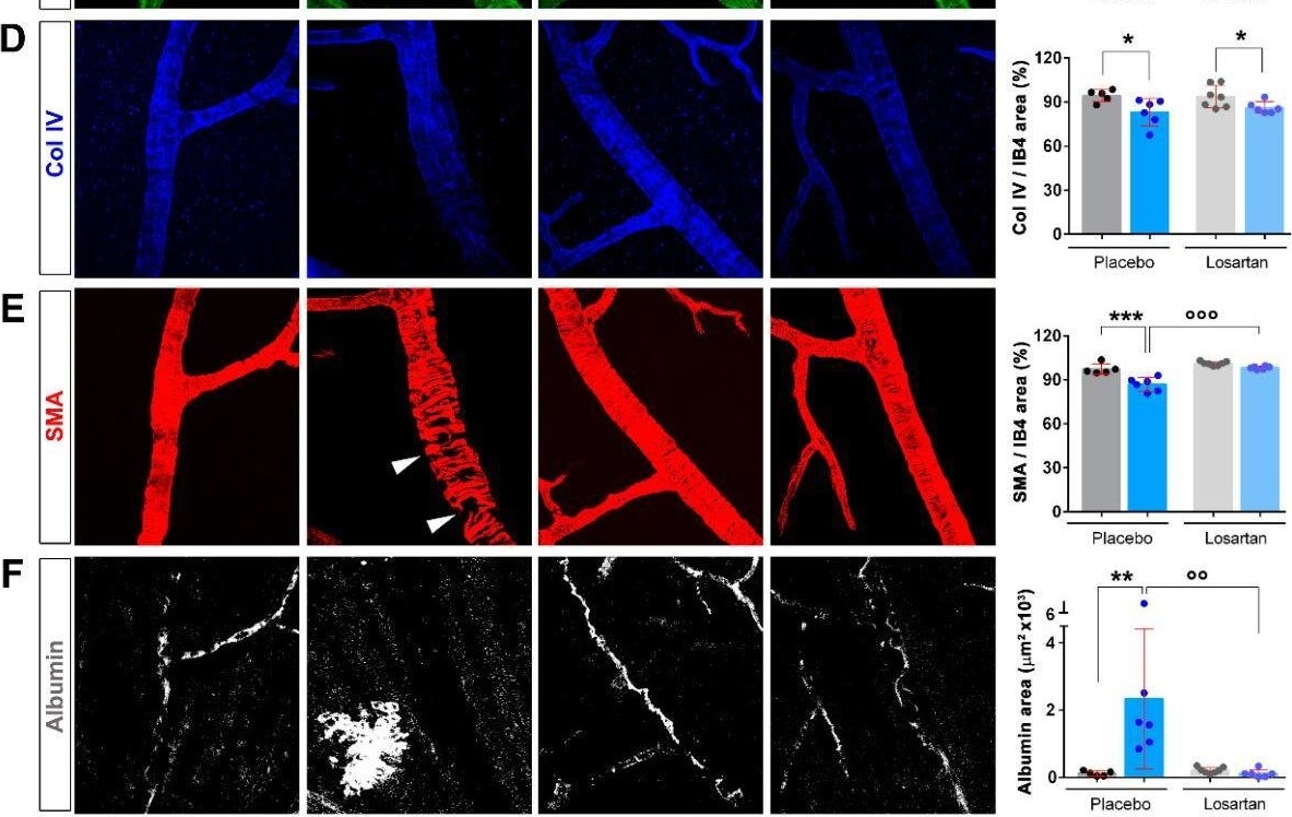
Detection of Serum Albumin by Immunohistochemistry
Late losartan treatment rescues the functional defects in the retinal arteriole of adult Fbn1C1041G/+ mice. (A) Schematic representation of the protocol of preventive losartan treatment in WT and Fbn1C1041G/+ animals. The treatment was initiated at 3 months for a duration of 6 months. (B) IB4 (green), alpha SMA (red), serum albumin (white), and Col IV (blue) stained retinal arteriole from 9-month-old WT and Fbn1C1041G/+ mice treated or not with losartan. (C) Images of the single IB4 channel (green) from the merged pictures shown in (B). Quantification of the arteriolar diameter, measured at the first bifurcation (red line) is shown (n = 5 H2O-treated WT; 6 H2O-treated Fbn1C1041G/+; 7 losartan-treated WT; 6 losartan-treated Fbn1C1041G/+ mice). D. Images of the single Col IV channel (blue) from the merged pictures shown in (B). Quantification of the Col IV positive area over the IB4 positive area is shown.). (E,F) Same experimental setup as in (D) for alpha SMA (red) in (E) and for albumin (white) in (F). White arrowheads—focal loss of VSMCs. A—arteriole. * p < 0.05; ** p < 0.01; *** p < 0.001 vs. respective WT mice. °° p < 0.01; °°° p < 0.001 vs. respective untreated mice (one-way ANOVA followed by Tukey’s post-test). Image collected and cropped by CiteAb from the following open publication (https://pubmed.ncbi.nlm.nih.gov/36291539), licensed under a CC-BY license. Not internally tested by R&D Systems.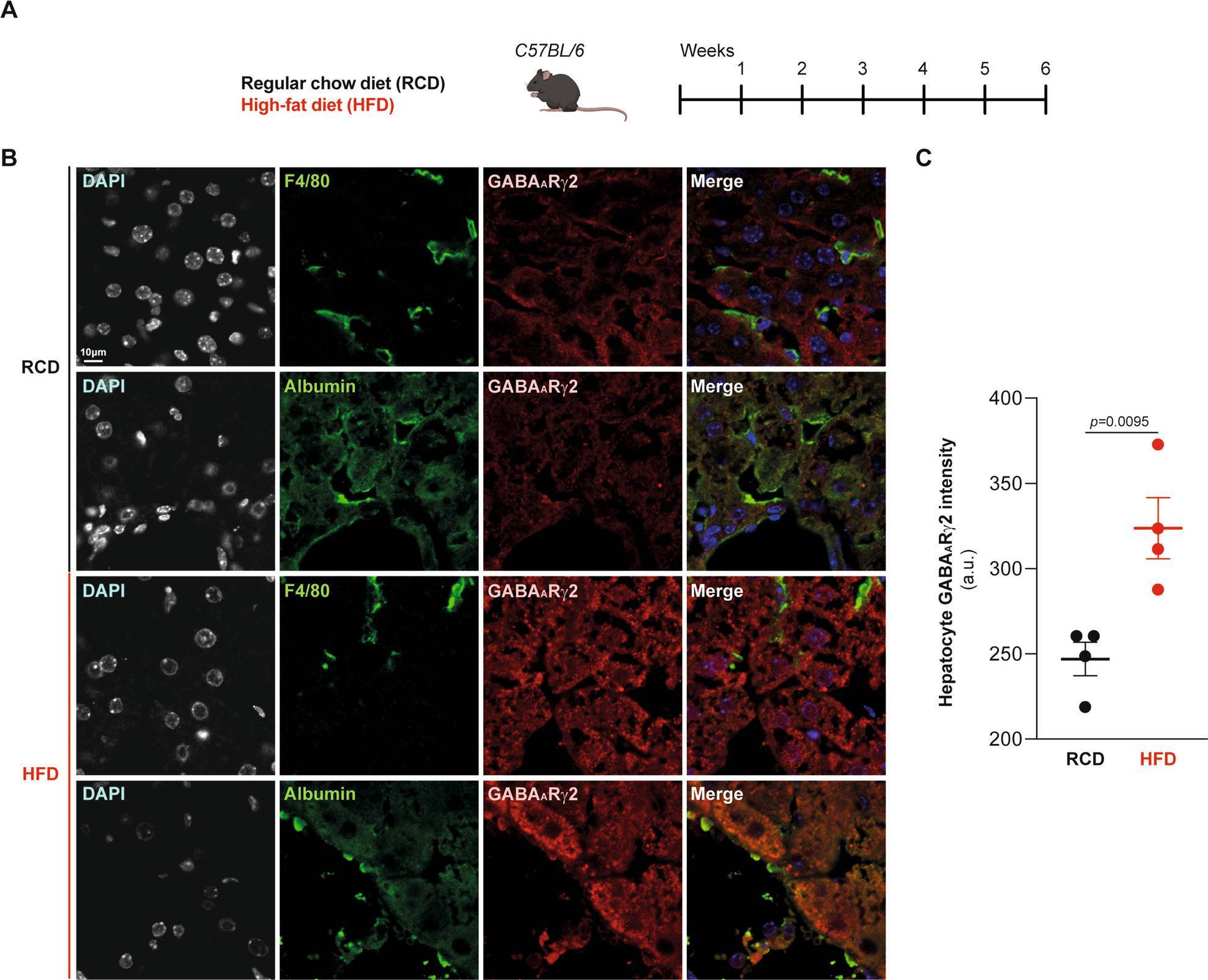
Detection of Serum Albumin by Immunocytochemistry/ Immunofluorescence
Hepatocyte-specific GABAAR gamma 2 upregulation in response to obesogenic diets determined by immunofluorescence.A Experimental design. C57BL/6 male mice were fed with RCD or HFD over the course of 6 weeks. B Representative confocal microscopy images and quantification (C) of GABAAR gamma 2 levels in mice fed with RCD or HFD (n = 4 mice per condition). F4/80 and albumin are markers specific for murine macrophages and hepatocytes, respectively. For statistical analyses, p values are calculated by two-tailed unpaired Mann–Whitney U-test. Scale bar: 10 μm. a.u.: arbitrary units. Image collected and cropped by CiteAb from the following open publication (https://www.nature.com/articles/s41419-024-06633-6), licensed under a CC-BY license. Not internally tested by R&D Systems.Applications for Serum Albumin Antibody
Application
Recommended Usage
Immunocytochemistry
5-15 µg/mL
Sample: Immersion fixed HepG2 human hepatocellular carcinoma cell line
Sample: Immersion fixed HepG2 human hepatocellular carcinoma cell line
Simple Western
5-10 µg/mL
Sample: Human liver tissue, mouse liver tissue, hepatocyte differentiated iBJ6 human induced pluripotent stem cell line, and human serum
Sample: Human liver tissue, mouse liver tissue, hepatocyte differentiated iBJ6 human induced pluripotent stem cell line, and human serum
Western Blot
0.1 µg/mL
Sample: Human liver tissue and mouse liver tissue
Sample: Human liver tissue and mouse liver tissue
Formulation, Preparation, and Storage
Purification
Antigen Affinity-purified
Reconstitution
Reconstitute at 0.2 mg/mL in sterile PBS. For liquid material, refer to CoA for concentration.
Loading...
Formulation
Lyophilized from a 0.2 μm filtered solution in PBS with Trehalose. *Small pack size (SP) is supplied either lyophilized or as a 0.2 µm filtered solution in PBS.
Shipping
Lyophilized product is shipped at ambient temperature. Liquid small pack size (-SP) is shipped with polar packs. Upon receipt, store immediately at the temperature recommended below.
Stability & Storage
Use a manual defrost freezer and avoid repeated freeze-thaw cycles.
- 12 months from date of receipt, -20 to -70 °C as supplied.
- 1 month, 2 to 8 °C under sterile conditions after reconstitution.
- 6 months, -20 to -70 °C under sterile conditions after reconstitution.
Calculators
Background: Albumin
Additional Albumin Products
Product Documents for Serum Albumin Antibody
Certificate of Analysis
To download a Certificate of Analysis, please enter a lot or batch number in the search box below.
Note: Certificate of Analysis not available for kit components.
Product Specific Notices for Serum Albumin Antibody
For research use only
Related Research Areas
Citations for Serum Albumin Antibody
 Powered by Bioz
Customer Reviews for Serum Albumin Antibody
There are currently no reviews for this product. Be the first to review Serum Albumin Antibody and earn rewards!
Have you used Serum Albumin Antibody?
Submit a review and receive an Amazon gift card!
$25/€18/£15/$25CAN/¥2500 for a review with an image
$10/€7/£6/$10CAN/¥1110 for a review without an image
Submit a review
Protocols
Find general support by application which include: protocols, troubleshooting, illustrated assays, videos and webinars.
- Appropriate Fixation of IHC/ICC Samples
- Cellular Response to Hypoxia Protocols
- ClariTSA™ Fluorophore Kits
- Detection & Visualization of Antibody Binding
- ICC Cell Smear Protocol for Suspension Cells
- ICC Immunocytochemistry Protocol Videos
- ICC for Adherent Cells
- Immunocytochemistry (ICC) Protocol
- Immunocytochemistry Troubleshooting
- Immunofluorescence of Organoids Embedded in Cultrex Basement Membrane Extract
- Immunohistochemistry (IHC) and Immunocytochemistry (ICC) Protocols
- Preparing Samples for IHC/ICC Experiments
- Preventing Non-Specific Staining (Non-Specific Binding)
- Primary Antibody Selection & Optimization
- Protocol for VisUCyte™ HRP Polymer Detection Reagent
- Protocol for the Fluorescent ICC Staining of Cell Smears - Graphic
- Protocol for the Fluorescent ICC Staining of Cultured Cells on Coverslips - Graphic
- Protocol for the Preparation and Fluorescent ICC Staining of Cells on Coverslips
- Protocol for the Preparation and Fluorescent ICC Staining of Non-adherent Cells
- Protocol for the Preparation and Fluorescent ICC Staining of Stem Cells on Coverslips
- Protocol for the Preparation of a Cell Smear for Non-adherent Cell ICC - Graphic
- R&D Systems Quality Control Western Blot Protocol
- TUNEL and Active Caspase-3 Detection by IHC/ICC Protocol
- The Importance of IHC/ICC Controls
- Troubleshooting Guide: Western Blot Figures
- Western Blot Conditions
- Western Blot Protocol
- Western Blot Protocol for Cell Lysates
- Western Blot Troubleshooting
- Western Blot Troubleshooting Guide
- View all Protocols, Troubleshooting, Illustrated assays and Webinars
Loading...
Associated Pathways
