ONECUT2 (one "cut" domain family member 2; also OC-2 and HNF6 beta ) is a monomeric 54 kDa (predicted) member of the One Cut-domain containing class of homeodomain proteins. It is expressed in embryonic neural crest plus hepatic, pancreatic, and intestinal tissue. In the adult, it is found in hepatocytes and small intestine epithelial cells. ONECUT2 is a transcriptional regulator that is known to impact the expression of Ngn3, OPN and Thbs4. This likely contributes to embryonic cell differentiation and migration. Human ONECUT2 is 504 amino acids (aa) in length. It contains two DNA binding regions, one classified as a CUT domain (aa 331‑410) and another as a homeobox domain (aa 427-481). There is one potential alternative start site at Met20. Over aa 185-326, human ONECUT2 shares 98% aa identity with mouse ONECUT2.

Key Product Details
Species Reactivity
Validated:
Human
Cited:
Human, Mouse, Avian - Chicken, Transgenic Mouse
Applications
Validated:
Immunocytochemistry
Cited:
Immunohistochemistry, Immunohistochemistry-Paraffin, Immunohistochemistry-Frozen, Flow Cytometry, Immunocytochemistry, Immunoprecipitation
Label
Unconjugated
Antibody Source
Polyclonal Sheep IgG
Loading...
Product Specifications
Immunogen
E. coli-derived recombinant human ONECUT2/OC-2
Gln185-Thr326
Accession # O95948
Gln185-Thr326
Accession # O95948
Specificity
Detects human ONECUT2/OC-2 in direct ELISAs.
Clonality
Polyclonal
Host
Sheep
Isotype
IgG
Scientific Data Images for Human ONECUT2/OC-2 Antibody
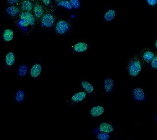
ONECUT2/OC-2 in HepG2 Human Cell Line.
ONECUT2/OC-2 was detected in immersion fixed HepG2 human hepatocellular carcinoma cell line using Human ONECUT2/OC-2 Antigen Affinity-purified Polyclonal Antibody (Catalog # AF6294) at 10 µg/mL for 3 hours at room temperature. Cells were stained using the NorthernLights™ 557-conjugated Anti-Sheep IgG Secondary Antibody (red, upper panel; NL010) and counterstained with DAPI (blue, lower panel). Specific staining was localized to nuclei. View our protocol for Fluorescent ICC Staining of Cells on Coverslips.
ONECUT2/OC‑2 in HeLa Human Cell Line.
ONECUT2/OC‑2 was detected in immersion fixed HeLa human cervical epithelial carcinoma cell line using Sheep Anti-Human ONECUT2/OC‑2 Antigen Affinity-purified Polyclonal Antibody (Catalog # AF6294) at 5 µg/mL for 3 hours at room temperature. Cells were stained using the NorthernLights™ 557-conjugated Anti-Sheep IgG Secondary Antibody (red; NL010) and counterstained with DAPI (blue). Specific staining was localized to cell nuclei. Staining was performed using our protocol for Fluorescent ICC Staining of Non-adherent Cells.Applications for Human ONECUT2/OC-2 Antibody
Application
Recommended Usage
Immunocytochemistry
5-15 µg/mL
Sample: Immersion fixed HepG2 human hepatocellular carcinoma cell line and HeLa human cervical epithelial carcinoma cell line
Sample: Immersion fixed HepG2 human hepatocellular carcinoma cell line and HeLa human cervical epithelial carcinoma cell line
Reviewed Applications
Read 1 review rated 5 using AF6294 in the following applications:
Formulation, Preparation, and Storage
Purification
Antigen Affinity-purified
Reconstitution
Sterile PBS to a final concentration of 0.2 mg/mL. For liquid material, refer to CoA for concentration.
Loading...
Formulation
Lyophilized from a 0.2 μm filtered solution in PBS with Trehalose. *Small pack size (SP) is supplied either lyophilized or as a 0.2 µm filtered solution in PBS.
Shipping
Lyophilized product is shipped at ambient temperature. Liquid small pack size (-SP) is shipped with polar packs. Upon receipt, store immediately at the temperature recommended below.
Stability & Storage
Use a manual defrost freezer and avoid repeated freeze-thaw cycles.
- 12 months from date of receipt, -20 to -70 °C as supplied.
- 1 month, 2 to 8 °C under sterile conditions after reconstitution.
- 6 months, -20 to -70 °C under sterile conditions after reconstitution.
Calculators
Background: ONECUT2/OC-2
Long Name
One Cut Homeobox 2
Alternate Names
HNF-6B, OC-2, OC2
Gene Symbol
ONECUT2
UniProt
Additional ONECUT2/OC-2 Products
Product Documents for Human ONECUT2/OC-2 Antibody
Certificate of Analysis
To download a Certificate of Analysis, please enter a lot or batch number in the search box below.
Note: Certificate of Analysis not available for kit components.
Product Specific Notices for Human ONECUT2/OC-2 Antibody
For research use only
Related Research Areas
Citations for Human ONECUT2/OC-2 Antibody
 Powered by Bioz
Customer Reviews for Human ONECUT2/OC-2 Antibody (1)
5 out of 5
1 Customer Rating
Have you used Human ONECUT2/OC-2 Antibody?
Submit a review and receive an Amazon gift card!
$25/€18/£15/$25CAN/¥2500 Yen for a review with an image
$10/€7/£6/$10CAN/¥1110 Yen for a review without an image
Submit a review
Customer Images
Showing
1
-
1 的
1 review
Showing All
Filter By:
-
Application: Immunocytochemistry/ImmunofluorescenceSample Tested: OVCAR-3 human ovarian carcinoma cell lineSpecies: HumanVerified Customer | Posted 09/10/2021

There are no reviews that match your criteria.
Protocols
Find general support by application which include: protocols, troubleshooting, illustrated assays, videos and webinars.
- Appropriate Fixation of IHC/ICC Samples
- Cellular Response to Hypoxia Protocols
- ClariTSA™ Fluorophore Kits
- Detection & Visualization of Antibody Binding
- ICC Cell Smear Protocol for Suspension Cells
- ICC Immunocytochemistry Protocol Videos
- ICC for Adherent Cells
- Immunocytochemistry (ICC) Protocol
- Immunocytochemistry Troubleshooting
- Immunofluorescence of Organoids Embedded in Cultrex Basement Membrane Extract
- Immunohistochemistry (IHC) and Immunocytochemistry (ICC) Protocols
- Preparing Samples for IHC/ICC Experiments
- Preventing Non-Specific Staining (Non-Specific Binding)
- Primary Antibody Selection & Optimization
- Protocol for VisUCyte™ HRP Polymer Detection Reagent
- Protocol for the Fluorescent ICC Staining of Cell Smears - Graphic
- Protocol for the Fluorescent ICC Staining of Cultured Cells on Coverslips - Graphic
- Protocol for the Preparation and Fluorescent ICC Staining of Cells on Coverslips
- Protocol for the Preparation and Fluorescent ICC Staining of Non-adherent Cells
- Protocol for the Preparation and Fluorescent ICC Staining of Stem Cells on Coverslips
- Protocol for the Preparation of a Cell Smear for Non-adherent Cell ICC - Graphic
- TUNEL and Active Caspase-3 Detection by IHC/ICC Protocol
- The Importance of IHC/ICC Controls
- View all Protocols, Troubleshooting, Illustrated assays and Webinars
Loading...
Associated Pathways
