
Focal adhesion kinase 1 (FAK) is a ubiquitously expressed non-receptor protein tyrosine kinase that is concentrated in the focal adhesions that form between cells growing in the presence of extracellular matrix constituents. This cellular localization is directed by a "Focal Adhesion Targeting" (FAT) sequence, a 125 amino acid sequence at the C-terminus. FAK plays an important role in migration, cell spreading, differentiation, cytoskeleton protein phosphorylation, apoptosis and acceleration of the G1 to S phase transition of the cell cycle. It associates with several different signaling proteins such as Src-family PTKs, p130Cas, Shc, Grb2, PI 3-kinase, and paxillin. This enables FAK to function within a network of integrin-stimulated signaling pathways leading to the activation of targets such as the ERK and JNK/mitogen-activated protein kinase pathways. FAK is also linked to oncogenes at biochemical and functional levels. Increased expression and/or activity of FAK in various tumors has been correlated with enhanced migration and invasiveness of human tumor cells in addition to promoting increased cell proliferation.
Human phospho-FAK (Y397) Antibody (820755)
R&D Systems | Catalog # MAB4528

Scientific images may be resized, cropped, or adjusted for brightness or contrast for presentation purposes. These updates are limited to presentation and do not affect the underlying scientific interpretation of the data. Where available, additional source data may be provided upon request.
Key Product Details
Validated by
Species Reactivity
Validated:
Cited:
Applications
Validated:
Cited:
Label
Antibody Source
Product Specifications
Immunogen
Accession # Q05397
Specificity
Clonality
Host
Isotype
Scientific Data Images for Human phospho-FAK (Y397) Antibody (820755)
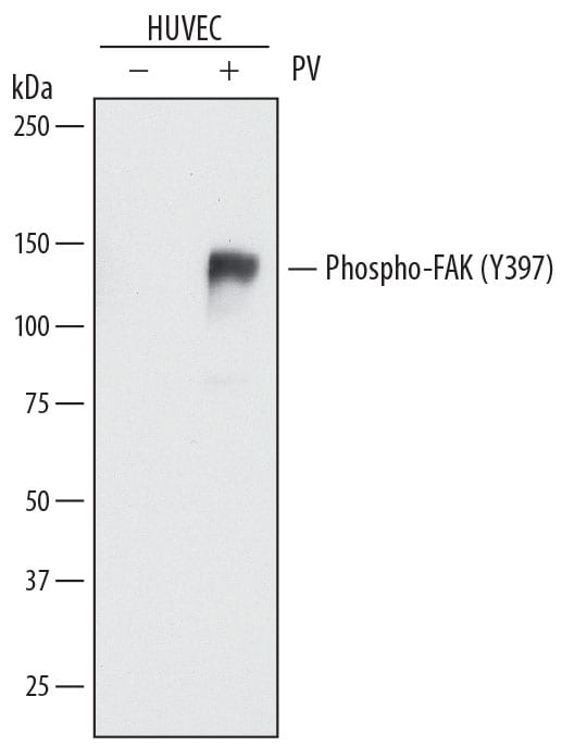
Detection of Human Phospho-FAK (Y397) by Western Blot.
Western blot shows lysates of HUVEC human umbilical vein endothelial cells untreated (-) or treated (+) with 0.1 mM Pervanadate (PV) for 10 minutes. PVDF membrane was probed with 0.5 µg/mL of Rat Anti-Human Phospho-FAK (Y397) Monoclonal Antibody (Catalog # MAB4528) followed by HRP-conjugated Anti-Rat IgG Secondary Antibody (Catalog # HAF005). A specific band was detected for Phospho-FAK (Y397) at approximately 150 kDa (as indicated). This experiment was conducted under reducing conditions and using Immunoblot Buffer Group 1.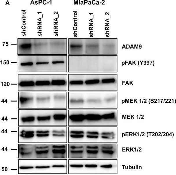
Detection of Human Phospho-FAK (Y397) by Western Blot
ADAM9 affects auto‐/paracrine MEK/ERK signaling. (A) Western blot of phosphorylation of MEK1/2 (S217/221) and ERK1/2 (T202/204) in AsPC‐1 and MiaPaCa‐2 cells following ADAM9 expression silencing. In AsPC‐1 cells, we observed decreased pFAK (Y397), which was absent in MiaPaCa‐2 cells. Quantitation of pFAK, pMEK1/2, and pERK1/2 in AsPc‐1 and MiaPaCa‐2 cells from three independent experiments. (B) Western blot analysis shows decreased phosphorylation of MEK1/2 (S217/221) and ERK1/2 (T202/204) in HUVEC cells following 30 min of incubation with cancer cell conditioned medium from both AsPC‐1 and MiaPaCa‐2 cells. Image collected and cropped by CiteAb from the following open publication (https://pubmed.ncbi.nlm.nih.gov/30556643), licensed under a CC-BY license. Not internally tested by R&D Systems.Applications for Human phospho-FAK (Y397) Antibody (820755)
Western Blot
Sample: HUVEC human umbilical vein endothelial cells treated with Pervanadate (PV)
Reviewed Applications
Read 3 reviews rated 4 using MAB4528 in the following applications:
Formulation, Preparation, and Storage
Purification
Reconstitution
Sterile PBS to a final concentration of 0.5 mg/mL. For liquid material, refer to CoA for concentration.
Formulation
Shipping
Stability & Storage
- 12 months from date of receipt, -20 to -70 °C as supplied.
- 1 month, 2 to 8 °C under sterile conditions after reconstitution.
- 6 months, -20 to -70 °C under sterile conditions after reconstitution.
Calculators
Background: FAK
Long Name
Alternate Names
Gene Symbol
UniProt
Additional FAK Products
Product Documents for Human phospho-FAK (Y397) Antibody (820755)
Certificate of Analysis
To download a Certificate of Analysis, please enter a lot or batch number in the search box below.
Note: Certificate of Analysis not available for kit components.
Product Specific Notices for Human phospho-FAK (Y397) Antibody (820755)
For research use only
Related Research Areas
Citations for Human phospho-FAK (Y397) Antibody (820755)
 Powered by Bioz
Customer Reviews for Human phospho-FAK (Y397) Antibody (820755) (3)
Have you used Human phospho-FAK (Y397) Antibody (820755)?
Submit a review and receive an Amazon gift card!
$25/€18/£15/$25CAN/¥2500 for a review with an image
$10/€7/£6/$10CAN/¥1110 for a review without an image
Submit a review
Customer Images
-
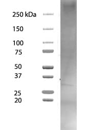
Application: Western BlotSample Tested: MDA-MB-231 human breast cancer cell lineSpecies: HumanVerified Customer | Posted 05/28/2026No band in MDA-MB-231 cells total protein10 ug of total protein from MDA-MB-231 cells was loaded and antibody used at 1ug/mL as recommended
 Bio-Techne ResponseThank you for reviewing our product. We are sorry to hear that this product did not perform as expected. We have been in touch with the customer to resolve this issue according to our Product Guarantee and to the customer’s satisfaction.
 -
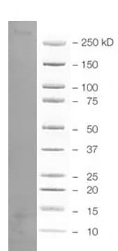
Application: Western BlotSample Tested: HCT-116 human colorectal carcinoma cell lineSpecies: HumanVerified Customer | Posted 05/28/2026No band in HCT116 cells proteinTotal protein of HCT116 cells with recommended dilution
Bio-Techne ResponseThank you for reviewing our product. We are sorry to hear that this product did not perform as expected. We have been in touch with the customer to resolve this issue according to our Product Guarantee and to the customer’s satisfaction.
-
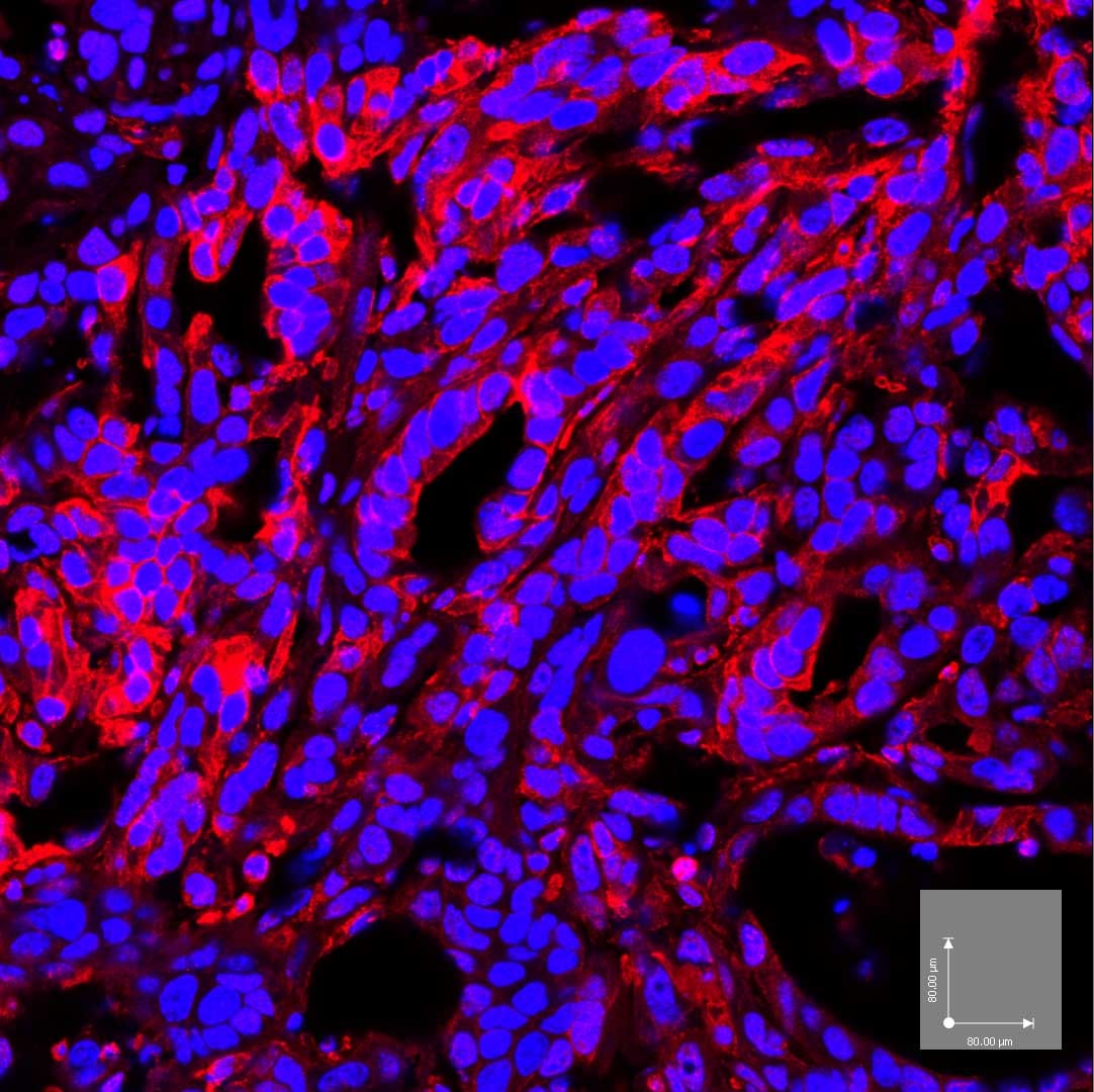
Application: ImmunohistochemistrySample Tested: oct embedded section from mouse pancreati tumor and oct embedded section from mouse pancreatic tumorSpecies: MouseVerified Customer | Posted 04/18/2018Mouse pancreatic cancer cells were implanted in C57 BL/6 syngeneic host for 1 month Tumor tissue was fixed in 4% PFA in PBS overnight in cold Then equilibriated in 30% sucrose in PBS in cold overnight Embedded in OCT and sectioned at 10um Stain with antibody for 2h after blocking with Fc recerptor Counterstained with DAPI
There are no reviews that match your criteria.
Protocols
Find general support by application which include: protocols, troubleshooting, illustrated assays, videos and webinars.
- Cellular Response to Hypoxia Protocols
- R&D Systems Quality Control Western Blot Protocol
- Troubleshooting Guide: Western Blot Figures
- Western Blot Conditions
- Western Blot Protocol
- Western Blot Protocol for Cell Lysates
- Western Blot Troubleshooting
- Western Blot Troubleshooting Guide
- View all Protocols, Troubleshooting, Illustrated assays and Webinars
Associated Pathways
