Vesicle-associated membrane protein 7 (VAMP-7) is a 25 kDa, widely expressed, type IV transmembrane protein and member of the synaptobrevin family. Mature human VAMP-7 consists of a 187 aa cytoplasmic domain, a 21 aa transmembrane region, and an 11 aa vesicular region. The cytoplasmic domain contains a longin domain (aa 7‑110) and a v-SNARE coiled-coil homology domain (aa 125‑185). Two splicing variants produce three isoforms for human VAMP-7. Isoform 2 has a 116 aa substitution for aa 145‑220 found in isoform 1, and isoform 3 is missing the residues corresponding to aa 28‑68 in isoform 1. Human VAMP-7 shares 99%, 97%, and 95% aa sequence identity with bovine, mouse, and rat VAMP-7, respectively.

Key Product Details
Validated by
Knockout/Knockdown
Species Reactivity
Validated:
Human
Cited:
Human, Xenopus
Applications
Validated:
Immunohistochemistry, Western Blot
Cited:
Immunohistochemistry, Western Blot
Label
Unconjugated
Antibody Source
Monoclonal Mouse IgG2A Clone # 549115
Loading...
Product Specifications
Immunogen
E. coli-derived recombinant human VAMP‑7
Ala2-Met140
Accession # P51809
Ala2-Met140
Accession # P51809
Specificity
Detects human VAMP‑7 in direct ELISAs and Western blots.
Clonality
Monoclonal
Host
Mouse
Isotype
IgG2A
Scientific Data Images for Human VAMP-7 Antibody (549115)
Detection of Human VAMP‑7 by Western Blot.
Western blot shows lysates of A172 human glioblastoma cell line and K562 human chronic myelogenous leukemia cell line. PVDF Membrane was probed with 1 µg/mL of Mouse Anti-Human VAMP-7 Monoclonal Antibody (Catalog # MAB6117) followed by HRP-conjugated Anti-Mouse IgG Secondary Antibody (Catalog # HAF007). A specific band was detected for VAMP-7 at approximately 25 kDa (as indicated). This experiment was conducted under reducing conditions and using Immunoblot Buffer Group 1.
Detection of Human VAMP-7 by Western Blot
Amphisome/lysosome fusion is not required for autophagy-mediated exosomal secretion of ANXA2. (a) Cells with VAMP7 knockdown and control knockdown were transfected with a GFP-LC3 plasmid and treated with or without 500 U/ml IFN-gamma for 24 h. Cells were then fixed, permeabilized, and stained for ANXA2 (red) and LAMP1 (blue). The colocalization of ANXA2, GFP-LC3 and LAMP1 was observed by confocal microscopy. Scale bar: 10 μm. The arrow and dotted inset mark an autolysosome. (b) Line tracing analysis of fluorescence signal from image in (a) of VAMP7 knockdown and control knockdown cells after IFN-gamma stimulation is shown. (c) VAMP7 knockdown efficiency was detected by western blotting. Control and VAMP7-silenced cells were treated with or without 500 U/ml IFN-gamma for 48 h. The exosome pellets were collected. ANXA2, alpha -tubulin, Tsg101 and calnexin from exosome pellets and total cell lysates were detected by western blotting. kD, molecular weight as kDa. (d) A549 cells were incubated with 500 U/ml IFN-gamma in the presence or absence of 5 nM bafilomycin A1 for 48 h. ANXA2 and alpha -tubulin from cultured supernatant and total cell lysate were analyzed by western blotting. kD, molecular weight as kDa. Image collected and cropped by CiteAb from the following publication (https://pubmed.ncbi.nlm.nih.gov/28720835), licensed under a CC-BY license. Not internally tested by R&D Systems.Applications for Human VAMP-7 Antibody (549115)
Application
Recommended Usage
Immunohistochemistry
8-25 µg/mL
Sample: Immersion-fixed paraffin-embedded sections of human breast and human breast cancer tissue
Sample: Immersion-fixed paraffin-embedded sections of human breast and human breast cancer tissue
Western Blot
1 µg/mL
Sample: A172 human glioblastoma cell line and K562 human chronic myelogenous leukemia cell line
Sample: A172 human glioblastoma cell line and K562 human chronic myelogenous leukemia cell line
Reviewed Applications
Read 2 reviews rated 5 using MAB6117 in the following applications:
Formulation, Preparation, and Storage
Purification
Protein A or G purified from hybridoma culture supernatant
Reconstitution
Reconstitute at 0.5 mg/mL in sterile PBS. For liquid material, refer to CoA for concentration.
Loading...
Formulation
Lyophilized from a 0.2 μm filtered solution in PBS with Trehalose. *Small pack size (SP) is supplied either lyophilized or as a 0.2 µm filtered solution in PBS.
Shipping
Lyophilized product is shipped at ambient temperature. Liquid small pack size (-SP) is shipped with polar packs. Upon receipt, store immediately at the temperature recommended below.
Stability & Storage
Use a manual defrost freezer and avoid repeated freeze-thaw cycles.
- 12 months from date of receipt, -20 to -70 °C as supplied.
- 1 month, 2 to 8 °C under sterile conditions after reconstitution.
- 6 months, -20 to -70 °C under sterile conditions after reconstitution.
Calculators
Background: VAMP-7
Long Name
Vesicle-Associated Membrane Protein 7
Alternate Names
SYBL1, TI-VAMP, VAMP7
Gene Symbol
VAMP7
UniProt
Additional VAMP-7 Products
Product Documents for Human VAMP-7 Antibody (549115)
Certificate of Analysis
To download a Certificate of Analysis, please enter a lot or batch number in the search box below.
Note: Certificate of Analysis not available for kit components.
Product Specific Notices for Human VAMP-7 Antibody (549115)
For research use only
Related Research Areas
Citations for Human VAMP-7 Antibody (549115)
 Powered by Bioz
Customer Reviews for Human VAMP-7 Antibody (549115) (2)
5 out of 5
2 Customer Ratings
Have you used Human VAMP-7 Antibody (549115)?
Submit a review and receive an Amazon gift card!
$25/€18/£15/$25CAN/¥2500 Yen for a review with an image
$10/€7/£6/$10CAN/¥1110 Yen for a review without an image
Submit a review
Customer Images
Showing
1
-
2 的
2 reviews
Showing All
Filter By:
-
Application: Flow CytometrySample Tested: Peripheral blood mononuclear cells (PBMCs)Species: HumanVerified Customer | Posted 11/27/2024Used for intracellular staining with Flow Cytometry/CyTOF
-
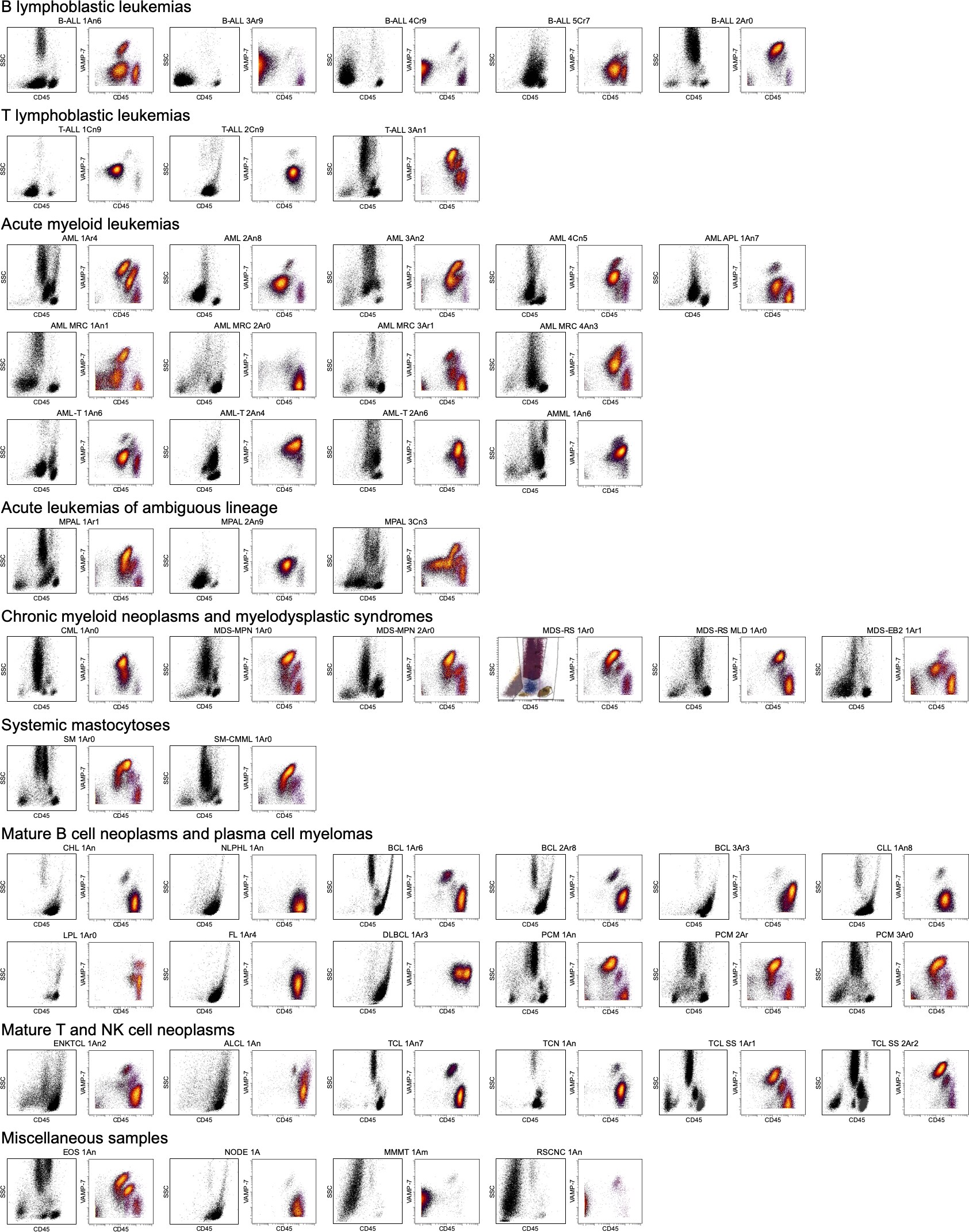
Application: CyTOFSample Tested: bone marrow and Peripheral bloodSpecies: HumanVerified Customer | Posted 03/18/2020VAMP-7 by mass cytometry recapitulates side scatter by flow cytometry. Nat Med. 2020. 26:408-417.

There are no reviews that match your criteria.
Protocols
Find general support by application which include: protocols, troubleshooting, illustrated assays, videos and webinars.
- Antigen Retrieval Protocol (PIER)
- Antigen Retrieval for Frozen Sections Protocol
- Appropriate Fixation of IHC/ICC Samples
- Cellular Response to Hypoxia Protocols
- Chromogenic IHC Staining of Formalin-Fixed Paraffin-Embedded (FFPE) Tissue Protocol
- Chromogenic Immunohistochemistry Staining of Frozen Tissue
- ClariTSA™ Fluorophore Kits
- Detection & Visualization of Antibody Binding
- Fluorescent IHC Staining of Frozen Tissue Protocol
- Graphic Protocol for Heat-induced Epitope Retrieval
- Graphic Protocol for the Preparation and Fluorescent IHC Staining of Frozen Tissue Sections
- Graphic Protocol for the Preparation and Fluorescent IHC Staining of Paraffin-embedded Tissue Sections
- Graphic Protocol for the Preparation of Gelatin-coated Slides for Histological Tissue Sections
- IHC Sample Preparation (Frozen sections vs Paraffin)
- Immunofluorescent IHC Staining of Formalin-Fixed Paraffin-Embedded (FFPE) Tissue Protocol
- Immunohistochemistry (IHC) and Immunocytochemistry (ICC) Protocols
- Immunohistochemistry Frozen Troubleshooting
- Immunohistochemistry Paraffin Troubleshooting
- Preparing Samples for IHC/ICC Experiments
- Preventing Non-Specific Staining (Non-Specific Binding)
- Primary Antibody Selection & Optimization
- Protocol for Heat-Induced Epitope Retrieval (HIER)
- Protocol for Making a 4% Formaldehyde Solution in PBS
- Protocol for VisUCyte™ HRP Polymer Detection Reagent
- Protocol for the Preparation & Fixation of Cells on Coverslips
- Protocol for the Preparation and Chromogenic IHC Staining of Frozen Tissue Sections
- Protocol for the Preparation and Chromogenic IHC Staining of Frozen Tissue Sections - Graphic
- Protocol for the Preparation and Chromogenic IHC Staining of Paraffin-embedded Tissue Sections
- Protocol for the Preparation and Chromogenic IHC Staining of Paraffin-embedded Tissue Sections - Graphic
- Protocol for the Preparation and Fluorescent IHC Staining of Frozen Tissue Sections
- Protocol for the Preparation and Fluorescent IHC Staining of Paraffin-embedded Tissue Sections
- Protocol for the Preparation of Gelatin-coated Slides for Histological Tissue Sections
- R&D Systems Quality Control Western Blot Protocol
- TUNEL and Active Caspase-3 Detection by IHC/ICC Protocol
- The Importance of IHC/ICC Controls
- Troubleshooting Guide: Immunohistochemistry
- Troubleshooting Guide: Western Blot Figures
- Western Blot Conditions
- Western Blot Protocol
- Western Blot Protocol for Cell Lysates
- Western Blot Troubleshooting
- Western Blot Troubleshooting Guide
- View all Protocols, Troubleshooting, Illustrated assays and Webinars
Loading...