Mouse CCL2 is a member of the beta (C-C) subfamily of chemokines. The murine CCL2 gene was initially identified as a platelet-derived growth factor-inducible gene in murine fibroblasts. Mouse CCL2 cDNA encodes a 148 amino acid (aa) residue with a putative 23 aa signal peptide that is cleaved to generate the mature protein. Mouse CCL2 shares 82% amino acid sequence identity with rat CCL2. Mouse CCL2 also shares 55% amino acid sequence identity with human MCP-1. Compared to human MCP-1, mouse CCL2 has a 49 aa residue extension at the carboxy-terminus. Mouse CCL2 has full activity on human cells while human MCP-1 has limited activity on mouse cells.

Scientific images may be resized, cropped, or adjusted for brightness or contrast for presentation purposes. These updates are limited to presentation and do not affect the underlying scientific interpretation of the data. Where available, additional source data may be provided upon request.
Key Product Details
Species Reactivity
Validated:
Mouse
Cited:
Human, Mouse, Rat, Transgenic Mouse
Applications
Validated:
Western Blot, ELISA Capture (Matched Antibody Pair), Neutralization
Cited:
Immunohistochemistry, Immunohistochemistry-Paraffin, Immunohistochemistry-Frozen, Western Blot, Neutralization, Flow Cytometry, Immunocytochemistry, Bioassay, Dot Blot, ELISA Development, ELISA Development (Capture), In vivo assay, Luminex Development
Label
Unconjugated
Antibody Source
Polyclonal Goat IgG
Loading...
Product Specifications
Immunogen
E. coli-derived recombinant mouse CCL2/JE/MCP-1
Gln24-Arg96
Accession # Q5SVU3
Gln24-Arg96
Accession # Q5SVU3
Specificity
Detects mouse CCL2/JE/MCP-1 in ELISAs and Western blots.
Clonality
Polyclonal
Host
Goat
Isotype
IgG
Endotoxin Level
<0.10 EU per 1 μg of the antibody by the LAL method.
Scientific Data Images for Mouse CCL2/JE/MCP-1 Antibody
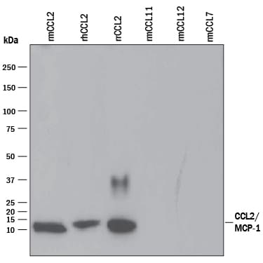
Detection of Recombinant Mouse CCL2/JE/MCP‑1 by Western Blot.
Western blot shows 25 ng of Recombinant Mouse CCL2/JE/MCP-1 (479-JE), Recombinant Human CCL2/JE/MCP-1 (Catalog # 279-MC), Recombinant Rat CCL2/JE/MCP 3144-JE), Recombinant Mouse CCL11/Eotaxin (Catalog # 420-ME), Recombinant Mouse CCL12/MCP-5 (Catalog # 428-P5), and Recombinant Mouse CCL7/MCP-3/MARC (456-MC). PVDF Membrane was probed with 0.1 µg/mL of Goat Anti-Mouse CCL2/JE/MCP-1 Antigen Affinity-purified Polyclonal Antibody (Catalog # AF-479-NA) followed by HRP-conjugated Anti-Goat IgG Secondary Antibody (HAF109). A specific band was detected for CCL2/JE/ MCP-1 at approximately 10 kDa (as indicated). This experiment was conducted under reducing conditions and using Immunoblot Buffer Group 3.
Chemotaxis Induced by CCL2/JE and Neutralization by Mouse CCL2/JE Antibody.
Recombinant Mouse CCL2/JE (Catalog # 479-JE) chemoattracts the BaF3 mouse pro-B cell line transfected with human CCR2A in a dose-dependent manner (orange line). The amount of cells that migrated through to the lower chemotaxis chamber was measured by Resazurin (Catalog # AR002). Chemotaxis elicited by Recombinant Mouse CCL2/JE (0.04 µg/mL) is neutralized (green line) by increasing concentrations of Goat Anti-Mouse CCL2/JE Antigen Affinity-purified Polyclonal Antibody (Catalog # AF-479-NA). The ND50 is typically 0.5-2.5 µg/mL.
Mouse CCL2 / JE / MCP-1 ELISA Standard Curve
Recombinant Mouse CCL2/JE/MCP‑1 (Catalog # 479-JE) was serially diluted and captured by Goat Anti-Mouse CCL2/JE/MCP‑1 Antigen Affinity-purified Polyclonal Antibody (Catalog # AF-479-NA) coated on a Clear Polystyrene Microplate (Catalog # DY990). Goat Anti-Mouse CCL2/JE/MCP‑1 Antigen Affinity-purified Polyclonal Antibody (Catalog # AF-479-NA) was biotinylated and incubated with the protein captured on the plate. Detection of the standard curve was achieved by incubating Streptavidin-HRP (Catalog # DY998)
Mouse CCL2 / JE / MCP-1 ELISA Standard Curve
Recombinant Mouse CCL2/JE/MCP‑1 (Catalog # 479-JE) was serially diluted and captured by Goat Anti-Mouse CCL2/JE/MCP‑1 Antigen Affinity-purified Polyclonal Antibody (Catalog # AF-479-NA) coated on a Clear Polystyrene Microplate (Catalog # DY990). Goat Anti-Mouse CCL2/JE/MCP‑1 Antigen Affinity-purified Polyclonal Antibody (Catalog # AF-479-NA) was biotinylated and incubated with the protein captured on the plate. Detection of the standard curve was achieved by incubating Streptavidin-HRP (Catalog # DY998)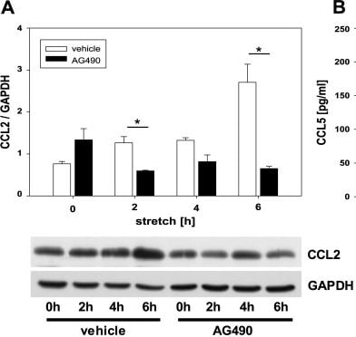
Detection of CCL2/JE/MCP-1 by Western Blot
Tyrphostin AG490 reduced chemokine secretion and cell cycle arrest.Tyrphostin decreased CC-chemokine ligand 2 (CCL2) expression in stretched tubular PKSV-PR cells (a). Tubular cells were grown on flexible membranes, pretreated with Tyrphostin AG490 or vehicle for 24hrs before treatment with cyclic stretch for 2, 4, or 6 hrs. Cell lysates were subjected to immunoblot analysis with specific antibodies against CCL2 or GAPDH. (b) Tyrphostin AG490 blocked CC-chemokine ligand 5 (CCL5) secretion. Tubular cells were stretched for 2, 4, or 6 hrs and pretreated with Tyrphostin AG490 or vehicle. Cell supernatants were analyzed for CCL5 using ELISA. Tyrphostin AG490 reduced cell cycle arrest in vivo(c) and in vitro(d). Protein expression of cyclin B1, cyclin D1, and GAPDH was measured in obstructed kidneys (c). G2/M arrest is indicated by the cyclin B1/ D1 ratio. Tubular cells were stretched for 2, 4, or 6 hrs and pretreated with Tyrphostin AG490 or vehicle (d). Cell lysates were analyzed for expression of cyclin B1, cyclin D1, and GAPDH. Representative immunoblots are shown. Values are means + s.e. of at least three independent experiments *P <0.05. Image collected and cropped by CiteAb from the following open publication (https://pubmed.ncbi.nlm.nih.gov/31846485), licensed under a CC-BY license. Not internally tested by R&D Systems.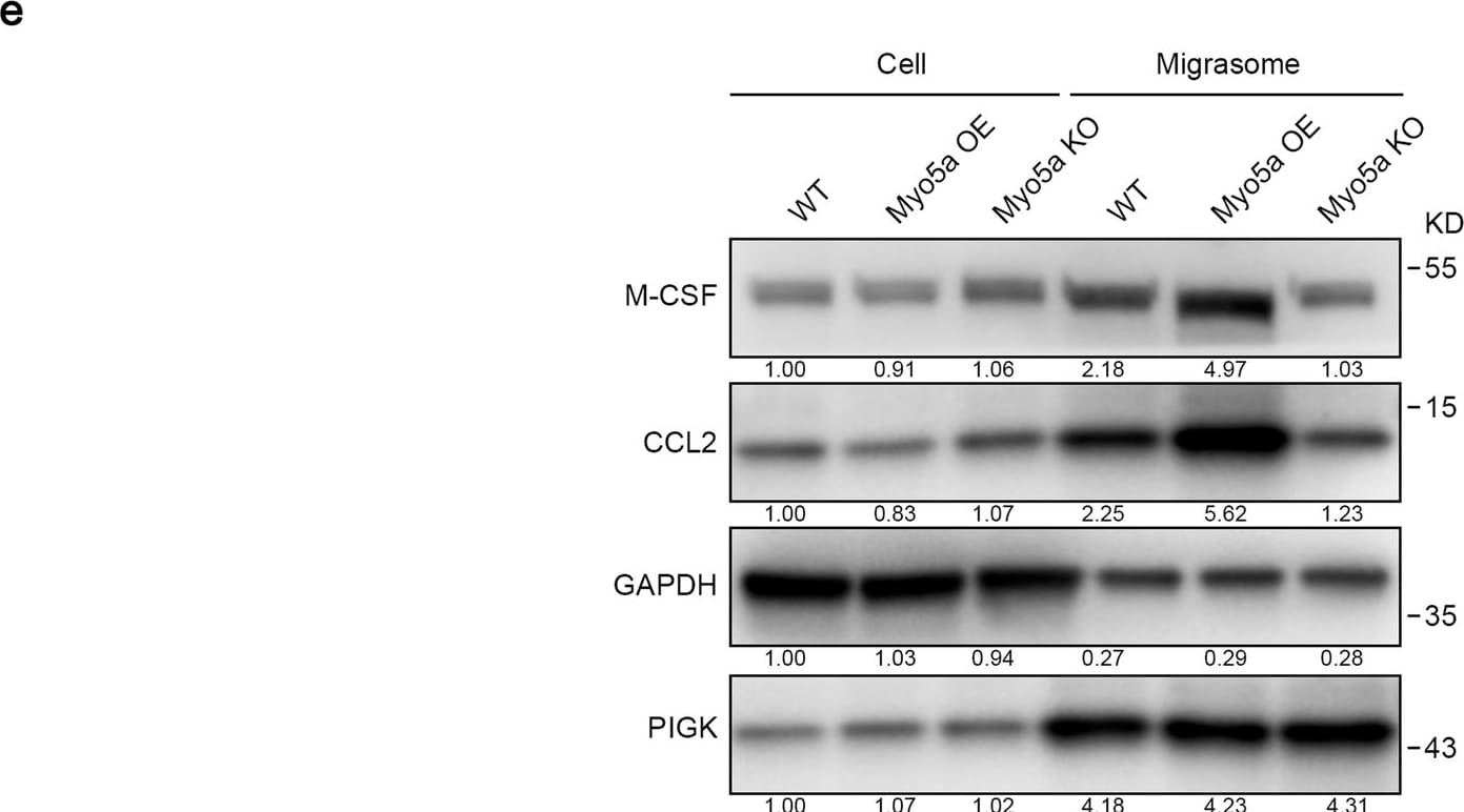
Detection of CCL2/JE/MCP-1 by Western Blot
Migrasomes are enriched with cytokines.a, b L929-GFP-VAMP2 cells were stained with M-CSF (a) or CCL2 (b) antibody and then visualized. Scale bar, 20 μm. The right panels show enlarged migrasomes. Scale bar, 2 μm. c, d Immunostaining of endogenous M-CSF (c) or CCL2 (d) in WT, Myo5a OE and Myo5a KO L929 cells. Scale bar, 20 µm. Right panels, enlarged ROI. Scale bar, 2 µm. Statistical analysis of the number of M-CSF (c) and CCL2 (d) puncta in migrasomes per cell is shown as the means ± SEM. n > 100 cells from three independent experiments were analyzed using the two-tailed unpaired t-test (right panel). **P < 0.01, ***P < 0.001. e Migrasomes were purified from the indicated L929 cells. Cell lysates and migrasomes were normalized with total protein and subjected to western blot analysis using the indicated antibodies. PIGK was used as a migrasome marker in L929 cells. Representative densitometry analysis of western blot gray values is shown. Three independent experiments were conducted. Image collected and cropped by CiteAb from the following open publication (https://pubmed.ncbi.nlm.nih.gov/38918584), licensed under a CC-BY license. Not internally tested by R&D Systems.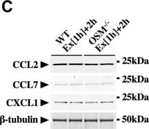
Detection of CCL2/JE/MCP-1 by Western Blot
Effects of OSM on the protein expression of exercise-induced chemokines in the skeletal muscle.A, Western blot analysis of CCL2, CCL7, and CXCL1 in the skeletal muscle of sedentary mice (Sed [1 h] + 2 h) and mice at 2 h after the exercise (Ex [1 h] + 2 h). The apparent molecular weights are indicated on the right. B, quantitative analysis of the protein expression of CCL2, CCL7, and CXCL1. The band intensities of CCL2, CCL7, and CXCL1 were normalized to beta -tubulin and are represented as the fold induction relative to the intensities of sedentary mice in the bar graph. C, Western blot analysis of CCL2, CCL7, and CXCL1 in the skeletal muscle of WT and OSM−/− mice at 2 h after the exercise. The apparent molecular weights are indicated on the right. D, quantitative analysis of the protein expression of CCL2, CCL7, and CXCL1. The band intensities of CCL2, CCL7, and CXCL1 were normalized to beta -tubulin and are represented as the fold induction relative to the intensities of WT mice in the bar graph. E, Western blot analysis of CCL2, CCL7, and CXCL1 in the skeletal muscle of mice at 2 h after the injection of vehicle (Veh) or OSM. F, quantitative analysis of the protein expression of CCL2, CCL7, and CXCL1. The band intensities of CCL2, CCL7, and CXCL1 were normalized to beta -tubulin and are represented as the fold induction relative to the intensities of Veh group in the bar graph. Data are expressed as mean ± SD; n = 5 per group. ∗p < 0.05, ∗∗p < 0.01, and ∗∗∗p < 0.001. Student’s t test. CCL, CC chemokine ligand; CXCL1, CXC chemokine ligand 1; OSM, oncostatin M. Image collected and cropped by CiteAb from the following open publication (https://linkinghub.elsevier.com/retrieve/pii/S0021925822011292), licensed under a CC-BY license. Not internally tested by R&D Systems.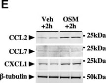
Detection of CCL2/JE/MCP-1 by Western Blot
Effects of OSM on the protein expression of exercise-induced chemokines in the skeletal muscle.A, Western blot analysis of CCL2, CCL7, and CXCL1 in the skeletal muscle of sedentary mice (Sed [1 h] + 2 h) and mice at 2 h after the exercise (Ex [1 h] + 2 h). The apparent molecular weights are indicated on the right. B, quantitative analysis of the protein expression of CCL2, CCL7, and CXCL1. The band intensities of CCL2, CCL7, and CXCL1 were normalized to beta -tubulin and are represented as the fold induction relative to the intensities of sedentary mice in the bar graph. C, Western blot analysis of CCL2, CCL7, and CXCL1 in the skeletal muscle of WT and OSM−/− mice at 2 h after the exercise. The apparent molecular weights are indicated on the right. D, quantitative analysis of the protein expression of CCL2, CCL7, and CXCL1. The band intensities of CCL2, CCL7, and CXCL1 were normalized to beta -tubulin and are represented as the fold induction relative to the intensities of WT mice in the bar graph. E, Western blot analysis of CCL2, CCL7, and CXCL1 in the skeletal muscle of mice at 2 h after the injection of vehicle (Veh) or OSM. F, quantitative analysis of the protein expression of CCL2, CCL7, and CXCL1. The band intensities of CCL2, CCL7, and CXCL1 were normalized to beta -tubulin and are represented as the fold induction relative to the intensities of Veh group in the bar graph. Data are expressed as mean ± SD; n = 5 per group. ∗p < 0.05, ∗∗p < 0.01, and ∗∗∗p < 0.001. Student’s t test. CCL, CC chemokine ligand; CXCL1, CXC chemokine ligand 1; OSM, oncostatin M. Image collected and cropped by CiteAb from the following open publication (https://linkinghub.elsevier.com/retrieve/pii/S0021925822011292), licensed under a CC-BY license. Not internally tested by R&D Systems.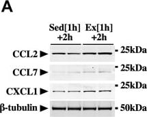
Detection of CCL2/JE/MCP-1 by Western Blot
Effects of OSM on the protein expression of exercise-induced chemokines in the skeletal muscle.A, Western blot analysis of CCL2, CCL7, and CXCL1 in the skeletal muscle of sedentary mice (Sed [1 h] + 2 h) and mice at 2 h after the exercise (Ex [1 h] + 2 h). The apparent molecular weights are indicated on the right. B, quantitative analysis of the protein expression of CCL2, CCL7, and CXCL1. The band intensities of CCL2, CCL7, and CXCL1 were normalized to beta -tubulin and are represented as the fold induction relative to the intensities of sedentary mice in the bar graph. C, Western blot analysis of CCL2, CCL7, and CXCL1 in the skeletal muscle of WT and OSM−/− mice at 2 h after the exercise. The apparent molecular weights are indicated on the right. D, quantitative analysis of the protein expression of CCL2, CCL7, and CXCL1. The band intensities of CCL2, CCL7, and CXCL1 were normalized to beta -tubulin and are represented as the fold induction relative to the intensities of WT mice in the bar graph. E, Western blot analysis of CCL2, CCL7, and CXCL1 in the skeletal muscle of mice at 2 h after the injection of vehicle (Veh) or OSM. F, quantitative analysis of the protein expression of CCL2, CCL7, and CXCL1. The band intensities of CCL2, CCL7, and CXCL1 were normalized to beta -tubulin and are represented as the fold induction relative to the intensities of Veh group in the bar graph. Data are expressed as mean ± SD; n = 5 per group. ∗p < 0.05, ∗∗p < 0.01, and ∗∗∗p < 0.001. Student’s t test. CCL, CC chemokine ligand; CXCL1, CXC chemokine ligand 1; OSM, oncostatin M. Image collected and cropped by CiteAb from the following open publication (https://linkinghub.elsevier.com/retrieve/pii/S0021925822011292), licensed under a CC-BY license. Not internally tested by R&D Systems.Applications for Mouse CCL2/JE/MCP-1 Antibody
Application
Recommended Usage
Western Blot
0.1 µg/mL
Sample: Recombinant Mouse CCL2/JE/MCP‑1 (Catalog # 479-JE)
Sample: Recombinant Mouse CCL2/JE/MCP‑1 (Catalog # 479-JE)
Neutralization
Measured by its ability to neutralize CCL2/JE/MCP‑1-induced chemotaxis in the BaF3 mouse pro‑B cell line transfected with human CCR2A. The Neutralization Dose (ND50) is typically 0.5-2.5 µg/mL in the presence of 0.04 µg/mL Recombinant Mouse CCL2/JE/MCP‑1.
Mouse CCL2/JE/MCP-1 Sandwich Immunoassay
Please Note: Optimal dilutions of this antibody should be experimentally determined.
Reviewed Applications
Read 4 reviews rated 4.3 using AF-479-NA in the following applications:
Formulation, Preparation, and Storage
Purification
Antigen Affinity-purified
Reconstitution
Reconstitute at 0.2 mg/mL in sterile PBS. For liquid material, refer to CoA for concentration.
Loading...
Formulation
Lyophilized from a 0.2 μm filtered solution in PBS with Trehalose. *Small pack size (SP) is supplied either lyophilized or as a 0.2 µm filtered solution in PBS.
Shipping
Lyophilized product is shipped at ambient temperature. Liquid small pack size (-SP) is shipped with polar packs. Upon receipt, store immediately at the temperature recommended below.
Stability & Storage
Use a manual defrost freezer and avoid repeated freeze-thaw cycles.
- 12 months from date of receipt, -20 to -70 °C as supplied.
- 1 month, 2 to 8 °C under sterile conditions after reconstitution.
- 6 months, -20 to -70 °C under sterile conditions after reconstitution.
Calculators
Background: CCL2/JE/MCP-1
Alternate Names
GDCF-2, HC11, HSMCR30, MCAF, MCP-1, SMC-CF
Gene Symbol
CCL2
UniProt
Additional CCL2/JE/MCP-1 Products
Product Documents for Mouse CCL2/JE/MCP-1 Antibody
Certificate of Analysis
To download a Certificate of Analysis, please enter a lot or batch number in the search box below.
Note: Certificate of Analysis not available for kit components.
Product Specific Notices for Mouse CCL2/JE/MCP-1 Antibody
For research use only
Citations for Mouse CCL2/JE/MCP-1 Antibody
 Powered by Bioz
Customer Reviews for Mouse CCL2/JE/MCP-1 Antibody (4)
4.3 out of 5
4 Customer Ratings
Have you used Mouse CCL2/JE/MCP-1 Antibody?
Submit a review and receive an Amazon gift card!
$25/€18/£15/$25CAN/¥2500 for a review with an image
$10/€7/£6/$10CAN/¥1110 for a review without an image
Submit a review
Customer Images
Showing
1
-
4 的
4 reviews
Showing All
Filter By:
-
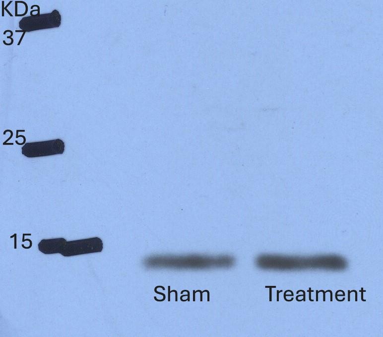
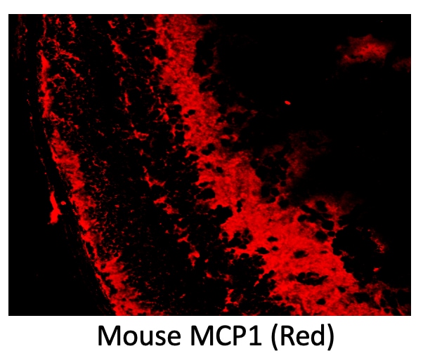
Application: Western BlotSample Tested: Lung tissueSpecies: MouseVerified Customer | Posted 11/13/2024

-
Application: Immunocytochemistry/ImmunofluorescenceSample Tested: Retina (outer nuclear layer)Species: MouseVerified Customer | Posted 07/25/2023
-
Application: Immunohistochemistry-ParaffinSample Tested: See PMID 23746482Species: MouseVerified Customer | Posted 01/08/2015
-
Application: Immunohistochemistry-FrozenSample Tested: See PMID 23748430Species: MouseVerified Customer | Posted 01/08/2015
There are no reviews that match your criteria.
Protocols
Find general support by application which include: protocols, troubleshooting, illustrated assays, videos and webinars.
- Cellular Response to Hypoxia Protocols
- R&D Systems Quality Control Western Blot Protocol
- Troubleshooting Guide: Western Blot Figures
- Western Blot Conditions
- Western Blot Protocol
- Western Blot Protocol for Cell Lysates
- Western Blot Troubleshooting
- Western Blot Troubleshooting Guide
- View all Protocols, Troubleshooting, Illustrated assays and Webinars