
CD27 is a lymphocyte-specific member of the tumor necrosis factor receptor superfamily (TNFRSF) and is designated TNFRSF7 (1, 2). Mouse CD27 cDNA encodes a 250 amino acid (aa) residue type I transmembrane protein with a 20 aa putative signal peptide, a 162 aa extracellular region containing three TNFR cysteine-rich repeats, a 21 aa transmembrane domain and a 47 aa cytoplasmic region (3). Mouse and human CD27 share approximately 65% amino acid identity. CD27 exists as homodimers on the cell surface via an extracellular disulfide bond in the membrane-proximal region. A soluble form of CD27 is also produced during the immune response and is found in various body fluids (4). CD27 is expressed on subsets of T and B cells. The expression of CD27 is upregulated upon T-cell activation. Although CD27 appears to be a marker for human memory B cells, it is only expressed in a small population of mouse B cells in germinal centers and at sites of B cell stimulation, suggesting that mouse CD27 may be a marker for activated B cells (5). CD27 interacts with CD27 ligand (also named CD70 and TNFSF7), which is a member of the TNF ligand superfamily. Ligation of CD27 on T cells provides costimulatory signals that are required for T cell proliferation, clonal expansion and the promotion of effector T cell formation (1, 2). Ligation of CD27 on B cells has been shown to inhibit terminal differentiation of activated mouse B cells into plasma cells and enhances commitment to memory B cell responses (5).
Mouse CD27/TNFRSF7 Antibody (137910)
R&D Systems | Catalog # MAB574

Scientific images may be resized, cropped, or adjusted for brightness or contrast for presentation purposes. These updates are limited to presentation and do not affect the underlying scientific interpretation of the data. Where available, additional source data may be provided upon request.
Key Product Details
Species Reactivity
Validated:
Cited:
Applications
Validated:
Cited:
Label
Antibody Source
Product Specifications
Immunogen
Thr21-Arg182
Accession # P41272
Specificity
Clonality
Host
Isotype
Scientific Data Images for Mouse CD27/TNFRSF7 Antibody (137910)
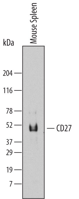
Detection of Mouse CD27/TNFRSF7 by Western Blot.
Western blot shows lysates of mouse spleen tissue. PVDF Membrane was probed with 2 µg/mL of Mouse CD27/TNFRSF7 Monoclonal Antibody (Catalog # MAB574) followed by HRP-conjugated Anti-Rat IgG Secondary Antibody (Catalog # HAF005). A specific band was detected for CD27/TNFRSF7 at approximately 45 kDa (as indicated). This experiment was conducted under reducing conditions and using Immunoblot Buffer Group 7.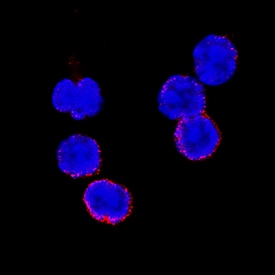
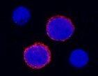
CD27/TNFRSF7 in Mouse Splenocytes.
CD27/TNFRSF7 was detected in immersion fixed mouse splenocytes using Rat Anti-Mouse CD27/TNFRSF7 Monoclonal Antibody (Catalog # MAB574) at 8 µg/mL for 3 hours at room temperature. Cells were stained using the NorthernLights™ 557-conjugated Anti-Rat IgG Secondary Antibody (red; Catalog # NL013) and counterstained with DAPI (blue). Specific staining was localized to plasma membrane. View our protocol for Fluorescent ICC Staining of Non-adherent Cells.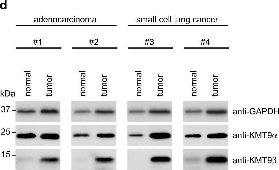
Detection of CD27/TNFRSF7 by Western Blot
KMT9 is expressed in lung cancer tissue and cell lines. a Dynamics of KMT9 alpha expression in matched normal and stage 1a lung adenocarcinoma tissue from eleven patients that underwent curative lobectomy. Normal samples were taken at 6 cm distance from macroscopic tumor sites. Data were extracted from (GSE83213). Red lines indicate increased expression of KMT9 alpha in tumor (n = 8), green lines indicate decreased expression of KMT9 alpha in tumor (n = 3). b TCGA data comparing KMT9 alpha expression in n = 515 lung adenocarcinoma with non-matched normal lung tissue (n = 59). Data represent interquartile range including minimum, 25th percentile, median, 75th percentile and maximum values. Significance was accessed by t test. c Kaplan–Meier survival analysis of patients with adenocarcinoma expressing high (n = 58) and low (n = 57) KMT9 alpha. Data were extracted from GSE26939. HR = hazard ratio. d Western blots of matched tissue from normal and tumor samples from patients with adenocarcinoma (#1 and #2) or SCLC (#3 and #4). Western blots were performed with the indicated antibodies. e Expression levels of KMT9 alpha and KMT9 beta in human cell lines from SCLC (GLC-2 and NCI-H82), adenocarcinoma (A549, PC-9 and NCI-H2087) and human immortalized normal lung fibroblasts (CRL-7000 and IMR-90) were analyzed by western blot using the indicated antibodies. f In A549 cells, KMT9 alpha and KMT9 beta are present in both nuclear and cytoplasmic compartments. Western blots were performed with the indicated antibodies. g Levels of H4K12me1 in SCLC (GLC-2 and NCI-H82) and adenocarcinoma (A549, PC-9 and NCI-H2087) cells were analyzed by western blotting using the indicated antibodies Image collected and cropped by CiteAb from the following open publication (https://pubmed.ncbi.nlm.nih.gov/32095117), licensed under a CC-BY license. Not internally tested by R&D Systems.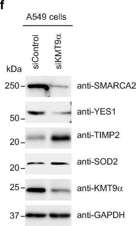
Detection of CD27/TNFRSF7 by Western Blot
KMT9 controls expression of genes involved in the organization of organelles, cells death and cell proliferation. a Venn diagram showing overlap and number of genes/proteins in A549 cells that are differentially expressed upon RNAi mediated knock-down of KMT9 alpha (log2(fold-change) > ± 0.26). In total, 460 targets are concomitantly up- or down-regulated on RNA and protein level upon knock-down of KMT9 alpha. Enriched GO_cellular components b and GO_biological processes c gene sets obtained for the indicated 460 KMT9 alpha -regulated target genes. d Heat map displaying mRNA levels of the 460 KMT9 alpha -regulated genes involved in cell proliferation (GO:0042127) in A549 cells treated with siControl or siKMT9 alpha #1. e RNA sequencing reads (left panel) and mass spectrometry volcano plot (right panel) for the indicated genes and proteins are represented exemplarily. f Western blot displaying expression of the target proteins indicated in e upon knock-down of KMT9 alpha in A549 cells. The indicated antibodies were used. g Quantitative real-time PCR analysis of the mRNA expression of the target genes displayed in e after knock-down of KMT9 alpha. Data represent means + standard deviation. Significance was accessed by two-tailed t test, n = 3 (TIMP2 n = 6) Image collected and cropped by CiteAb from the following open publication (https://pubmed.ncbi.nlm.nih.gov/32095117), licensed under a CC-BY license. Not internally tested by R&D Systems.Applications for Mouse CD27/TNFRSF7 Antibody (137910)
Immunocytochemistry
Sample: Immersion fixed mouse splenocytes
Western Blot
Sample: Mouse spleen tissue
Reviewed Applications
Read 1 review rated 5 using MAB574 in the following applications:
Formulation, Preparation, and Storage
Purification
Reconstitution
Reconstitute at 0.5 mg/mL in sterile PBS. For liquid material, refer to CoA for concentration.
Formulation
Shipping
Stability & Storage
- 12 months from date of receipt, -20 to -70 °C as supplied.
- 1 month, 2 to 8 °C under sterile conditions after reconstitution.
- 6 months, -20 to -70 °C under sterile conditions after reconstitution.
Calculators
Background: CD27/TNFRSF7
References
- Croft, M. (2003) Nature Reviews Immunol. 3:609.
- Croft, M. (2003) Cytokine and Growth Factor Reviews 14:265.
- Gravestein, L.A. et al. (1993) Eur. J. Immunol. 23:943.
- Lens, S.M. et al. (1998) Semin. Immunol. 10:491.
- Raman, V.S. et al. (2003) J. Immunol. 171:5876.
Alternate Names
Gene Symbol
UniProt
Additional CD27/TNFRSF7 Products
Product Documents for Mouse CD27/TNFRSF7 Antibody (137910)
Certificate of Analysis
To download a Certificate of Analysis, please enter a lot or batch number in the search box below.
Note: Certificate of Analysis not available for kit components.
Product Specific Notices for Mouse CD27/TNFRSF7 Antibody (137910)
For research use only
Related Research Areas
Citations for Mouse CD27/TNFRSF7 Antibody (137910)
 Powered by Bioz
Customer Reviews for Mouse CD27/TNFRSF7 Antibody (137910) (1)
Have you used Mouse CD27/TNFRSF7 Antibody (137910)?
Submit a review and receive an Amazon gift card!
$25/€18/£15/$25CAN/¥2500 for a review with an image
$10/€7/£6/$10CAN/¥1110 for a review without an image
Submit a review
Customer Images
-
Application: Immunocytochemistry/ImmunofluorescenceSample Tested: SplenocytesSpecies: MouseVerified Customer | Posted 01/25/2022

There are no reviews that match your criteria.
Protocols
Find general support by application which include: protocols, troubleshooting, illustrated assays, videos and webinars.
- Appropriate Fixation of IHC/ICC Samples
- Cellular Response to Hypoxia Protocols
- ClariTSA™ Fluorophore Kits
- Detection & Visualization of Antibody Binding
- ICC Cell Smear Protocol for Suspension Cells
- ICC Immunocytochemistry Protocol Videos
- ICC for Adherent Cells
- Immunocytochemistry (ICC) Protocol
- Immunocytochemistry Troubleshooting
- Immunofluorescence of Organoids Embedded in Cultrex Basement Membrane Extract
- Immunohistochemistry (IHC) and Immunocytochemistry (ICC) Protocols
- Preparing Samples for IHC/ICC Experiments
- Preventing Non-Specific Staining (Non-Specific Binding)
- Primary Antibody Selection & Optimization
- Protocol for VisUCyte™ HRP Polymer Detection Reagent
- Protocol for the Fluorescent ICC Staining of Cell Smears - Graphic
- Protocol for the Fluorescent ICC Staining of Cultured Cells on Coverslips - Graphic
- Protocol for the Preparation and Fluorescent ICC Staining of Cells on Coverslips
- Protocol for the Preparation and Fluorescent ICC Staining of Non-adherent Cells
- Protocol for the Preparation and Fluorescent ICC Staining of Stem Cells on Coverslips
- Protocol for the Preparation of a Cell Smear for Non-adherent Cell ICC - Graphic
- R&D Systems Quality Control Western Blot Protocol
- TUNEL and Active Caspase-3 Detection by IHC/ICC Protocol
- The Importance of IHC/ICC Controls
- Troubleshooting Guide: Western Blot Figures
- Western Blot Conditions
- Western Blot Protocol
- Western Blot Protocol for Cell Lysates
- Western Blot Troubleshooting
- Western Blot Troubleshooting Guide
- View all Protocols, Troubleshooting, Illustrated assays and Webinars
Associated Pathways
