


Mouse IL-3 Antibody (MP28F8)

R&D Systems | Catalog # MAB403

R&D Systems

Key Product Details

Species Reactivity

Validated:

Mouse

Cited:

Mouse

Applications

Validated:

Western Blot, ELISA Capture (Matched Antibody Pair), Neutralization

Cited:

Neutralization, Luminex Development

Label

Unconjugated

Antibody Source

Monoclonal Rat IgG1 Clone # MP28F8
Loading...

Product Specifications

Immunogen

COS-7 African green monkey SV40 transformed kidney fibroblast-like cell line-derived recombinant mouse IL-3
Accession # P01586

Specificity

Detects mouse IL-3 in direct ELISAs and Western blots. In Western blots, no cross‑reactivity with recombinant human (rh) IL‑3 or recombinant rat IL-3 is observed. Does not neutralize the biological activity of rhIL-3.

Clonality

Monoclonal

Host

Rat

Isotype

IgG1

Endotoxin Level

<0.10 EU per 1 μg of the antibody by the LAL method.

Scientific Data Images for Mouse IL-3 Antibody (MP28F8)

Cell Proliferation Induced by IL‑3 and Neutralization by Mouse IL‑3 Antibody.

Cell Proliferation Induced by IL‑3 and Neutralization by Mouse IL‑3 Antibody.

Recombinant Mouse IL-3 (Catalog # 403-ML) stimulates proliferation in the NFS60 mouse myeloid cell line in a dose-dependent manner (orange line). Proliferation elicited by Recombinant Mouse IL-3 (0.5 ng/mL) is neutralized (green line) by increasing concentrations of Rat Anti-Mouse IL-3 Monoclonal Antibody (Catalog # MAB403). The ND50 is typically 0.05-0.15 µg/mL.
Detection of Mouse IL-3 by Block/Neutralize

Detection of Mouse IL-3 by Block/Neutralize

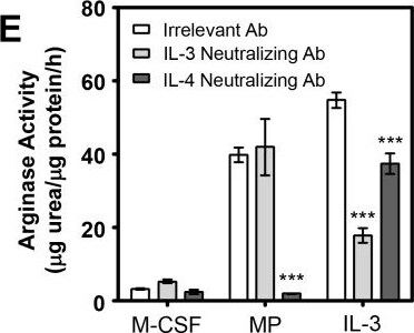
MP-induced M2 skewing of SHIP-/- BM progenitors requires IL-4 and basophils but not IL-3.A) Western blots of MФs derived from BM for 6 days with M-CSF ± IL-3 (10 ng/mL) or MP (5%). B) Arginase activity of MФs from SHIP-/- BM derived for 6 days with 10 ng/mL M-CSF ± IL-3 (10 ng/mL) or MP (5%) with 2.5 μg/mL of either an irrelevant or IL-4-neutralizing Ab. Data (mean ± SD) are representative of 3 independent experiments performed in triplicate. * p < 0.05 compared to all other conditions. C) Western blot of SHIP-/- MФs from (B) probed for Ym1, using SHC as a loading control. Dashed lines indicate where irrelevant lanes have been cropped out. All lanes are part of the same time-exposed film on the same gel. D) Western blot of SHIP-/- basophil-depleted (DX5-) or basophils (DX5+) + DX5- BM derived with M-CSF ± MP (5%). E) Arginase activity of SHIP-/- MФs derived from BM for 6 days with M-CSF ± MP (5%) or IL-3 (10 ng/mL) ± neutralizing Ab to IL-3 (2.5 μg/mL) or IL-4 (2.5 μg/mL). F) Western blot of cells corresponding to panel (E). *** p < 0.001 compared to relevant control. Image collected and cropped by CiteAb from the following publication (https://pubmed.ncbi.nlm.nih.gov/27977740), licensed under a CC-BY license. Not internally tested by R&D Systems.
Mouse IL-3 ELISA Standard Curve

Mouse IL-3 ELISA Standard Curve

Recombinant Mouse IL‑3 (Catalog # 403-ML) was serially diluted and captured by Rat Anti-Mouse IL‑3 Monoclonal Antibody (Catalog # MAB403) coated on a Clear Polystyrene Microplate (Catalog # DY990). Goat Anti-Mouse IL‑3 Antigen Affinity-purified Polyclonal Antibody (Catalog # AF-403-NA) was biotinylated and incubated with the protein captured on the plate. Detection of the standard curve was achieved by incubating Streptavidin-HRP (Catalog # DY998)
Detection of IL-3 by Western Blot

Detection of IL-3 by Western Blot

Intrathecal injection of IL‐3‐neutralizing antibody obstacles lipid droplet clearance after SCI. (A) Timeline of the intrathecal injection. (B) Immunofluorescent staining of MAC2 (red) and BODIPY (green) in sagittal sections of the IgG and Anti‐IL‐3 groups at 14 dpi. (C) Immunofluorescent staining of CD68 (red) and BODIPY (green) in sagittal sections of the IgG and Anti‐IL‐3 groups at 14 dpi. The region of interest (ROI) represents the boxed region on the left. Asterisks indicate the injured core. Scale bars: 200 μm (left panel in B, C) and 20 μm (right panel in B, C). n = 4–5 animals per group. (D–F) Quantification of the percentage of the BODIPY+ area (D), MAC2+ area (E) and CD68+ area (F) in the spinal cord segment spanning the injured core at 14 dpi. ****p < 0.0001 by Student's t test. Image collected and cropped by CiteAb from the following open publication (https://pubmed.ncbi.nlm.nih.gov/39697159), licensed under a CC-BY license. Not internally tested by R&D Systems.
Detection of IL-3 by Immunohistochemistry

Detection of IL-3 by Immunohistochemistry

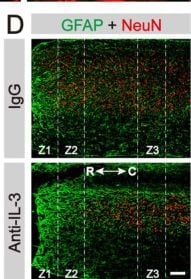
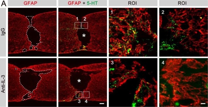
Intrathecal injection of IL‐3‐neutralizing antibody contributes to neurological decline and hinders motor function recovery. (A) Immunofluorescent staining of GFAP (green) and 5‐HT (red) in sagittal sections of the IgG and Anti‐IL‐3 groups at 28 dpi. The region of interest (ROI) represents the boxed region on the left and shows the astrocytic scar boundary. Asterisks indicate the injured core. (B) Quantification of the percentage of GFAP− area in the spinal cord segment spanning the injured core at 28 dpi. (C) Quantification of the lesion distance to the 5‐HT axon tip. *p < 0.05 and **p < 0.01 by Student's t test, n = 3–4 animals per group. (D) Immunofluorescent staining of GFAP (green) and NeuN (red) in sagittal sections of the IgG and Anti‐IL‐3 groups at 28 dpi. Scale bars: 200 μm (left panel in A) and D, 20 μm (right panel in A). (E) Quantification of NeuN+ cells in Z1–Z3 zones adjacent to the lesion core at 28 dpi. Anti‐IL‐3 group compared with IgG group, NS, no significance, *p < 0.05 and ****p < 0.0001 by Student's t test, n = 3 animals per group. (F) Representative images of footprint analysis in the PBS and IL‐3 groups at 28 dpi. The front paws are shown in green dyes, and the hind paws are shown in red dyes. (G–I) Quantification of the paw rotation, stride width and stride length at 28 dpi. ***p < 0.001 and ****p < 0.0001 by Student's t test, n = 5 animals per group. Image collected and cropped by CiteAb from the following open publication (https://pubmed.ncbi.nlm.nih.gov/39697159), licensed under a CC-BY license. Not internally tested by R&D Systems.
Detection of IL-3 by Immunocytochemistry/ Immunofluorescence

Detection of IL-3 by Immunocytochemistry/ Immunofluorescence

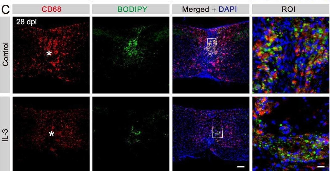
In situ injection of IL‐3 promotes the clearance of lipid droplets after SCI. (A) Timeline of the in situ injection. (B) Immunofluorescent staining of MAC2 (red) and BODIPY (green) in sagittal sections of the control and IL‐3 groups at 28 dpi. (C) Immunofluorescent staining of CD68 (red) and BODIPY (green) in sagittal sections of the control and IL‐3 groups at 28 dpi. The region of interest (ROI) represents the boxed region on the left. Asterisks indicate the injured core. Scale bars: 200 μm (left panel in B, C) and 20 μm (right panel in B, C). n = 5 animals per group. (D–F) Quantification of the percentage of BODIPY+ area (D), MAC2+ area (E), and CD68+ area (F) in the spinal cord segment spanning the injured core at 28 dpi. ****p < 0.0001 by Student's t test. Image collected and cropped by CiteAb from the following open publication (https://pubmed.ncbi.nlm.nih.gov/39697159), licensed under a CC-BY license. Not internally tested by R&D Systems.
Detection of IL-3 by Immunohistochemistry

Detection of IL-3 by Immunohistochemistry

Intrathecal injection of IL‐3‐neutralizing antibody contributes to neurological decline and hinders motor function recovery. (A) Immunofluorescent staining of GFAP (green) and 5‐HT (red) in sagittal sections of the IgG and Anti‐IL‐3 groups at 28 dpi. The region of interest (ROI) represents the boxed region on the left and shows the astrocytic scar boundary. Asterisks indicate the injured core. (B) Quantification of the percentage of GFAP− area in the spinal cord segment spanning the injured core at 28 dpi. (C) Quantification of the lesion distance to the 5‐HT axon tip. *p < 0.05 and **p < 0.01 by Student's t test, n = 3–4 animals per group. (D) Immunofluorescent staining of GFAP (green) and NeuN (red) in sagittal sections of the IgG and Anti‐IL‐3 groups at 28 dpi. Scale bars: 200 μm (left panel in A) and D, 20 μm (right panel in A). (E) Quantification of NeuN+ cells in Z1–Z3 zones adjacent to the lesion core at 28 dpi. Anti‐IL‐3 group compared with IgG group, NS, no significance, *p < 0.05 and ****p < 0.0001 by Student's t test, n = 3 animals per group. (F) Representative images of footprint analysis in the PBS and IL‐3 groups at 28 dpi. The front paws are shown in green dyes, and the hind paws are shown in red dyes. (G–I) Quantification of the paw rotation, stride width and stride length at 28 dpi. ***p < 0.001 and ****p < 0.0001 by Student's t test, n = 5 animals per group. Image collected and cropped by CiteAb from the following open publication (https://pubmed.ncbi.nlm.nih.gov/39697159), licensed under a CC-BY license. Not internally tested by R&D Systems.

Applications for Mouse IL-3 Antibody (MP28F8)

Application
Recommended Usage

Western Blot

1 µg/mL
Sample: Recombinant Mouse IL‑3 (Catalog # 403-ML)

Neutralization

Measured by its ability to neutralize IL‑3-induced proliferation in the NFS60 mouse myeloid cell line. The Neutralization Dose (ND50) is typically 0.05 - 0.15 µg/mL in the presence of 0.5 ng/mL Recombinant Mouse IL‑3.

Mouse IL-3 Sandwich Immunoassay

ELISA Capture (Matched Antibody Pair)
Recommended Concentration: 2-8 µg/mL
Use in combination with these reagents:
  • Detection Reagent: Mouse IL‑3 Biotinylated Antibody (Catalog # BAF403)
  • Standard: Recombinant Mouse IL-3 Protein (Catalog # 403-ML)
Please Note: Optimal dilutions of this antibody should be experimentally determined.

Reviewed Applications

Read 2 reviews rated 4 using MAB403 in the following applications:

Formulation, Preparation, and Storage

Purification

Protein A or G purified from hybridoma culture supernatant

Reconstitution

Reconstitute at 0.5 mg/mL in sterile PBS. For liquid material, refer to CoA for concentration.


Loading...

Formulation

Lyophilized from a 0.2 μm filtered solution in PBS with Trehalose. *Small pack size (SP) is supplied either lyophilized or as a 0.2 µm filtered solution in PBS.

Shipping

Lyophilized product is shipped at ambient temperature. Liquid small pack size (-SP) is shipped with polar packs. Upon receipt, store immediately at the temperature recommended below.

Stability & Storage

Use a manual defrost freezer and avoid repeated freeze-thaw cycles.
  • 12 months from date of receipt, -20 to -70 °C as supplied.
  • 1 month, 2 to 8 °C under sterile conditions after reconstitution.
  • 6 months, -20 to -70 °C under sterile conditions after reconstitution.

Calculators

The reconstitution calculator allows you to quickly calculate the volume of a reagent to reconstitute your vial. Simply enter the mass of reagent and the target concentration and the calculator will determine the rest.

=
÷

Background: IL-3

Interleukin 3 is a pleiotropic factor produced primarily by activated T cells that can stimulate the proliferation and differentiation of pluripotent hematopoietic stem cells as well as various lineage committed progenitors. In addition, IL-3 also affects the functional activity of mature mast cells, basophils, eosinophils and macrophages. Because of its multiple functions and targets, it was originally studied under different names, including mast cell growth factor P-cell stimulating factor, burst promoting activity, multi-colony stimulating factor, thy-1 inducing factor and WEHI-3 growth factor. In addition to activated T cells, other cell types such as human thymic epithelial cells, activated mouse mast cells, mouse keratinocytes and neurons/astrocytes can also produce IL-3. At the amino acid sequence level, mature human and mouse IL-3 share only 29% sequence identity. Consistent with this lack of homology, IL-3 activity is highly species-specific and human IL-3 does not show activity on mouse cells.

IL-3 exerts its biological activities through binding to specific cell surface receptors. The high affinity receptor responsible for IL-3 signaling is composed of alpha and beta  subunits. The IL-3 R alpha is a member of the cytokine receptor super family and binds IL-3 with low affinity. Two distinct beta  subunits, AIC2A ( beta IL-3) and AIC2B ( beta c) are present in mouse cells. beta IL-3 also binds IL-3 with low affinity and forms a high affinity receptor with the alpha  subunit. The beta c subunits does not bind any cytokine but forms functional high affinity receptors with the alpha  subunit of the IL-3, IL-5 and GM-CSF receptors. Receptors for IL-3 are present on bone marrow progenitors, macrophages, mast cells, eosinophils, megakaryocytes, basophils and various myeloid leukemic cells.

References

  1. Yokota, T. et al. (1984) Proc. Natl. Acad. Sci. USA 81:1070.
  2. Fung, M.C. et al. (1984) Nature 307:233.
  3. Miyatake, S. et al. (1985) Proc. Natl. Acad. Sci. USA 82:316.

Long Name

Interleukin 3

Alternate Names

IL3, MCGF, MULTI-CSF

Entrez Gene IDs

3562 (Human); 16187 (Mouse); 24495 (Rat)

Gene Symbol

IL3

UniProt

Additional IL-3 Products

Product Documents for Mouse IL-3 Antibody (MP28F8)

Certificate of Analysis

To download a Certificate of Analysis, please enter a lot or batch number in the search box below.

Note: Certificate of Analysis not available for kit components.

Product Specific Notices for Mouse IL-3 Antibody (MP28F8)

For research use only

Citations for Mouse IL-3 Antibody (MP28F8)

Customer Reviews for Mouse IL-3 Antibody (MP28F8) (2)

4 out of 5
2 Customer Ratings
5 Stars
0%
4 Stars
100%
3 Stars
0%
2 Stars
0%
1 Stars
0%

Have you used Mouse IL-3 Antibody (MP28F8)?

Submit a review and receive an Amazon gift card!

$25/€18/£15/$25CAN/¥2500 for a review with an image

$10/€7/£6/$10CAN/¥1110 for a review without an image

Submit a review
Amazon Gift Card
Showing  1 - 2 的 2 reviews Showing All
Filter By:
  • Mouse IL-3 Antibody
    Name: Anonymous
    Application: Functional Assay
    Sample Tested: Splenocytes
    Species: Mouse
    Verified Customer | Posted 07/27/2016
  • Mouse IL-3 Antibody
    Name: Anonymous
    Application: Flow Cytometry
    Sample Tested: T cells
    Species: Mouse
    Verified Customer | Posted 07/27/2016

There are no reviews that match your criteria.

Showing  1 - 2 的 2 reviews Showing All

FAQs

No product specific FAQs exist for this product.

View all FAQs for Antibodies
