The Toll-like family of molecules are a group of integral membrane proteins that serve as pattern recognition receptors for microbial pathogens. There are at least eleven mouse and ten human members that activate the innate immune system following exposure to a variety of microbial species (1‑4). All Toll-like receptors (TLRs) are type I transmembrane (TM) proteins that exist either in the plasma membrane or in the membranes of endosomal structures (where they bind intracellular microbial nucleic acids). All TLRs also contain a large number of extracellular leucine-rich repeats (LRRs) and a cytoplasmic tail with a Toll/IL-1 receptor (TIR) domain. The mouse TLR6 cDNA encodes a 795 amino acid (aa) precursor that includes a 27 aa signal sequence, a 557 aa extracellular domain (ECD), a 21 aa transmembrane segment, and a 190 aa cytoplasmic domain. The ECD contains 14 Leu-rich repeats, and the cytoplasmic region contains one TIR domain (5). Within the ECD, mouse TLR6 shares 59% aa sequence identity with mouse TLR1 and 20‑27% aa sequence identitity with mouse TLR2, -3, -4, -5, -7, -8, -9, -11, -12, and -13. It shares 71%, 72%, and 86% aa sequence identity with bovine, human, and rat TLR6, respectively. TLR6 is expressed on the cell surface of macrophages, monocytes, neutrophils, and dermal endothelial cells in ligand-independent association with TLR2 (6‑9). TLR2 also associates with TLR1, a functional complex with specificity for distinct but related microbial ligands (6‑8). TLR6 and TLR2 cooperate in the recognition of acylated bacterial and mycoplasma lipopeptides, peptidoglycan, and glycosylphosphatidylinositols (7‑14). The cytoplasmic TIR domain is necessary and sufficient to initiate signal transduction which leads to activation of NF kappa B (7, 15).

Key Product Details
Species Reactivity
Validated:
Mouse
Cited:
Mouse
Applications
Validated:
Flow Cytometry, CyTOF-ready
Cited:
Western Blot, Flow Cytometry, Immunocytochemistry
Label
Unconjugated
Antibody Source
Monoclonal Rat IgG2A Clone # 418601
Loading...
Product Specifications
Immunogen
HEK293 human embryonic kidney cell line transfected with mouse TLR6
Phe39-Thr806
Accession # BAA78632
Phe39-Thr806
Accession # BAA78632
Specificity
Detects mouse TLR6. Stains mouse TLR6 transfectants and not irrelevant transfectants.
Clonality
Monoclonal
Host
Rat
Isotype
IgG2A
Scientific Data Images for Mouse TLR6 Antibody (418601)
Detection of TLR6 in RAW 264.7 Mouse Cell Line by Flow Cytometry.
RAW 264.7 mouse monocyte/ macrophage cell line was stained with Rat Anti-Mouse TLR6 Monoclonal Antibody (Catalog # MAB1533, filled histogram) or isotype control antibody (Catalog # MAB006, open histogram), followed by Phycoerythrin-conjugated Anti-Rat IgG F(ab')2Secondary Antibody (Catalog # F0105B).Applications for Mouse TLR6 Antibody (418601)
Application
Recommended Usage
CyTOF-ready
Ready to be labeled using established conjugation methods. No BSA or other carrier proteins that could interfere with conjugation.
Flow Cytometry
0.25 µg/106 cells
Sample: RAW 264.7 mouse monocyte/macrophage cell line
Sample: RAW 264.7 mouse monocyte/macrophage cell line
Reviewed Applications
Read 2 reviews rated 4 using MAB1533 in the following applications:
Flow Cytometry Panel Builder
Bio-Techne Knows Flow Cytometry
Save time and reduce costly mistakes by quickly finding compatible reagents using the Panel Builder Tool.
Advanced Features
- Spectra Viewer - Custom analysis of spectra from multiple fluorochromes
- Spillover Popups - Visualize the spectra of individual fluorochromes
- Antigen Density Selector - Match fluorochrome brightness with antigen density

Formulation, Preparation, and Storage
Purification
Protein A or G purified from hybridoma culture supernatant
Reconstitution
Reconstitute at 0.5 mg/mL in sterile PBS. For liquid material, refer to CoA for concentration.
Loading...
Formulation
Lyophilized from a 0.2 μm filtered solution in PBS with Trehalose. *Small pack size (SP) is supplied either lyophilized or as a 0.2 µm filtered solution in PBS.
Shipping
Lyophilized product is shipped at ambient temperature. Liquid small pack size (-SP) is shipped with polar packs. Upon receipt, store immediately at the temperature recommended below.
Stability & Storage
Use a manual defrost freezer and avoid repeated freeze-thaw cycles.
- 12 months from date of receipt, -20 to -70 °C as supplied.
- 1 month, 2 to 8 °C under sterile conditions after reconstitution.
- 6 months, -20 to -70 °C under sterile conditions after reconstitution.
Calculators
Background: TLR6
References
- Hopkins, P.A. and S. Sriskandan (2005) Clin. Exp. Immunol. 140:395.
- Roeder, A. et al. (2004) Med. Mycol. 42:485.
- Netea, M. et al. (2004) J. Leukoc. Biol. 75:749.
- Wetzler, L.M. (2003) Vaccine 21:S55.
- Takeuchi, O. et al. (1999) Gene 231:59.
- Hajjar, A.M. et al. (2001) J. Immunol. 166:15.
- Ozinsky, A. et al. (2000) Proc. Natl. Acad. Sci. USA 97:13766.
- Lee, J.Y. et al. (2004) J. Biol. Chem. 279:16971.
- Nakao, Y. et al. (2005) J. Immunol. 174:1566.
- Bulut, Y. et al. (2001) J. Immunol. 167:987.
- Takeuchi, O. et al. (2001) Int. Immunol. 13:933.
- Morr, M. et al. (2002) Eur. J. Immunol. 32:3337.
- Krishnegowda, G. et al. (2005) J. Biol. Chem. 280:8606.
- Omueti, K.O. et al. (2005) J. Biol. Chem. 280:36616.
- Nishiya, T. and A.L. DeFranco (2004) J. Biol. Chem. 279:19008.
Long Name
Toll-like Receptor 6
Alternate Names
CD286
Gene Symbol
TLR6
UniProt
Additional TLR6 Products
Product Documents for Mouse TLR6 Antibody (418601)
Certificate of Analysis
To download a Certificate of Analysis, please enter a lot or batch number in the search box below.
Note: Certificate of Analysis not available for kit components.
Product Specific Notices for Mouse TLR6 Antibody (418601)
For research use only
Citations for Mouse TLR6 Antibody (418601)
 Powered by Bioz
Customer Reviews for Mouse TLR6 Antibody (418601) (2)
4 out of 5
2 Customer Ratings
Have you used Mouse TLR6 Antibody (418601)?
Submit a review and receive an Amazon gift card!
$25/€18/£15/$25CAN/¥2500 Yen for a review with an image
$10/€7/£6/$10CAN/¥1110 Yen for a review without an image
Submit a review
Customer Images
Showing
1
-
2 的
2 reviews
Showing All
Filter By:
-
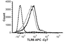
Application: Flow CytometrySample Tested: Bone marrow-derived macrophages and alveolar macrophagesSpecies: MouseVerified Customer | Posted 03/18/2016Acute silica exposure reduced TLR 6 expression on F4-80+CD11c+ alveolar macrophages. C57Bl/6 wild-type mice were exposed to saline (25 μl, black line) or silica (1 mg, gray line) through intranasal aspiration. After 4 h, whole lungs were lavaged, cells immunostained, and surface markers assessed by flow symmetry using a BD FACS Aria II. Data shown as histograms relative to unstained control (dashed line).

-
Application: Flow CytometrySample Tested: See PMID 22633994Species: MouseVerified Customer | Posted 02/10/2015
There are no reviews that match your criteria.
Protocols
Find general support by application which include: protocols, troubleshooting, illustrated assays, videos and webinars.
- 7-Amino Actinomycin D (7-AAD) Cell Viability Flow Cytometry Protocol
- Extracellular Membrane Flow Cytometry Protocol
- Flow Cytometry Protocol for Cell Surface Markers
- Flow Cytometry Protocol for Staining Membrane Associated Proteins
- Flow Cytometry Staining Protocols
- Flow Cytometry Troubleshooting Guide
- Intracellular Flow Cytometry Protocol Using Alcohol (Methanol)
- Intracellular Flow Cytometry Protocol Using Detergents
- Intracellular Nuclear Staining Flow Cytometry Protocol Using Detergents
- Intracellular Staining Flow Cytometry Protocol Using Alcohol Permeabilization
- Intracellular Staining Flow Cytometry Protocol Using Detergents to Permeabilize Cells
- Propidium Iodide Cell Viability Flow Cytometry Protocol
- Protocol for Liperfluo
- Protocol for the Characterization of Human Th22 Cells
- Protocol for the Characterization of Human Th9 Cells
- Protocol: Annexin V and PI Staining by Flow Cytometry
- Protocol: Annexin V and PI Staining for Apoptosis by Flow Cytometry
- Troubleshooting Guide: Fluorokine Flow Cytometry Kits
- View all Protocols, Troubleshooting, Illustrated assays and Webinars


