NARF Antibody - BSA Free
Novus Biologicals | Catalog # NBP2-55660
![Immunocytochemistry/ Immunofluorescence: NARF Antibody [NBP2-55660]](https://resources.rndsystems.com/images/products/NARF-Antibody-Immunocytochemistry-Immunofluorescence-NBP2-55660-img0005.jpg "Immunocytochemistry/ Immunofluorescence: NARF Antibody [NBP2-55660]")
Loading...
Key Product Details
Validated by
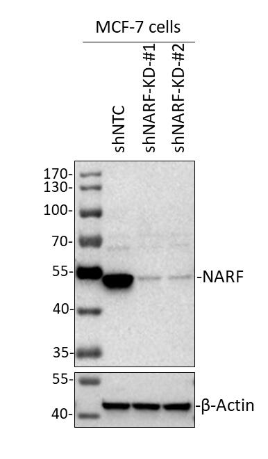
Knockout/Knockdown
Species Reactivity
Validated:
Human
Cited:
Human
Predicted:
Rat (93%). Backed by our 100% Guarantee.
Applications
Validated:
Western Blot, Immunocytochemistry/ Immunofluorescence, Knockdown Validated
Cited:
Western Blot, Immunocytochemistry/ Immunofluorescence, Immunoprecipitation
Label
Unconjugated
Antibody Source
Polyclonal Rabbit IgG
Format
BSA Free
Loading...
Product Specifications
Immunogen
This antibody was developed against a recombinant protein corresponding to the following amino acid sequence: KTDDQENVSADAPSPAQENGEKGEFHKLADAKIFLSDCLACDSCMTAEEGVQLSQQNAKDFFRVLNLNKKCDTSKHKVLVVSVCPQSLPYFAAKFNLSVTDASRRLCGFLKSLG
Reactivity Notes
Mouse 84%
Clonality
Polyclonal
Host
Rabbit
Isotype
IgG
Scientific Data Images for NARF Antibody - BSA Free
Immunocytochemistry/ Immunofluorescence: NARF Antibody [NBP2-55660]
Immunocytochemistry/Immunofluorescence: NARF Antibody [NBP2-55660] - Human MCF7 cells was stained with primary NARF antibody (1:50), followed by Goat anti-Rabbit IgG (H+L) Secondary Antibody [Texas Red] [NB120-7088] (1:1000). Image added from verified customer review.![Immunocytochemistry/ Immunofluorescence: NARF Antibody [NBP2-55660]](https://resources.rndsystems.com/images/products/NARF-Antibody-Immunocytochemistry-Immunofluorescence-NBP2-55660-img0001.jpg "Immunocytochemistry/ Immunofluorescence: NARF Antibody [NBP2-55660]")
Immunocytochemistry/ Immunofluorescence: NARF Antibody [NBP2-55660]
Immunocytochemistry/Immunofluorescence: NARF Antibody [NBP2-55660] - ICC/IF of human cell line MCF7 shows localization to nucleus & nucleoli. Antibody staining is in green.![Immunocytochemistry/ Immunofluorescence: NARF Antibody [NBP2-55660]](https://resources.rndsystems.com/images/products/NARF-Antibody-Immunocytochemistry-Immunofluorescence-NBP2-55660-img0004.jpg "Immunocytochemistry/ Immunofluorescence: NARF Antibody [NBP2-55660]")
Immunocytochemistry/ Immunofluorescence: NARF Antibody [NBP2-55660]
Immunocytochemistry/Immunofluorescence: NARF Antibody [NBP2-55660] - SUM-159PT human breast cancer cell line. Antibody was used at 1:50 for labeling and microscopy analysis of the distribution of NARF proteins in SUM-159PT breast cancer cells. ICC/IF image submitted by a verified customer review.![Knockdown Validated: NARF Antibody [NBP2-55660]](https://resources.rndsystems.com/images/products/NARF-Antibody-Western-Blot-NBP2-55660-img0003.jpg "Western Blot: NARF Antibody [NBP2-55660]")
Western Blot: NARF Antibody [NBP2-55660]
Western Blot: NARF Antibody [NBP2-55660] - Cell lysate from MCF7 cells grown in DMEM medium. 30 ug cell lysate per lane. 10% SDS-PAGE. NARF antibody at 1:1000 dilution. HRP-conjugated secondary donkey anti-rabbit IgG polyclonal antibody at 1:2000 dilution. WB image submitted by a verified customer review.
Western Blot: NARF Antibody [NBP2-55660] -
Western Blot: NARF Antibody [NBP2-55660] - Analysis in human cell line MOLT-4.Applications for NARF Antibody - BSA Free
Application
Recommended Usage
Immunocytochemistry/ Immunofluorescence
0.25 - 2 ug/mL
Knockdown Validated
Validated from a verified customer review
Western Blot
0.04 - 0.4 ug/mL
Application Notes
ICC/IF Fixation Permeabilization: Use PFA/Triton X-100.
Reviewed Applications
Read 2 reviews rated 5 using NBP2-55660 in the following applications:
Formulation, Preparation, and Storage
Purification
Affinity purified
Formulation
PBS (pH 7.2) and 40% Glycerol
Format
BSA Free
Preservative
0.02% Sodium Azide
Concentration
Concentrations vary lot to lot. See vial label for concentration. If unlisted please contact technical services.
Shipping
The product is shipped with polar packs. Upon receipt, store it immediately at the temperature recommended below.
Stability & Storage
Store at 4C short term. Aliquot and store at -20C long term. Avoid freeze-thaw cycles.
Background: NARF
Alternate Names
FLJ10067, IOP2DKFZp434G0420, Iron-only hydrogenase-like protein 2, nuclear prelamin A recognition factor, prenyl-dependent prelamin A binding protein
Gene Symbol
NARF
Additional NARF Products
Product Documents for NARF Antibody - BSA Free
Certificate of Analysis
To download a Certificate of Analysis, please enter a lot or batch number in the search box below.
Product Specific Notices for NARF Antibody - BSA Free
This product is for research use only and is not approved for use in humans or in clinical diagnosis. Primary Antibodies are guaranteed for 1 year from date of receipt.
Citations for NARF Antibody - BSA Free
 Powered by Bioz
Customer Reviews for NARF Antibody - BSA Free (2)
5 out of 5
2 Customer Ratings
Have you used NARF Antibody - BSA Free?
Submit a review and receive an Amazon gift card!
$25/€18/£15/$25CAN/¥2500 Yen for a review with an image
$10/€7/£6/$10CAN/¥1110 Yen for a review without an image
Submit a review
Customer Images

Showing
1
-
2 的
2 reviews
Showing All
Filter By:
-
Application: ImmunofluorescenceSample Tested: SUM-159PT human breast cancer cell lineSpecies: HumanVerified Customer | Posted 03/17/2021Immunofluorescence: NARF Antibody [NBP2-55660, 1:50 dilution] was used for labeling and microscopy analysis of the distribution of NARF proteins in SUM-159PT breast cancer cells.

-
Application: Western BlotSample Tested: Cell lysate from MCF7 cells grown in DMEM mediumSpecies: HumanVerified Customer | Posted 08/16/201930ug MCF-7 cell lysate/lane, 10% SDS-PAGE; Primary antibody (NARF Antibody (NBP2-55660), Lot: R70828): 1:1000 diluted in 5% milk incubate at 4℃ for overnight; Secondary antibody: NA934V, Donkey Anti-Rabbit IgG Polyclonal Antibody (HRP (Horseradish Peroxidase)- GE Healthcare (Lot: 16908227) (1:2000 diluted in 5% milk). Incubate at room temperature for 1 hour.
There are no reviews that match your criteria.
Protocols
Find general support by application which include: protocols, troubleshooting, illustrated assays, videos and webinars.
- Appropriate Fixation of IHC/ICC Samples
- Cellular Response to Hypoxia Protocols
- ClariTSA™ Fluorophore Kits
- Detection & Visualization of Antibody Binding
- ICC Cell Smear Protocol for Suspension Cells
- ICC Immunocytochemistry Protocol Videos
- ICC for Adherent Cells
- Immunocytochemistry (ICC) Protocol
- Immunocytochemistry Troubleshooting
- Immunofluorescence of Organoids Embedded in Cultrex Basement Membrane Extract
- Immunohistochemistry (IHC) and Immunocytochemistry (ICC) Protocols
- Preparing Samples for IHC/ICC Experiments
- Preventing Non-Specific Staining (Non-Specific Binding)
- Primary Antibody Selection & Optimization
- Protocol for VisUCyte™ HRP Polymer Detection Reagent
- Protocol for the Fluorescent ICC Staining of Cell Smears - Graphic
- Protocol for the Fluorescent ICC Staining of Cultured Cells on Coverslips - Graphic
- Protocol for the Preparation and Fluorescent ICC Staining of Cells on Coverslips
- Protocol for the Preparation and Fluorescent ICC Staining of Non-adherent Cells
- Protocol for the Preparation and Fluorescent ICC Staining of Stem Cells on Coverslips
- Protocol for the Preparation of a Cell Smear for Non-adherent Cell ICC - Graphic
- R&D Systems Quality Control Western Blot Protocol
- TUNEL and Active Caspase-3 Detection by IHC/ICC Protocol
- The Importance of IHC/ICC Controls
- Troubleshooting Guide: Western Blot Figures
- Western Blot Conditions
- Western Blot Protocol
- Western Blot Protocol for Cell Lysates
- Western Blot Troubleshooting
- Western Blot Troubleshooting Guide
- View all Protocols, Troubleshooting, Illustrated assays and Webinars
Loading...