
Neurofibromin 1 Antibody - BSA Free
Novus Biologicals | Catalog # NB100-418

Scientific images may be resized, cropped, or adjusted for brightness or contrast for presentation purposes. These updates are limited to presentation and do not affect the underlying scientific interpretation of the data. Where available, additional source data may be provided upon request.
Key Product Details
Validated by
Species Reactivity
Validated:
Cited:
Predicted:
Applications
Validated:
Cited:
Label
Antibody Source
Format
Product Specifications
Immunogen
Reactivity Notes
Clonality
Host
Isotype
Scientific Data Images for Neurofibromin 1 Antibody - BSA Free
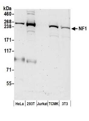
Western Blot: Neurofibromin 1 Antibody [NB100-418]
Western Blot: Neurofibromin 1 Antibody [NB100-418] - Samples: Whole cell lysate (50 ug) from HeLa, 293T, Jurkat, mouse TCMK-1, and mouse NIH3T3 cells prepared using NETN lysis buffer. Antibodies: Affinity purified rabbit anti-NF1 antibody used for WB at 0.1 ug/ml. Detection: Chemiluminescence with an exposure time of 75 seconds.
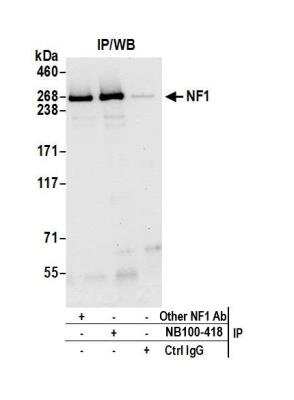
Immunoprecipitation: Neurofibromin 1 Antibody [NB100-418]
Immunoprecipitation: Neurofibromin 1 Antibody [NB100-418] - Detection of human NF1 by western blot of immunoprecipitates. Samples: Whole cell lysate (0.5 or 1.0 mg per IP reaction; 20% of IP loaded) from HeLa cells prepared using NETN lysis buffer. Antibodies: Affinity purified rabbit anti-NF1 antibody NB100-418 used for IP at 6 ug per reaction. NF1 was also immunoprecipitated by another rabbit anti-NF1 antibody. For blotting immunoprecipitated NF1, NB100-418 was used at 1 ug/ml. Detection: Chemiluminescence with an exposure time of 30 seconds.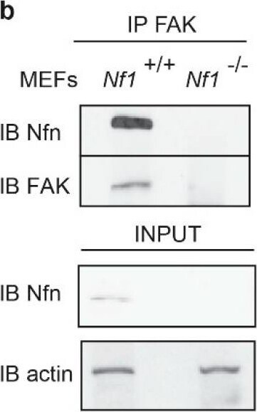
Western Blot: Neurofibromin 1 Antibody - BSA Free [NB100-418] -
Neurofibromin loss sensitizes MEF cells to growth factor and matrix stimulation: (a) MEF cells isolated and immortalized from wild-type and knockout mice for Nf1 gene were analyzed by Western blotting with anti-neurofibromin (Nfn) antibodies; (b) focal adhesion kinase FAK binds to Nfn. FAK was immunoprecipitated (IP) from cell lysates of wild-type and knockout MEFs (Nf1+/+, Nf1−/−); following SDS-PAGE electrophoresis, co-immunoprecipitated Nfn were assayed by Western blotting analysis (IB) with anti-FAK and anti-Nfn antibodies. Total protein loaded on the IP is also shown as INPUT ((b), lower panel) and examined by anti-Nfn and anti-actin antibodies; (c) transient phosphorylation kinetics of MEFs stimulated with PDGF-BB (10 ng/mL), [38,39] for increasing time periods. Following SDS-PAGE, cell lysates were incubated with anti-phospho-Erk (1/2) (T202/Y204, T185/Y187) MAP kinase, phospho-FAK (Y397) and phospho-Src (Y416) antibodies and anti-GAPDH; (d,e) colony formation assay of MEFs fully embedded by low growth factor containing Matrigel in absence (upper panel) or presence (lower panel) of insoluble matrix proteins Collagen I/Fibronectin. Cells were seeded in the Matrigel and 24 h later treated or not with PDGF-BB ligand for 8 days (representative microscope pictures (10×)). Where indicated, MEF cells were additionally treated with FAK and MEK inhibitors named Y10 (0.62 μM) and Selumetinib (6.62 μM), respectively; the area of colonies was calculated in five fields for each well, and mean value and SD calculated and plotted in the right histograms. n = 8; * p < 0.05 **; p < 0.001 by Student’s t-test. Image collected and cropped by CiteAb from the following open publication (https://pubmed.ncbi.nlm.nih.gov/34066061), licensed under a CC-BY license. Not internally tested by Novus Biologicals.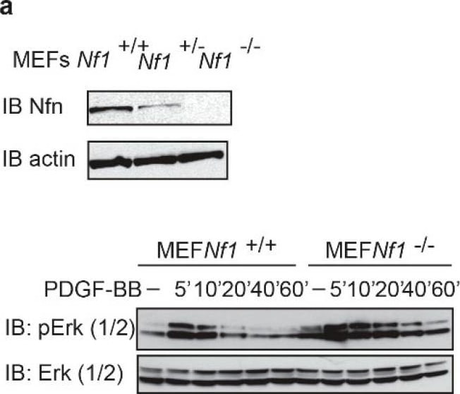
Western Blot: Neurofibromin 1 Antibody - BSA Free [NB100-418] -
Neurofibromin loss sensitizes MEF cells to growth factor and matrix stimulation: (a) MEF cells isolated and immortalized from wild-type and knockout mice for Nf1 gene were analyzed by Western blotting with anti-neurofibromin (Nfn) antibodies; (b) focal adhesion kinase FAK binds to Nfn. FAK was immunoprecipitated (IP) from cell lysates of wild-type and knockout MEFs (Nf1+/+, Nf1−/−); following SDS-PAGE electrophoresis, co-immunoprecipitated Nfn were assayed by Western blotting analysis (IB) with anti-FAK and anti-Nfn antibodies. Total protein loaded on the IP is also shown as INPUT ((b), lower panel) and examined by anti-Nfn and anti-actin antibodies; (c) transient phosphorylation kinetics of MEFs stimulated with PDGF-BB (10 ng/mL), [38,39] for increasing time periods. Following SDS-PAGE, cell lysates were incubated with anti-phospho-Erk (1/2) (T202/Y204, T185/Y187) MAP kinase, phospho-FAK (Y397) and phospho-Src (Y416) antibodies and anti-GAPDH; (d,e) colony formation assay of MEFs fully embedded by low growth factor containing Matrigel in absence (upper panel) or presence (lower panel) of insoluble matrix proteins Collagen I/Fibronectin. Cells were seeded in the Matrigel and 24 h later treated or not with PDGF-BB ligand for 8 days (representative microscope pictures (10×)). Where indicated, MEF cells were additionally treated with FAK and MEK inhibitors named Y10 (0.62 μM) and Selumetinib (6.62 μM), respectively; the area of colonies was calculated in five fields for each well, and mean value and SD calculated and plotted in the right histograms. n = 8; * p < 0.05 **; p < 0.001 by Student’s t-test. Image collected and cropped by CiteAb from the following open publication (https://pubmed.ncbi.nlm.nih.gov/34066061), licensed under a CC-BY license. Not internally tested by Novus Biologicals.Applications for Neurofibromin 1 Antibody - BSA Free
Immunoprecipitation
Western Blot
Formulation, Preparation, and Storage
Purification
Formulation
Format
Preservative
Concentration
Shipping
Stability & Storage
Background: Neurofibromin 1
Alternate Names
Entrez Gene IDs
Gene Symbol
UniProt
Additional Neurofibromin 1 Products
Product Documents for Neurofibromin 1 Antibody - BSA Free
Certificate of Analysis
To download a Certificate of Analysis, please enter a lot or batch number in the search box below.
Product Specific Notices for Neurofibromin 1 Antibody - BSA Free
This product is for research use only and is not approved for use in humans or in clinical diagnosis. Primary Antibodies are guaranteed for 1 year from date of receipt.
Citations for Neurofibromin 1 Antibody - BSA Free
 Powered by Bioz
Customer Reviews for Neurofibromin 1 Antibody - BSA Free
There are currently no reviews for this product. Be the first to review Neurofibromin 1 Antibody - BSA Free and earn rewards!
Have you used Neurofibromin 1 Antibody - BSA Free?
Submit a review and receive an Amazon gift card!
$25/€18/£15/$25CAN/¥2500 for a review with an image
$10/€7/£6/$10CAN/¥1110 for a review without an image
Submit a review
Protocols
Find general support by application which include: protocols, troubleshooting, illustrated assays, videos and webinars.
- Appropriate Fixation of IHC/ICC Samples
- Cellular Response to Hypoxia Protocols
- ClariTSA™ Fluorophore Kits
- Detection & Visualization of Antibody Binding
- ICC Cell Smear Protocol for Suspension Cells
- ICC Immunocytochemistry Protocol Videos
- ICC for Adherent Cells
- Immunocytochemistry (ICC) Protocol
- Immunocytochemistry Troubleshooting
- Immunofluorescence of Organoids Embedded in Cultrex Basement Membrane Extract
- Immunohistochemistry (IHC) and Immunocytochemistry (ICC) Protocols
- Immunoprecipitation Protocol
- Preparing Samples for IHC/ICC Experiments
- Preventing Non-Specific Staining (Non-Specific Binding)
- Primary Antibody Selection & Optimization
- Protocol for VisUCyte™ HRP Polymer Detection Reagent
- Protocol for the Fluorescent ICC Staining of Cell Smears - Graphic
- Protocol for the Fluorescent ICC Staining of Cultured Cells on Coverslips - Graphic
- Protocol for the Preparation and Fluorescent ICC Staining of Cells on Coverslips
- Protocol for the Preparation and Fluorescent ICC Staining of Non-adherent Cells
- Protocol for the Preparation and Fluorescent ICC Staining of Stem Cells on Coverslips
- Protocol for the Preparation of a Cell Smear for Non-adherent Cell ICC - Graphic
- R&D Systems Quality Control Western Blot Protocol
- TUNEL and Active Caspase-3 Detection by IHC/ICC Protocol
- The Importance of IHC/ICC Controls
- Troubleshooting Guide: Western Blot Figures
- Western Blot Conditions
- Western Blot Protocol
- Western Blot Protocol for Cell Lysates
- Western Blot Troubleshooting
- Western Blot Troubleshooting Guide
- View all Protocols, Troubleshooting, Illustrated assays and Webinars