
Redefine the Limits of Western Blotting with Automated RePlex Assays on Simple Western
More Quantitative Data from Every Sample
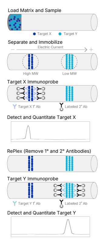
Simple Western technology is a capillary-based immunoassay that automates and modernizes protein expression analysis, offering precise, reliable data with minimal hands-on time. The RePlex™ Assay builds on this advancement, enabling you to perform two sequential immunoassays (IA) or an IA + Total Protein Assay (TPA) within the same capillary.
RePlex is More Than Stripping and Reprobing
RePlex is not your traditional stripping and reprobing. Because samples are covalently bound to the capillary wall, RePlex efficiently removes antibodies between cycles without loss of signal intensity, preserving protein integrity and delivering reproducible, quantitative results. With RePlex, you get more data points from every sample, save time, and confidently move your research forward.
How can RePlex help you?
- More Data from Every Sample: Analyze two targets sequentially on the same 3 µL sample in a single capillary without wasting limited sample volume.
- Reproducible Antibody Removal: Achieve efficient antibody removal between cycles without loss of signal intensity thanks to covalent sample bonding to the capillary wall.
- Multi-target Detection: Quantify expressed phosphorylated target and total target levels from the same sample for comprehensive protein characterization.
- Confident Normalization: Normalize with ease and precision by utilizing a second detection cycle in the same capillary dedicated to total protein analysis.
- Fully Automated Workflow: Simple Western systems automate every step of the RePlex process, reducing manual effort and minimizing human error
How does RePlex Work?
Run two sequential immunoassays with RePlex
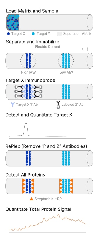
Run an immunoassays and total protein normalization with RePlex
Efficient Antibody Removal Between Probing Cycles
Because the samples are covalently bound to the capillary wall, RePlex completely and reproducibly removes antibodies between cycles without loss of signal intensity.
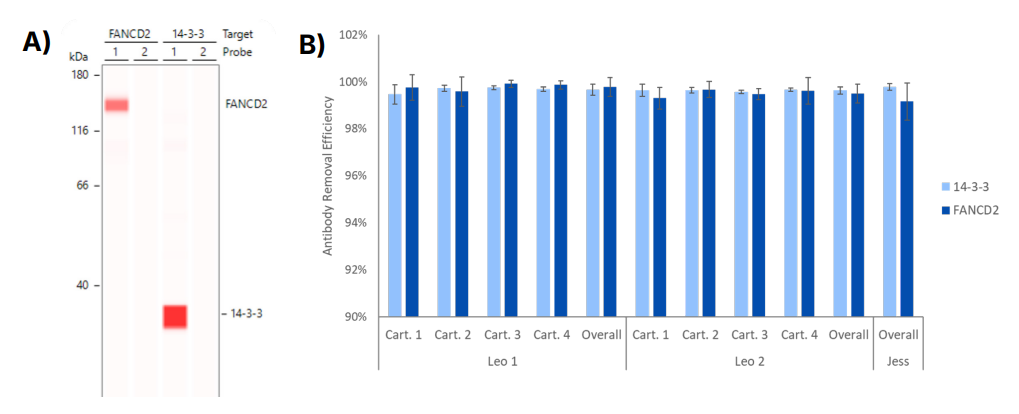
First antibody probes are efficiently removed during the RePlex assay on Jess and Leo. (A) More than 99% of the signal from Probe 1 was removed for antibodies targeting 14-3-3 and FANCD2. (A) Lane view shows the immunoassay signal for Probe 1 using 14-3-3 and FANCD2 antibodies and no residual signal for Probe 2 when probed with the secondary antibody only. (B) Removal efficiencies (%) for these targets were calculated as (Peak area Probe 1 – Peak area Probe 2)/Peak area Probe 1 x 100. Error bars represent standard deviations of the means.
Choose the Probing Order with RePlex
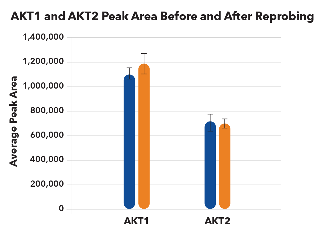
Regardless of the order in which each protein was probed, AKT1 and AKT2 in MCF7 lysates detected in Probe 1 or Probe 2 in a RePlex assay show excellent reproducibility and similar signal intensity across both probing cycles of a RePlex. Protein integrity and re-probing are not compromised by antibody removal in RePlex assays.
RePlex Enables Simultaneous Phospho / Total Detection in the Same Capillary
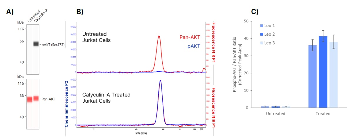
Total and phosphorylated isoforms of the signaling protein AKT in untreated and Calyculin-A treated Jurkat cells. A) Lane view showing total AKT detected in Probe 1 and phosphorylated AKT detected in Probe 2 of RePlex, with clear phospho-AKT upregulation in treated cells. B) Electropherograms show how the overlapping molecular weights of total AKT and pAKT can be distinguished using two detection channels (Chemi and NIR), which simultaneously confirms the specificity of each immunoassay. (C) Quantification of phospho-to-total AKT peak area ratio shows a 40-fold increase in treated samples. This study was performed across 3 Leo instruments using a single lot of reagents and split samples. The overlapping dynamic ranges of the NIR and chemiluminescence channels allow for the detection of both the highly abundant total AKT and lower abundant pAKT in the same sample since. Combining these capabilities provides relative quantitation through multiplexing without any additional sample prep, saving time, increasing accuracy, while providing a much richer dataset.
RePlex Enables Immunoassay and Total Protein Detection in the Same Capillary
The second cycle of RePlex can be dedicated to total protein detection so that you can normalize your data with confidence. All the steps of RePlex are automatically performed with Simple Western platform, providing more data, and lowering the cost of reagents and consumables per result.
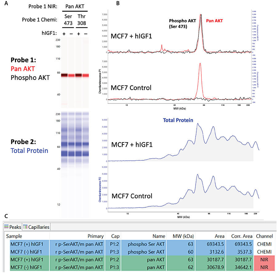
The figure shows AKT phosphorylation in MCF7 lysates untreated and activated with h-IGF1. (A-B) Phospho-AKT and pan AKT were detected in Probe 1 using chemiluminescence and NIR fluorescence, respectively, while total protein signal was detected in Probe 2. (C) The Peak Table in Compass for Simple Western shows automated normalization of phosphorylated and pan AKT signal to total protein signal, demonstrating quantitation of target protein expression.
Application Note: ReProbe with RePlex on Jess

Speak with an Expert

RePlex Module


"By switching my assays over to Simple Western, I am able to increase my throughput, allowing for full dose responses to be generated in each experiment. Utilizing the RePlex module even further increases this throughput as I often probe for 5-6 proteins per capillary in a single run including phosphorylated/total protein pairs in the same lane."
- Joseph Klebba, Ph.D., Scientist, Vividion Therapeutics