Recombinant Mouse alpha-Synuclein Active, Pre-formed Fibrils, (Type 1) Protein
Novus Biologicals | Catalog # NBP2-61596
![Immunohistochemistry: Recombinant Mouse alpha-Synuclein Active, Pre-formed Fibrils, (Type 1) Protein [NBP2-61596]](https://resources.rndsystems.com/images/products/Recombinant-Mouse-alpha-Synuclein-Active-Pre-formed-Fibrils-Type-1-Protein-Immunohistochemistry-NBP2-61596-img0003.jpg "Immunohistochemistry: Recombinant Mouse alpha-Synuclein Active, Pre-formed Fibrils, (Type 1) Protein [NBP2-61596]")
Loading...
Key Product Details
Source
E. coli
Applications
Immunohistochemistry, Western Blot, Bioactivity, Functional Assay, Microscopy, SDS-PAGE
Loading...
Product Specifications
Description
Active Mouse Recombinant Alpha Synuclein Protein Aggregate (pre-formed fibrils, Type 1). NCBI Accession #: NP_001035916.1.
Source: E. coli
Amino Acid Sequence:
MDVFMKGLSKAKEGVVAAAEKTKQGVAEAAGKTKEGVLYVGSKTKEGVVHGVTTVAEKTKEQVTNVGGAVVTGVTAVAQKTVEGAGNIAAATGFVKKDQMGKGEEGYPQE GILEDMPVDPGSEAYEMPSEEGYQDYEPEA
Source: E. coli
Amino Acid Sequence:
MDVFMKGLSKAKEGVVAAAEKTKQGVAEAAGKTKEGVLYVGSKTKEGVVHGVTTVAEKTKEQVTNVGGAVVTGVTAVAQKTVEGAGNIAAATGFVKKDQMGKGEEGYPQE GILEDMPVDPGSEAYEMPSEEGYQDYEPEA
Purity
Ion exchange chromatography
Predicted Molecular Mass
14.46 kDa.
Disclaimer note: The observed molecular weight of the protein may vary from the listed predicted molecular weight due to post translational modifications, post translation cleavages, relative charges, and other experimental factors.
Disclaimer note: The observed molecular weight of the protein may vary from the listed predicted molecular weight due to post translational modifications, post translation cleavages, relative charges, and other experimental factors.
Activity
Endogenous alpha-synuclein phosphorylation. 100 uM alpha synuclein protein monomer (NBP2-61595) seeded with 10 nM alpha synuclein protein PFF (NBP2-61596) in 25 uM Thioflavin T (PBS pH 7.4, 100 ul reaction volume) generated an increased fluorescence intensity after incubation at 37C with shaking at 600 rpm for 24 hours. Fluorescence was measured by excitation at 450 nm and emission at 485 nm on a Molecular Devices Gemini XPS microplate reader.
Protein / Peptide Type
Recombinant Protein
Scientific Data Images
Immunohistochemistry: Recombinant Mouse alpha-Synuclein Active, Pre-formed Fibrils, (Type 1) Protein [NBP2-61596]
Immunohistochemistry: Recombinant Mouse alpha-Synuclein Active, Pre-formed Fibrils, (Type 1) Protein [NBP2-61596] - Immunohistochemistry analysis of rat brain injected with Type 1 mouse alpha synuclein PFFs (NBP2-61596). Species: Female Sprague-Dawley Rat. Rat was injected with 16g Type 1 mouse alpha synuclein PFFs (NBP2-61596) in each of 2 injection sites: AP+1.6, ML+2.4, DV-4.2 from skull; and AP-1.4, ML+0.2, DV-2.8 from skull. 30 days post-injection. Fixation: Saline perfusion followed by 4% PFA fixation for 48 hrs. Primary antibody: rabbit monoclonal anti-pSer129 alpha synuclein. Secondary Antibody: Biotin-SP Donkey Anti-Rabbit IgG (H+L) at 1:500 for 2 hours in cold room with shaking. ABC signal amplification, DAB staining. Magnification: 20X. Alpha synuclein pathology is seen in the periform/insular cortex and the cingulate cortex on both the same (ipsi) and opposite (contra) sides as the injection sites.![In vitro assay: Recombinant Mouse alpha-Synuclein Active, Pre-formed Fibrils, (Type 1) Protein [NBP2-61596]](https://resources.rndsystems.com/images/products/Recombinant-Mouse-alpha-Synuclein-Active-Pre-formed-Fibrils-Type-1-Protein-In-vitro-assay-NBP2-61596-img0002.jpg "In vitro assay: Recombinant Mouse alpha-Synuclein Active, Pre-formed Fibrils, (Type 1) Protein [NBP2-61596]")
In vitro assay: Recombinant Mouse alpha-Synuclein Active, Pre-formed Fibrils, (Type 1) Protein [NBP2-61596]
In vitro assay: Recombinant Mouse alpha-Synuclein Active, Pre-formed Fibrils, (Type 1) Protein [NBP2-61596] - Active alpha synuclein aggregate seeds the formation of new alpha Synuclein aggregates from the pool of active monomers. Thioflavin T is a fluorescent dye that binds to beta sheet-rich structures, such as those in alpha Synuclein aggregates. Upon binding, the emission spectrum of the dye experiences a red-shift, and increased fluorescence intensity. Thioflavin T emission curves show increased fluorescence (correlated to alpha Synuclein protein aggregation) over time when 10 nM of active alpha Synuclein aggregate is combined with 100 uM of active alpha Synuclein monomer, as compared to active alpha Synuclein aggregate and active alpha Synuclein monomer alone. Thioflavin T excitation maximum = 450 nm; emission maximum = 485 nm.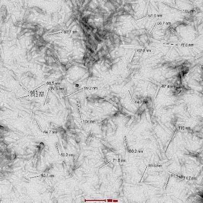
Electron Microscopy: Recombinant Mouse alpha-Synuclein Active, Pre-formed Fibrils, (Type 1) Protein [NBP2-61596] - TEM of Type 1 mouse alpha synuclein Pre-formed Fibrils (NBP2-61596). Fibrils were sonicated and image was taken at 100kx magnification.
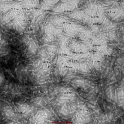
Electron Microscopy: Recombinant Mouse alpha-Synuclein Active, Pre-formed Fibrils, (Type 1) Protein [NBP2-61596] - TEM of Type 1 mouse alpha synuclein Pre-formed Fibrils (NBP2-61596). Image was taken at 100kx magnification.
 Protein [NBP2-61596]")
Immunohistochemistry: Recombinant Mouse alpha-Synuclein Active, Pre-formed Fibrils, (Type 1) Protein [NBP2-61596]
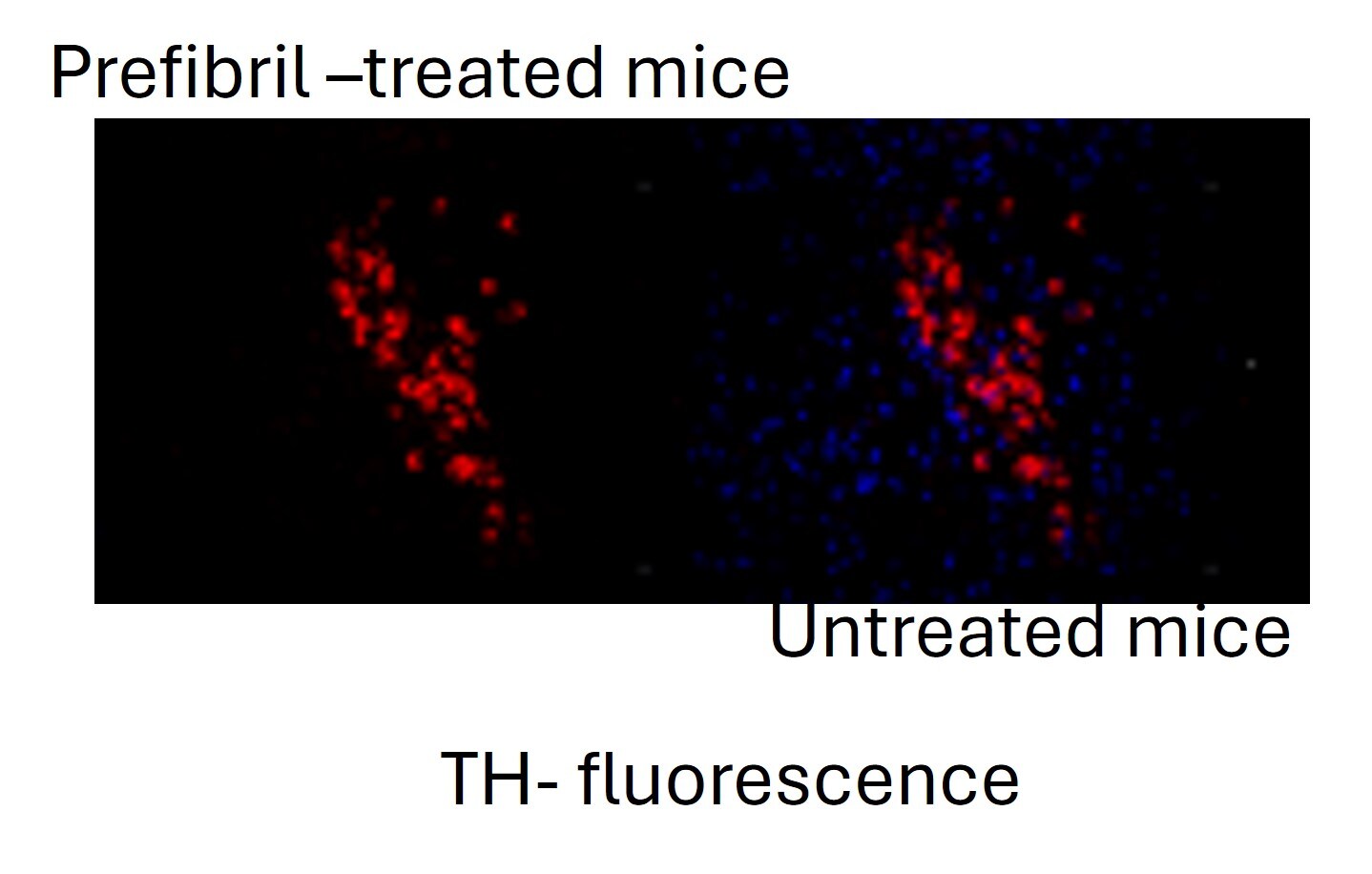
Immunohistochemistry: Recombinant Mouse alpha-Synuclein Active, Pre-formed Fibrils, (Type 1) Protein [NBP2-61596] - TH fluorescence in mouse brain. Image from a verified customer review.Formulation, Preparation, and Storage
NBP2-61596
| Formulation | PBS |
| Concentration | Please see the vial label for concentration. If unlisted please contact technical services. |
| Shipping | The product is shipped with dry ice or equivalent. Upon receipt, store it immediately at the temperature recommended below. |
| Stability & Storage | Store at -80C. Avoid freeze-thaw cycles. |
Background: alpha-Synuclein
A number of studies have revealed that alpha-synuclein aggregation is a hallmark feature in a number of neurodegenerative diseases, referred to as synucleinopathies (2-4). Alpha-synuclein protein aggregates are a large component of Lewy bodies that are present in Parkinson's disease (PD), Lewy body dementia (LBD), and multiple system atrophy (1-6). Research has shown phosphorylation of alpha-synuclein at Ser129 moves the protein from the nucleus to the cytoplasm and promotes fibril formation associated with synucleinopathies (1,2,5). Recent studies also suggest that alpha-synuclein accumulation can prevent mitochondrial import machinery causing mitochondrial dysfunction that is often observed in neurodegeneration (5). It is thought that preventing alpha-synuclein aggregation may prevent PD, thus alpha-synuclein is a target for many potential therapeutic interventions aimed at decreasing aggregate formation or increasing clearance (1,2,4-6).
References
1. Villar-Pique, A., Lopes da Fonseca, T., & Outeiro, T. F. (2016). Structure, function and toxicity of alpha-synuclein: the Bermuda triangle in synucleinopathies. Journal of neurochemistry. https://doi.org/10.1111/jnc.13249
2. Emamzadeh F. N. (2016). Alpha-synuclein structure, functions, and interactions. Journal of research in medical sciences : the official journal of Isfahan University of Medical Sciences. https://doi.org/10.4103/1735-1995.181989
3. Burre J. (2015). The Synaptic Function of alpha-Synuclein. Journal of Parkinson's disease. https://doi.org/10.3233/JPD-150642
4. Lashuel, H. A., Overk, C. R., Oueslati, A., & Masliah, E. (2013). The many faces of alpha-synuclein: from structure and toxicity to therapeutic target. Nature reviews. Neuroscience. https://doi.org/10.1038/nrn3406
5. Rocha, E. M., De Miranda, B., & Sanders, L. H. (2018). Alpha-synuclein: Pathology, mitochondrial dysfunction and neuroinflammation in Parkinson's disease. Neurobiology of disease. https://doi.org/10.1016/j.nbd.2017.04.004
6. O'Leary, E. I., & Lee, J. C. (2019). Interplay between alpha-synuclein amyloid formation and membrane structure. Biochimica et biophysica acta. Proteins and proteomics. https://doi.org/10.1016/j.bbapap.2018.09.012
Alternate Names
NACP, PARK1, PARK4, SNCA, Synuclein-alpha
Gene Symbol
SNCA
Additional alpha-Synuclein Products
Product Documents
Certificate of Analysis
To download a Certificate of Analysis, please enter a lot or batch number in the search box below.
Product Specific Notices
This product is for research use only and is not approved for use in humans or in clinical diagnosis. This product is guaranteed for 1 year from date of receipt.
Related Research Areas
Citations for Recombinant Mouse alpha-Synuclein Active, Pre-formed Fibrils, (Type 1) Protein
 Powered by Bioz
Customer Reviews (1)
5 out of 5
1 Customer Rating
Have you used Recombinant Mouse alpha-Synuclein Active, Pre-formed Fibrils, (Type 1) Protein?
Submit a review and receive an Amazon gift card!
$25/€18/£15/$25CAN/¥2500 Yen for a review with an image
$10/€7/£6/$10CAN/¥1110 Yen for a review without an image
Submit a review
Customer Images
Showing
1
-
1 of
1 review
Showing All
Filter By:
-
Verified Customer | Posted 05/12/2025TH fluorescence in mouse brainmice injected pre-fibril in brain

There are no reviews that match your criteria.
Protocols
Find general support by application which include: protocols, troubleshooting, illustrated assays, videos and webinars.
- Antigen Retrieval Protocol (PIER)
- Antigen Retrieval for Frozen Sections Protocol
- Appropriate Fixation of IHC/ICC Samples
- Cellular Response to Hypoxia Protocols
- Chromogenic IHC Staining of Formalin-Fixed Paraffin-Embedded (FFPE) Tissue Protocol
- Chromogenic Immunohistochemistry Staining of Frozen Tissue
- ClariTSA™ Fluorophore Kits
- Detection & Visualization of Antibody Binding
- Fluorescent IHC Staining of Frozen Tissue Protocol
- Graphic Protocol for Heat-induced Epitope Retrieval
- Graphic Protocol for the Preparation and Fluorescent IHC Staining of Frozen Tissue Sections
- Graphic Protocol for the Preparation and Fluorescent IHC Staining of Paraffin-embedded Tissue Sections
- Graphic Protocol for the Preparation of Gelatin-coated Slides for Histological Tissue Sections
- IHC Sample Preparation (Frozen sections vs Paraffin)
- Immunofluorescent IHC Staining of Formalin-Fixed Paraffin-Embedded (FFPE) Tissue Protocol
- Immunohistochemistry (IHC) and Immunocytochemistry (ICC) Protocols
- Immunohistochemistry Frozen Troubleshooting
- Immunohistochemistry Paraffin Troubleshooting
- Preparing Samples for IHC/ICC Experiments
- Preventing Non-Specific Staining (Non-Specific Binding)
- Primary Antibody Selection & Optimization
- Protocol for Heat-Induced Epitope Retrieval (HIER)
- Protocol for Making a 4% Formaldehyde Solution in PBS
- Protocol for VisUCyte™ HRP Polymer Detection Reagent
- Protocol for the Preparation & Fixation of Cells on Coverslips
- Protocol for the Preparation and Chromogenic IHC Staining of Frozen Tissue Sections
- Protocol for the Preparation and Chromogenic IHC Staining of Frozen Tissue Sections - Graphic
- Protocol for the Preparation and Chromogenic IHC Staining of Paraffin-embedded Tissue Sections
- Protocol for the Preparation and Chromogenic IHC Staining of Paraffin-embedded Tissue Sections - Graphic
- Protocol for the Preparation and Fluorescent IHC Staining of Frozen Tissue Sections
- Protocol for the Preparation and Fluorescent IHC Staining of Paraffin-embedded Tissue Sections
- Protocol for the Preparation of Gelatin-coated Slides for Histological Tissue Sections
- R&D Systems Quality Control Western Blot Protocol
- TUNEL and Active Caspase-3 Detection by IHC/ICC Protocol
- The Importance of IHC/ICC Controls
- Troubleshooting Guide: Immunohistochemistry
- Troubleshooting Guide: Western Blot Figures
- Western Blot Conditions
- Western Blot Protocol
- Western Blot Protocol for Cell Lysates
- Western Blot Troubleshooting
- Western Blot Troubleshooting Guide
- View all Protocols, Troubleshooting, Illustrated assays and Webinars
FAQs
Showing
1
-
1 of
1 FAQ
Showing All
-
Q: I'm looking for an alpha-Synuclein antibody with an epitope located in the first half (N-terminus) of the protein - preferably a monoclonal antibody. Can you help me with that?
A:
Please take a look at NB110-57475. It has been validated for human, rat and mouse and the applications ICC and WB and the epitope it detects is in the N terminal.
Loading...