CCL2/MCP1 Antibody - BSA Free
Novus Biologicals | Catalog # NBP1-07034
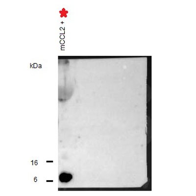
![Western Blot: CCL2/MCP1 AntibodyBSA Free [NBP1-07034]](https://resources.rndsystems.com/images/products/CCL2-MCP1-Antibody-Western-Blot-NBP1-07034-img0004.jpg "Western Blot: CCL2/MCP1 AntibodyBSA Free [NBP1-07034]")
Key Product Details
Species Reactivity
Validated:
Cited:
Predicted:
Applications
Validated:
Cited:
Label
Antibody Source
Format
Product Specifications
Immunogen
Reactivity Notes
Localization
Clonality
Host
Isotype
Theoretical MW
Disclaimer note: The observed molecular weight of the protein may vary from the listed predicted molecular weight due to post translational modifications, post translation cleavages, relative charges, and other experimental factors.
Scientific Data Images for CCL2/MCP1 Antibody - BSA Free
Western Blot: CCL2/MCP1 AntibodyBSA Free [NBP1-07034]
Western Blot: CCL2/MCP1 Antibody [NBP1-07034] - Analysis of recombinant mouse MCP1. Image provided via product review by verified customer.![Western Blot: CCL2/MCP1 AntibodyBSA Free [NBP1-07034]](https://resources.rndsystems.com/images/products/CCL2-MCP1-Antibody-Western-Blot-NBP1-07034-img0003.jpg "Western Blot: CCL2/MCP1 AntibodyBSA Free [NBP1-07034]")
Western Blot: CCL2/MCP1 AntibodyBSA Free [NBP1-07034]
Western Blot: CCL2/MCP1 Antibody [NBP1-07034] - Analysis of MCP1 in Hela whole cell lysates.Applications for CCL2/MCP1 Antibody - BSA Free
Western Blot
Reviewed Applications
Read 1 review rated 5 using NBP1-07034 in the following applications:
Formulation, Preparation, and Storage
Purification
Formulation
Format
Preservative
Concentration
Shipping
Stability & Storage
Background: CCL2/MCP1
Alternate Names
Gene Symbol
Additional CCL2/MCP1 Products
Product Documents for CCL2/MCP1 Antibody - BSA Free
Certificate of Analysis
To download a Certificate of Analysis, please enter a lot or batch number in the search box below.
Product Specific Notices for CCL2/MCP1 Antibody - BSA Free
This product is for research use only and is not approved for use in humans or in clinical diagnosis. Primary Antibodies are guaranteed for 1 year from date of receipt.
Citations for CCL2/MCP1 Antibody - BSA Free
 Powered by Bioz
Customer Reviews for CCL2/MCP1 Antibody - BSA Free (1)
Have you used CCL2/MCP1 Antibody - BSA Free?
Submit a review and receive an Amazon gift card!
$25/€18/£15/$25CAN/¥2500 Yen for a review with an image
$10/€7/£6/$10CAN/¥1110 Yen for a review without an image
Submit a review
Customer Images
-
Application: Western BlotSample Tested: Recombinant Mouse MPC1 proteinSpecies: MouseVerified Customer | Posted 06/07/2013Western Blot analysis of recombinant mouse MCP1

There are no reviews that match your criteria.
Protocols
View specific protocols for CCL2/MCP1 Antibody - BSA Free (NBP1-07034):
Procedure Guide for NBP1-07034 - MCP-1 Antibody
Western Blot Protocol
1. Perform SDS-PAGE (4-12% MOPS) on samples to be analyzed, loading 40 ug of total protein per lane.
2. Transfer proteins to Nitrocellulose according to the instructions provided by the manufacturer of the transfer
apparatus.
3. Rinse membrane with dH2O and then stain the blot using Ponceau S for 1-2 minutes to access the transfer of
proteins onto the nitrocellulose membrane. Rinse the blot in water to remove excess stain and mark the lane locations
and locations of molecular weight markers using a pencil.
4. Rinse the blot in TBS for approximately 5 minutes.
5. Block the membrane using 5% NFDM + 1% BSA in TBS + Tween, 1 hour at RT.
6. Rinse the membrane in dH2O and then wash the membrane in wash buffer [TBS + 0.1% Tween] 3 times for 10
minutes each.
7. Dilute the rabbit anti-MCP-1 primary antibody (NBP1-07034) in blocking buffer and incubate 1 hour at room
temperature.
8. Rinse the membrane in dH2O and then wash the membrane in wash buffer [TBS + 0.1% Tween] 3 times for 10
minutes each.
9. Apply the diluted rabbit-IgG HRP-conjugated secondary antibody in blocking buffer (as per manufacturers
instructions) and incubate 1 hour at room temperature.
10. Wash the blot in wash buffer [TBS + 0.1% Tween] 3 times for 10 minutes each (this step can be repeated as
required to reduce background).
11. Apply the detection reagent of choice in accordance with the manufacturers instructions (Pierce ECL).
Note: Tween-20 can be added to the blocking or antibody dilution buffer at a final concentration of 0.05-0.2%, provided
it does not interfere with antibody-antigen binding.
Find general support by application which include: protocols, troubleshooting, illustrated assays, videos and webinars.
- Cellular Response to Hypoxia Protocols
- R&D Systems Quality Control Western Blot Protocol
- Troubleshooting Guide: Western Blot Figures
- Western Blot Conditions
- Western Blot Protocol
- Western Blot Protocol for Cell Lysates
- Western Blot Troubleshooting
- Western Blot Troubleshooting Guide
- View all Protocols, Troubleshooting, Illustrated assays and Webinars
FAQs for CCL2/MCP1 Antibody - BSA Free
-
Q: Our customer would like to perform IHC with samples from Rat. She has questions regarding antibodies of choice: 1. Is it possible to use NB400-142 for IHC? 2. I am having difficulty of choosing between NBP1-77679 and NBP1-77680. Would you please help suggest? 3. Although it is described as IHC (-) on the product page of NBP1-07034, This antibody did not work in IHC with mouse tissue is also described. How about Rat samples? Is it possible that the antibody can work in IHC with rat tissues?
A: To answer your questions about NB400-142, this antibody has not been validated for IHC and we cannot guarantee that it will work. Both NBP1-77679 and NBP1-77680 have been validated for use in rat and IHC. The difference between these 2 antibodies is the immunogen as they were raised against different regions of the protein. Please point out the immunogen to your customer and have them decide what would be best for their research. NBP1-07034 does not work in IHC, it is only guaranteed for Western blot in human, mouse and rat. When an application is followed by a (-) I would not recommend using this application in any species.
-
Q: Would you please let me know what buffer I should use for WB with antibody (MCP1 Antibody)?
A: In western blot, we recommend diluting this antibody in 1% non-fat dry milk in TBST (0.1% tween-20). This is the same diluent we use for our secondary antibody. For blocking, we use 5% non-fat dry milk in TBST for 1 hour at room temperature.
-
Q: Our customer would like to perform IHC with samples from Rat. She has questions regarding antibodies of choice: 1. Is it possible to use NB400-142 for IHC? 2. I am having difficulty of choosing between NBP1-77679 and NBP1-77680. Would you please help suggest? 3. Although it is described as IHC (-) on the product page of NBP1-07034, This antibody did not work in IHC with mouse tissue is also described. How about Rat samples? Is it possible that the antibody can work in IHC with rat tissues?
A: To answer your questions about NB400-142, this antibody has not been validated for IHC and we cannot guarantee that it will work. Both NBP1-77679 and NBP1-77680 have been validated for use in rat and IHC. The difference between these 2 antibodies is the immunogen as they were raised against different regions of the protein. Please point out the immunogen to your customer and have them decide what would be best for their research. NBP1-07034 does not work in IHC, it is only guaranteed for Western blot in human, mouse and rat. When an application is followed by a (-) I would not recommend using this application in any species.
-
Q: Would you please let me know what buffer I should use for WB with antibody (MCP1 Antibody)?
A: In western blot, we recommend diluting this antibody in 1% non-fat dry milk in TBST (0.1% tween-20). This is the same diluent we use for our secondary antibody. For blocking, we use 5% non-fat dry milk in TBST for 1 hour at room temperature.