CD36 Antibody (D-2712) - BSA Free
Novus Biologicals | Catalog # NB110-59724
Key Product Details
Species Reactivity
Validated:
Mouse, Rat, Human (Negative)
Cited:
Human, Mouse, Rat
Applications
Validated:
Immunohistochemistry, Immunohistochemistry-Paraffin, Western Blot, Immunoprecipitation
Cited:
Immunohistochemistry, Immunohistochemistry-Paraffin, Western Blot, Proximity Ligation Assay, Westen Blot
Label
Unconjugated
Antibody Source
Monoclonal Mouse IgA Kappa Clone # D-2712
Format
BSA Free
Loading...
Product Specifications
Immunogen
This CD36 Antibody (D-2712) was developed against adenovirus expressing recombinant mouse CD36 [Uniprot: Q08857]
Reactivity Notes
Does not react with human.
Localization
Membrane
Marker
Endothelial Cell Marker
Clonality
Monoclonal
Host
Mouse
Isotype
IgA Kappa
Theoretical MW
110 kDa.
Disclaimer note: The observed molecular weight of the protein may vary from the listed predicted molecular weight due to post translational modifications, post translation cleavages, relative charges, and other experimental factors.
Disclaimer note: The observed molecular weight of the protein may vary from the listed predicted molecular weight due to post translational modifications, post translation cleavages, relative charges, and other experimental factors.
Scientific Data Images for CD36 Antibody (D-2712) - BSA Free
![Western Blot: CD36 Antibody (D-2712)BSA Free [NB110-59724]](https://resources.rndsystems.com/images/products/CD36-SR-B3-Antibody-D-2712-Western-Blot-NB110-59724-img0007.jpg "Western Blot: CD36 Antibody (D-2712)BSA Free [NB110-59724]")
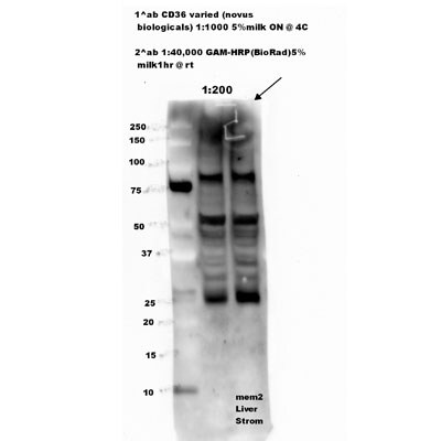
Western Blot: CD36 Antibody (D-2712)BSA Free [NB110-59724]
Western Blot: CD36 Antibody (D-2712) [NB110-59724] - Detection of CD36 in mouse adipose lysate.![Immunohistochemistry: CD36 Antibody (D-2712) - BSA Free [NB110-59724]](https://resources.rndsystems.com/images/products/CD36-SR-B3-Antibody-D-2712-Immunohistochemistry-NB110-59724-img0006.jpg "Immunohistochemistry: CD36 Antibody (D-2712) - BSA Free [NB110-59724]")
Immunohistochemistry: CD36 Antibody (D-2712) - BSA Free [NB110-59724]
Immunohistochemistry: CD36 Antibody (D-2712) [NB110-59724] - Staining of CD36 in mouse liver. - BSA Free [NB110-59724] -")
Western Blot: CD36 Antibody (D-2712) - BSA Free [NB110-59724] -
Effect of MSCs on cardiac expressions of proteins regulating cholesterol, triglycerides and FFA metabolism. (A) CS exposure caused significant elevation of cardiac expression of LDL-R, which was attenuated in both the BM-MSCs/CS and iPSC-MSCs/CS groups. (B) CS led to increased cardiac expression of HMGCR, which was attenuated in the iPSC-MSCs/CS group. (C) CS reduced cardiac expression of ABCA1, which was restored in the iPSC-MSCs/CS group. (D) CS exposure significantly reduced protein expression of LPL, which was restored in the iPSC-MSCs/CS group. (E) CS exposure led to a significant reduction in protein expression of cardiac CD36, which was significantly restored in the iPSC-MSCs/CS group. (F) Cardiac FAS protein expression was inhibited after CS exposure, which showed a trend of restoration but not reaching significant level in both the BM-MSCs/CS and iPSC-MSCs/CS groups. Cardiac levels of protein expression were normalized to GAPDH levels and expressed as fold change vs. relative control. Data are expressed as mean +/- SEM; n = 7–8. Data were analyzed by one-way ANOVA. ∗p < 0.05, ∗∗p < 0.01, ∗∗∗p < 0.001 for comparison between the CS and SA groups. #p < 0.05, ##p < 0.01 for comparison between the BM-MSCs/CS or iPSC-MSCs/CS groups and CS group, respectively. LDL-R, low density lipoprotein receptor; GAPDH, glyceraldehyde 3-phosphate dehydrogenase; HMGCR, 3-hydroxy-3-methylglutaryl coenzyme A reductase; ABCA1, ATP-binding cassette protein-A1; LPL, Lipoprotein lipase; CD36, fatty acid translocase (FAT)/CD36; FAS, fatty acid synthase. Image collected and cropped by CiteAb from the following open publication (https://pubmed.ncbi.nlm.nih.gov/28804458), licensed under a CC-BY license. Not internally tested by Novus Biologicals.Applications for CD36 Antibody (D-2712) - BSA Free
Application
Recommended Usage
Immunohistochemistry
1:100-1:250
Immunohistochemistry-Paraffin
1:100-1:250
Immunoprecipitation
10-20 ug
Western Blot
1:500
Application Notes
In Western blot, a band is observed at ~70-80 kDa. The theoretical molecular weight of CD36 is ~53 kDa. The difference in theoretical MW and actual MW, as seen in Western blot, is most likely due to the heavy glycosylation and palmitoylation of this protein.
Reviewed Applications
Read 1 review rated 3 using NB110-59724 in the following applications:
Formulation, Preparation, and Storage
Purification
Protein G purified
Formulation
PBS
Format
BSA Free
Preservative
0.05% Sodium Azide
Concentration
1.0 mg/ml
Shipping
The product is shipped with polar packs. Upon receipt, store it immediately at the temperature recommended below.
Stability & Storage
Store at 4C short term. Aliquot and store at -20C long term. Avoid freeze-thaw cycles.
Background: CD36
The expression of CD36 has been reported in platelets, erythrocytes, monocytes, differentiated adipocytes, skeletal muscle, mammary epithelial cells, spleen cells, some skin microdermal endothelial cells, and in cancer. Circulating levels of soluble CD36 (sCD36) has also been reported in chronic inflammatory disease such as type 2 diabetes and chronic kidney disease. CD36 participates in angiogenesis, innate immunity, and the clearance of apoptotic phagocytes. In lipid metabolism, CD36 functions as a macrophage receptor for oxidized LDL and as an adipocyte receptor/transporter for long-chain FFAs. Plasmodium falciparum, the parasite that causes malaria, binds CD36 via PfEMP1 proteins, tethering parasite-infected erythrocytes to endothelial receptors (5). Anti-CD36 isoantibodies have been detected in Type 1 CD36-deficient mothers and is implicated as the cause of fetal/neonatal alloimmune thrombocytopenia (6).
References
1) Febbraio, M., Hajjar, D. P., & Silverstein, R. L. (2001). CD36: a class B scavenger receptor involved in angiogenesis, atherosclerosis, inflammation, and lipid metabolism. The Journal of clinical investigation, 108(6), 785-791. PMID: 11560944
2) Silverstein RL, Febbraio M. (2000) CD36 and atherosclerosis. Curr Opin Lipidol. 2000 11(5):483-91. PMID: 11048891.
3) Endemann G, Stanton LW, Madden KS, Bryant CM, White RT, Protter AA. (1993) CD36 is a receptor for oxidized low density lipoprotein. J Biol Chem. 268(16):11811-6. PMID: 7685021.
4) Wang, J., & Li, Y. (2019). CD36 tango in cancer: signaling pathways and functions. Theranostics, 9(17), 4893-4908. PMID: 31410189
5) Hsieh FL, Turner L, Bolla JR, Robinson CV, Lavstsen T, Higgins MK. (2016) The structural basis for CD36 binding by the malaria parasite. Nat Commun. 7:12837. PMID: 27667267
6) Gruarin P, Ulliers D, Thorne RF, Alessio M. (2000) Methionine 156 in the immunodominant domain of CD36 contributes to define the epitope recognized by the NL07 MoAb. Mol Cell Biochem 214, 115-121. PMID: 11195795.
Alternate Names
BDPLT10, CD36 antigen (collagen type I receptor, thrombospondin receptor), CD36 molecule (thrombospondin receptor), CHDS7, cluster determinant 36, FAT, Fatty acid translocase, Glycoprotein IIIb, GP3B, GP4, GPIIIB, GPIV, leukocyte differentiation antigen CD36, PAS IV, PAS-4 protein, PASIV, platelet glycoprotein 4, platelet glycoprotein IV, SCARB3, scavenger receptor class B, member 3
Gene Symbol
CD36
Additional CD36 Products
Product Documents for CD36 Antibody (D-2712) - BSA Free
Certificate of Analysis
To download a Certificate of Analysis, please enter a lot or batch number in the search box below.
Product Specific Notices for CD36 Antibody (D-2712) - BSA Free
This product is for research use only and is not approved for use in humans or in clinical diagnosis. Primary Antibodies are guaranteed for 1 year from date of receipt.
Citations for CD36 Antibody (D-2712) - BSA Free
 Powered by Bioz
Customer Reviews for CD36 Antibody (D-2712) - BSA Free (1)
3 out of 5
1 Customer Rating
Have you used CD36 Antibody (D-2712) - BSA Free?
Submit a review and receive an Amazon gift card!
$25/€18/£15/$25CAN/¥2500 Yen for a review with an image
$10/€7/£6/$10CAN/¥1110 Yen for a review without an image
Submit a review
Customer Images
Showing
1
-
1 of
1 review
Showing All
Filter By:
-
Application: Western BlotSample Tested: Liver whole tissue homogenate, Sample Amount: 50ugSpecies: RatVerified Customer | Posted 11/28/2011

There are no reviews that match your criteria.
Protocols
View specific protocols for CD36 Antibody (D-2712) - BSA Free (NB110-59724):
CD36 Antibody (D-2712):
Antigen Unmasking:
Bring slides to a boil in 10mM sodium citrate buffer (pH 6.0) then maintain at a sub-boiling temperature for 10 minutes. Cool slides on bench top for 30 minutes.
Staining:
1. Wash sections in dH2O three times for 5 minutes each.
2. Wash section in wash buffer (1X PBS/0.1% Tween-20 (1X PBST)) for 5 minutes.
3. Block each section with 100-400 ul blocking solution (1X PBST, 5% goat serum) for 1 hour at room temperature.
4. Remove blocking solution and add 100-400 ul primary antibody diluted in 1X PBST, 5% goat serum to each section. Incubate overnight at 4C.
5. Remove antibody solution and wash sections in wash buffer three times for 5 minutes each.
6. Add 100-400 ul biotinylated secondary antibody, diluted in 1X PBST, 5% goat serum. Incubate 30 minutes at room temperature.
7. Remove secondary antibody solution and wash sections three times with wash buffer for 5 minutes each.
8. Add 100-400 ul Streptavidin-HRP reagent to each section and incubate for 30 minutes at room temperature.
9. Wash sections three times in wash buffer for 5 minutes each.
10. Add 100-400 ul DAB substrate to each section and monitor staining closely.
11. As soon as the sections develop, immerse slides in dH2O.
12. Counterstain sections in hematoxylin.
13. Wash sections in dH2O two times for 5 minutes each.
14. Dehydrate sections.
15. Mount the coverslips.
CD36 Antibody (D-2712):
1. Perform SDS-PAGE (4-12% MOPS) on samples to be analyzed, loading 30 ug of total protein per lane.
2. Transfer proteins to Nitrocellulose according to the instructions provided by the manufacturer of the transfer apparatus.
3. Rinse membrane with dH2O and then stain the blot using Ponceau S for 1-2 minutes to access the transfer of proteins onto the nitrocellulose membrane. Rinse the blot in water to remove excess stain and mark the lane locations and locations of molecular weight markers using a pencil.
4. Rinse the blot in TBS for approximately 5 minutes.
5. Block the membrane using 5% non-fat dry milk + 1% BSA in TBS, 1 hour at room temperature.
6. Rinse the membrane in dH2O and then wash the membrane in wash buffer [TBS + 0.1% Tween] 3 times for 10 minutes each.
7. Dilute the rabbit anti-CD36 primary antibody (NB 110-59724) in blocking buffer and incubate 2 hours at room temperature.
8. Rinse the membrane in dH2O and then wash the membrane in wash buffer [TBS + 0.1% Tween] 3 times for 10 minutes each.
9. Apply the diluted mouse-IgG HRP-conjugated secondary antibody in blocking buffer (as per manufacturer's instructions) and incubate 1 hour at room temperature.
10. Wash the blot in wash buffer [TBS + 0.1% Tween] 3 times for 10 minutes each (this step can be repeated as required to reduce background).
11. Apply the detection reagent of choice in accordance with the manufacturer's instructions (Pierce, ECL).
**Note: Tween-20 can be added to the blocking or antibody dilution buffer at a final concentration of 0.05-0.2%, provided it does not interfere with antibody-antigen binding.
Find general support by application which include: protocols, troubleshooting, illustrated assays, videos and webinars.
- Antigen Retrieval Protocol (PIER)
- Antigen Retrieval for Frozen Sections Protocol
- Appropriate Fixation of IHC/ICC Samples
- Cellular Response to Hypoxia Protocols
- Chromogenic IHC Staining of Formalin-Fixed Paraffin-Embedded (FFPE) Tissue Protocol
- Chromogenic Immunohistochemistry Staining of Frozen Tissue
- ClariTSA™ Fluorophore Kits
- Detection & Visualization of Antibody Binding
- Fluorescent IHC Staining of Frozen Tissue Protocol
- Graphic Protocol for Heat-induced Epitope Retrieval
- Graphic Protocol for the Preparation and Fluorescent IHC Staining of Frozen Tissue Sections
- Graphic Protocol for the Preparation and Fluorescent IHC Staining of Paraffin-embedded Tissue Sections
- Graphic Protocol for the Preparation of Gelatin-coated Slides for Histological Tissue Sections
- IHC Sample Preparation (Frozen sections vs Paraffin)
- Immunofluorescent IHC Staining of Formalin-Fixed Paraffin-Embedded (FFPE) Tissue Protocol
- Immunohistochemistry (IHC) and Immunocytochemistry (ICC) Protocols
- Immunohistochemistry Frozen Troubleshooting
- Immunohistochemistry Paraffin Troubleshooting
- Immunoprecipitation Protocol
- Preparing Samples for IHC/ICC Experiments
- Preventing Non-Specific Staining (Non-Specific Binding)
- Primary Antibody Selection & Optimization
- Protocol for Heat-Induced Epitope Retrieval (HIER)
- Protocol for Making a 4% Formaldehyde Solution in PBS
- Protocol for VisUCyte™ HRP Polymer Detection Reagent
- Protocol for the Preparation & Fixation of Cells on Coverslips
- Protocol for the Preparation and Chromogenic IHC Staining of Frozen Tissue Sections
- Protocol for the Preparation and Chromogenic IHC Staining of Frozen Tissue Sections - Graphic
- Protocol for the Preparation and Chromogenic IHC Staining of Paraffin-embedded Tissue Sections
- Protocol for the Preparation and Chromogenic IHC Staining of Paraffin-embedded Tissue Sections - Graphic
- Protocol for the Preparation and Fluorescent IHC Staining of Frozen Tissue Sections
- Protocol for the Preparation and Fluorescent IHC Staining of Paraffin-embedded Tissue Sections
- Protocol for the Preparation of Gelatin-coated Slides for Histological Tissue Sections
- R&D Systems Quality Control Western Blot Protocol
- TUNEL and Active Caspase-3 Detection by IHC/ICC Protocol
- The Importance of IHC/ICC Controls
- Troubleshooting Guide: Immunohistochemistry
- Troubleshooting Guide: Western Blot Figures
- Western Blot Conditions
- Western Blot Protocol
- Western Blot Protocol for Cell Lysates
- Western Blot Troubleshooting
- Western Blot Troubleshooting Guide
- View all Protocols, Troubleshooting, Illustrated assays and Webinars
Loading...