CD36 Antibody - BSA Free
Novus Biologicals | Catalog # NB400-145
![Immunohistochemistry: CD36 Antibody - BSA Free [NB400-145]](https://resources.rndsystems.com/images/products/CD36-Antibody-Immunohistochemistry-NB400-145-img0009.jpg "Immunohistochemistry: CD36 Antibody - BSA Free [NB400-145]")
Key Product Details
Species Reactivity
Validated:
Human, Mouse, Amphibian
Cited:
Human, Mouse, Amphibian
Applications
Validated:
Immunohistochemistry, Immunohistochemistry-Paraffin, Western Blot, Immunoblotting, Flow Cytometry, Immunocytochemistry/ Immunofluorescence, Simple Western, SDS-Page
Cited:
Western Blot, Immunoblotting, Flow Cytometry, Immunofluorescence, Immunocytochemistry/ Immunofluorescence, SDS-Page
Label
Unconjugated
Antibody Source
Polyclonal Rabbit IgG
Format
BSA Free
Loading...
Product Specifications
Immunogen
This CD36 Antibody was developed against a synthetic peptide mapping to a region of human CD36 between residues 300-400 [Uniprot# P16671]
Reactivity Notes
Amphibian reactivity reported in scientific literature (PMID: 31736973).
Localization
Integral membrane
Marker
Endothelial Cell Marker
Clonality
Polyclonal
Host
Rabbit
Isotype
IgG
Scientific Data Images for CD36 Antibody - BSA Free
Immunohistochemistry: CD36 Antibody - BSA Free [NB400-145]
CD36-Antibody-Immunohistochemistry-NB400-145-img0009.jpg![Simple Western: CD36 AntibodyBSA Free [NB400-145]](https://resources.rndsystems.com/images/products/CD36-Antibody-Simple-Western-NB400-145-img0007.jpg "Simple Western: CD36 AntibodyBSA Free [NB400-145]")
Simple Western: CD36 AntibodyBSA Free [NB400-145]
Simple Western: CD36 Antibody [NB400-145] - Image shows a specific band for CD36 in human adipose lysate. This experiment was performed under reducing conditions using the 12-230 kDa separation system.![Western Blot: CD36 AntibodyBSA Free [NB400-145]](https://resources.rndsystems.com/images/products/CD36-Antibody-Western-Blot-NB400-145-img0006.jpg "Western Blot: CD36 AntibodyBSA Free [NB400-145]")
Western Blot: CD36 AntibodyBSA Free [NB400-145]
Western Blot: CD36 Antibody [NB400-145] - Detection of CD36 in human adipocyte extract (30 ug). Lane 1: 0.5 ug/ml NB 400-145; lane 2: 2 ug/ml NB 400-145. ECL: 3 second exposure.
Western Blot: CD36 Antibody - BSA Free [NB400-145] -
Western Blot: CD36 Antibody - BSA Free [NB400-145] - CD36 ligand disrupts TLR2-CD36 interaction modulated TLR2 heterodimer-signaling. (A) Confocal imaging of central retina cryosection stained with CD11b (white), CD36 (red), TLR2 (green), & DAPI (blue) from blue light-challenged WT mice. Scale bar = 25 μm. (B) High magnification (3X) shows subretinal CD11b-positive cells (white) with the co-localisation of CD36 (red) & TLR2 (green). Scale bar = 5 μm. (C-F) Peritoneal MPs were stimulated with 300 ng/ml R-FSL1 in the presence of 10−7 M MPE-001 or vehicle. (C) MPE-001 disrupted the interaction between CD36 labeled with Cy5 (red) & TLR2 labeled with Cy3 (green) as assessed by FRET after 5 min stimulation with R-FSL1. (D) Percentage of energy transfer measured using LSM-700 confocal microscope (Zeiss). Data in B,C are representative of 3-4 independent experiments. (E) Phosphorylated & total Western blot density bands of IRAK4, IKK alpha beta & P65-NF kappa B, JNK & P38 in peritoneal MPs from CD36+/+ & CD36−/− mice stimulated with R-FSL1. (F) Quantification of P65-NF kappa B following stimulation of CD36+/+ & CD36−/− peritoneal MPs with R-FSL1 using ELISA-based assay. Data in C-F are representative of 3 independent experiments (n = 3/group). In (D,F) one-way ANOVA test with Newman-Keuls post-test for multiple comparison was performed. *P < 0.05, **P < 0.01 & ***P < 0.001 vs R-FSL1. Data are shown as mean ± S.E.M. Image collected & cropped by CiteAb from the following publication (https://pubmed.ncbi.nlm.nih.gov/31501473), licensed under a CC-BY license. Not internally tested by Novus Biologicals.
Immunocytochemistry/ Immunofluorescence: CD36 Antibody - BSA Free [NB400-145] -
Immunocytochemistry/ Immunofluorescence: CD36 Antibody - BSA Free [NB400-145] - CD36 ligand disrupts TLR2-CD36 interaction modulated TLR2 heterodimer-signaling. (A) Confocal imaging of central retina cryosection stained with CD11b (white), CD36 (red), TLR2 (green), & DAPI (blue) from blue light-challenged WT mice. Scale bar = 25 μm. (B) High magnification (3X) shows subretinal CD11b-positive cells (white) with the co-localisation of CD36 (red) & TLR2 (green). Scale bar = 5 μm. (C-F) Peritoneal MPs were stimulated with 300 ng/ml R-FSL1 in the presence of 10−7 M MPE-001 or vehicle. (C) MPE-001 disrupted the interaction between CD36 labeled with Cy5 (red) & TLR2 labeled with Cy3 (green) as assessed by FRET after 5 min stimulation with R-FSL1. (D) Percentage of energy transfer measured using LSM-700 confocal microscope (Zeiss). Data in B,C are representative of 3-4 independent experiments. (E) Phosphorylated & total Western blot density bands of IRAK4, IKK alpha beta & P65-NF kappa B, JNK & P38 in peritoneal MPs from CD36+/+ & CD36−/− mice stimulated with R-FSL1. (F) Quantification of P65-NF kappa B following stimulation of CD36+/+ & CD36−/− peritoneal MPs with R-FSL1 using ELISA-based assay. Data in C-F are representative of 3 independent experiments (n = 3/group). In (D,F) one-way ANOVA test with Newman-Keuls post-test for multiple comparison was performed. *P < 0.05, **P < 0.01 & ***P < 0.001 vs R-FSL1. Data are shown as mean ± S.E.M. Image collected & cropped by CiteAb from the following publication (https://pubmed.ncbi.nlm.nih.gov/31501473), licensed under a CC-BY license. Not internally tested by Novus Biologicals.
Immunohistochemistry-Paraffin: CD36 Antibody [NB400-145] - Staining of CD36 in human adipocytes.
Analysis of a FFPE tissue section of human adipose using 1:200 dilution of CD36 antibody (NB400-145). The staining was developed using HRP labeled anti-rabbit secondary antibody and DAB reagent, and nuclei of cells were counter-stained with hematoxylin.![CD36 Antibody - BSA Free Simple Western: CD36 Antibody - BSA Free [NB400-145] -](https://resources.rndsystems.com/images/products/nb300-327_rabbit-polyclonal-gapdh-antibody-simple-western-14112025740480.jpg "Simple Western: CD36 Antibody - BSA Free [NB400-145] -")
Simple Western: CD36 Antibody - BSA Free [NB400-145] -
The lack of fortilin in M? promotes oxLDL-induced apoptosis, slows cell proliferation, halts foam cell (FC) formation, and reduces CD36 expression. g JESS-based western blot analyses to assess the expression levels of CD36, an oxLDL receptor, with and without oxLDL stimulation, using THP1WT-fortilin and THP1KO-fortilin cells (n?=?3, 3). h Efferocytosis assay. Workflow of the assay ?, representative flow cytometry plots ?, and efferocytosis indices of WT and KO cells ? are shown. In (e), the scale bar equals 100 �m. Data are expressed as mean s.d., P values�determined by one-way ANOVA with Tukey�s comparison (g)�are shown. Image collected and cropped by CiteAb from the following publication (https://pubmed.ncbi.nlm.nih.gov/40640354/), licensed under a CC-BY license. Not internally tested by Novus Biologicals.Applications for CD36 Antibody - BSA Free
Application
Recommended Usage
Flow Cytometry
reported in scientific literature (PMID 32971872)
Immunoblotting
reported in scientific literature (PMID 31501473)
Immunocytochemistry/ Immunofluorescence
reported in scientific literature (PMID 27226602)
Immunohistochemistry
1:200 - 1:400
Immunohistochemistry-Paraffin
1:200 - 1:400
SDS-Page
reported in scientific literature (PMID 31501473)
Simple Western
1:40
Western Blot
1 - 2 ug/ml
Application Notes
In Western blot, a band is seen ~75-80 kDa. May see a very faint band ~130 kDa with a flash exposure and several bands with exposure times longer than 3 seconds. The theoretical molecular weight of CD36 is ~53 kDa. The difference in theoretical MW and actual MW as seen in Western blot is most likely due to the heavy glycosylation and palmitoylation of this protein.
Reviewed Applications
Read 1 review rated 4 using NB400-145 in the following applications:
Flow Cytometry Panel Builder
Bio-Techne Knows Flow Cytometry
Save time and reduce costly mistakes by quickly finding compatible reagents using the Panel Builder Tool.
Advanced Features
- Spectra Viewer - Custom analysis of spectra from multiple fluorochromes
- Spillover Popups - Visualize the spectra of individual fluorochromes
- Antigen Density Selector - Match fluorochrome brightness with antigen density

Formulation, Preparation, and Storage
Purification
Immunogen affinity purified
Formulation
PBS
Format
BSA Free
Preservative
0.02% Sodium Azide
Concentration
1 mg/ml
Shipping
The product is shipped with polar packs. Upon receipt, store it immediately at the temperature recommended below.
Stability & Storage
Store at 4C short term. Aliquot and store at -20C long term. Avoid freeze-thaw cycles.
Background: CD36
The expression of CD36 has been reported in platelets, erythrocytes, monocytes, differentiated adipocytes, skeletal muscle, mammary epithelial cells, spleen cells, some skin microdermal endothelial cells, and in cancer. Circulating levels of soluble CD36 (sCD36) has also been reported in chronic inflammatory disease such as type 2 diabetes and chronic kidney disease. CD36 participates in angiogenesis, innate immunity, and the clearance of apoptotic phagocytes. In lipid metabolism, CD36 functions as a macrophage receptor for oxidized LDL and as an adipocyte receptor/transporter for long-chain FFAs. Plasmodium falciparum, the parasite that causes malaria, binds CD36 via PfEMP1 proteins, tethering parasite-infected erythrocytes to endothelial receptors (5). Anti-CD36 isoantibodies have been detected in Type 1 CD36-deficient mothers and is implicated as the cause of fetal/neonatal alloimmune thrombocytopenia (6).
References
1) Febbraio, M., Hajjar, D. P., & Silverstein, R. L. (2001). CD36: a class B scavenger receptor involved in angiogenesis, atherosclerosis, inflammation, and lipid metabolism. The Journal of clinical investigation, 108(6), 785-791. PMID: 11560944
2) Silverstein RL, Febbraio M. (2000) CD36 and atherosclerosis. Curr Opin Lipidol. 2000 11(5):483-91. PMID: 11048891.
3) Endemann G, Stanton LW, Madden KS, Bryant CM, White RT, Protter AA. (1993) CD36 is a receptor for oxidized low density lipoprotein. J Biol Chem. 268(16):11811-6. PMID: 7685021.
4) Wang, J., & Li, Y. (2019). CD36 tango in cancer: signaling pathways and functions. Theranostics, 9(17), 4893-4908. PMID: 31410189
5) Hsieh FL, Turner L, Bolla JR, Robinson CV, Lavstsen T, Higgins MK. (2016) The structural basis for CD36 binding by the malaria parasite. Nat Commun. 7:12837. PMID: 27667267
6) Gruarin P, Ulliers D, Thorne RF, Alessio M. (2000) Methionine 156 in the immunodominant domain of CD36 contributes to define the epitope recognized by the NL07 MoAb. Mol Cell Biochem 214, 115-121. PMID: 11195795.
Alternate Names
BDPLT10, CD36 antigen (collagen type I receptor, thrombospondin receptor), CD36 molecule (thrombospondin receptor), CHDS7, cluster determinant 36, FAT, Fatty acid translocase, Glycoprotein IIIb, GP3B, GP4, GPIIIB, GPIV, leukocyte differentiation antigen CD36, PAS IV, PAS-4 protein, PASIV, platelet glycoprotein 4, platelet glycoprotein IV, SCARB3, scavenger receptor class B, member 3
Gene Symbol
CD36
Additional CD36 Products
Product Documents for CD36 Antibody - BSA Free
Certificate of Analysis
To download a Certificate of Analysis, please enter a lot or batch number in the search box below.
Product Specific Notices for CD36 Antibody - BSA Free
This product is for research use only and is not approved for use in humans or in clinical diagnosis. Primary Antibodies are guaranteed for 1 year from date of receipt.
Citations for CD36 Antibody - BSA Free
 Powered by Bioz
Customer Reviews for CD36 Antibody - BSA Free (1)
4 out of 5
1 Customer Rating
Have you used CD36 Antibody - BSA Free?
Submit a review and receive an Amazon gift card!
$25/€18/£15/$25CAN/¥2500 Yen for a review with an image
$10/€7/£6/$10CAN/¥1110 Yen for a review without an image
Submit a review
Customer Images
Showing
1
-
1 of
1 review
Showing All
Filter By:
-
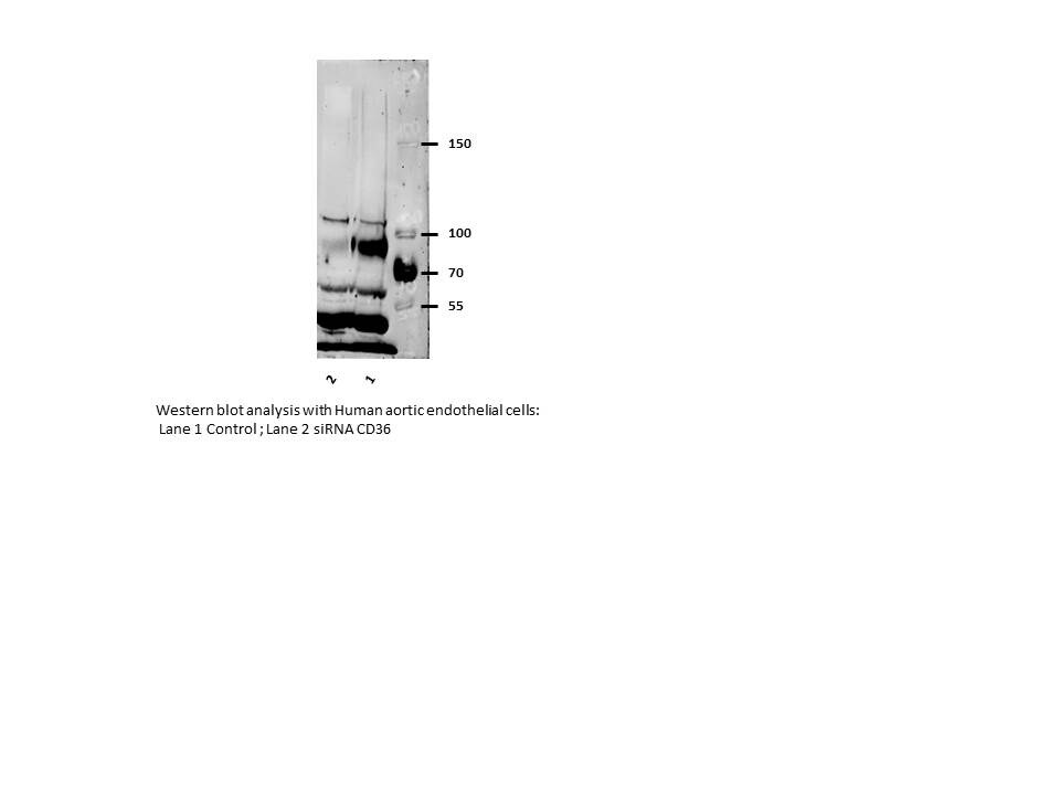
Application: Western BlotSample Tested: Human aortic endothelial cell lysate in RIPA bufferSpecies: HumanVerified Customer | Posted 07/06/2016Western blot analysis with Human aortic endothelial cells

There are no reviews that match your criteria.
Protocols
View specific protocols for CD36 Antibody - BSA Free (NB400-145):
Immunohistochemistry-Paraffin Embedded Sections
Antigen Unmasking:
Bring slides to a boil in 10 mM sodium citrate buffer (pH 6.0) then maintain at a sub-boiling temperature for 10 minutes. Cool slides on bench-top for 30 minutes (keep slides in the sodium citrate buffer at all times).
Staining:
1. Wash sections in deionized water three times for 5 minutes each.
2. Wash sections in PBS for 5 minutes.
3. Block each section with 100-400 ul blocking solution (1% BSA in PBS) for 1 hour at room temperature.
4. Remove blocking solution and add 100-400 ul diluted primary antibody. Incubate overnight at 4 C.
5. Remove antibody solution and wash sections in wash buffer three times for 5 minutes each.
6. Add 100-400 ul HRP polymer conjugated secondary antibody. Incubate 30 minutes at room temperature.
7. Wash sections three times in wash buffer for 5 minutes each.
8. Add 100-400 ul DAB substrate to each section and monitor staining closely.
9. As soon as the sections develop, immerse slides in deionized water.
10. Counterstain sections in hematoxylin.
11. Wash sections in deionized water two times for 5 minutes each.
12. Dehydrate sections.
13. Mount coverslips.
Western Blot Protocol
1. Perform SDS-PAGE on samples to be analyzed, loading 10-25 ug of total protein per lane.
2. Transfer proteins to PVDF membrane according to the instructions provided by the manufacturer of the membrane and transfer apparatus.
3. Stain the membrane with Ponceau S (or similar product) to assess transfer success, and mark molecular weight standards where appropriate.
4. Rinse the blot TBS -0.05% Tween 20 (TBST).
5. Block the membrane in 5% Non-fat milk in TBST (blocking buffer) for at least 1 hour.
6. Wash the membrane in TBST three times for 10 minutes each.
7. Dilute primary antibody in blocking buffer and incubate overnight at 4C with gentle rocking.
8. Wash the membrane in TBST three times for 10 minutes each.
9. Incubate the membrane in diluted HRP conjugated secondary antibody in blocking buffer (as per manufacturer's instructions) for 1 hour at room temperature.
10. Wash the blot in TBST three times for 10 minutes each (this step can be repeated as required to reduce background).
11. Apply the detection reagent of choice in accordance with the manufacturer's instructions.
Find general support by application which include: protocols, troubleshooting, illustrated assays, videos and webinars.
- 7-Amino Actinomycin D (7-AAD) Cell Viability Flow Cytometry Protocol
- Antigen Retrieval Protocol (PIER)
- Antigen Retrieval for Frozen Sections Protocol
- Appropriate Fixation of IHC/ICC Samples
- Cellular Response to Hypoxia Protocols
- Chromogenic IHC Staining of Formalin-Fixed Paraffin-Embedded (FFPE) Tissue Protocol
- Chromogenic Immunohistochemistry Staining of Frozen Tissue
- ClariTSA™ Fluorophore Kits
- Detection & Visualization of Antibody Binding
- Extracellular Membrane Flow Cytometry Protocol
- Flow Cytometry Protocol for Cell Surface Markers
- Flow Cytometry Protocol for Staining Membrane Associated Proteins
- Flow Cytometry Staining Protocols
- Flow Cytometry Troubleshooting Guide
- Fluorescent IHC Staining of Frozen Tissue Protocol
- Graphic Protocol for Heat-induced Epitope Retrieval
- Graphic Protocol for the Preparation and Fluorescent IHC Staining of Frozen Tissue Sections
- Graphic Protocol for the Preparation and Fluorescent IHC Staining of Paraffin-embedded Tissue Sections
- Graphic Protocol for the Preparation of Gelatin-coated Slides for Histological Tissue Sections
- ICC Cell Smear Protocol for Suspension Cells
- ICC Immunocytochemistry Protocol Videos
- ICC for Adherent Cells
- IHC Sample Preparation (Frozen sections vs Paraffin)
- Immunocytochemistry (ICC) Protocol
- Immunocytochemistry Troubleshooting
- Immunofluorescence of Organoids Embedded in Cultrex Basement Membrane Extract
- Immunofluorescent IHC Staining of Formalin-Fixed Paraffin-Embedded (FFPE) Tissue Protocol
- Immunohistochemistry (IHC) and Immunocytochemistry (ICC) Protocols
- Immunohistochemistry Frozen Troubleshooting
- Immunohistochemistry Paraffin Troubleshooting
- Intracellular Flow Cytometry Protocol Using Alcohol (Methanol)
- Intracellular Flow Cytometry Protocol Using Detergents
- Intracellular Nuclear Staining Flow Cytometry Protocol Using Detergents
- Intracellular Staining Flow Cytometry Protocol Using Alcohol Permeabilization
- Intracellular Staining Flow Cytometry Protocol Using Detergents to Permeabilize Cells
- Preparing Samples for IHC/ICC Experiments
- Preventing Non-Specific Staining (Non-Specific Binding)
- Primary Antibody Selection & Optimization
- Propidium Iodide Cell Viability Flow Cytometry Protocol
- Protocol for Heat-Induced Epitope Retrieval (HIER)
- Protocol for Liperfluo
- Protocol for Making a 4% Formaldehyde Solution in PBS
- Protocol for VisUCyte™ HRP Polymer Detection Reagent
- Protocol for the Characterization of Human Th22 Cells
- Protocol for the Characterization of Human Th9 Cells
- Protocol for the Fluorescent ICC Staining of Cell Smears - Graphic
- Protocol for the Fluorescent ICC Staining of Cultured Cells on Coverslips - Graphic
- Protocol for the Preparation & Fixation of Cells on Coverslips
- Protocol for the Preparation and Chromogenic IHC Staining of Frozen Tissue Sections
- Protocol for the Preparation and Chromogenic IHC Staining of Frozen Tissue Sections - Graphic
- Protocol for the Preparation and Chromogenic IHC Staining of Paraffin-embedded Tissue Sections
- Protocol for the Preparation and Chromogenic IHC Staining of Paraffin-embedded Tissue Sections - Graphic
- Protocol for the Preparation and Fluorescent ICC Staining of Cells on Coverslips
- Protocol for the Preparation and Fluorescent ICC Staining of Non-adherent Cells
- Protocol for the Preparation and Fluorescent ICC Staining of Stem Cells on Coverslips
- Protocol for the Preparation and Fluorescent IHC Staining of Frozen Tissue Sections
- Protocol for the Preparation and Fluorescent IHC Staining of Paraffin-embedded Tissue Sections
- Protocol for the Preparation of Gelatin-coated Slides for Histological Tissue Sections
- Protocol for the Preparation of a Cell Smear for Non-adherent Cell ICC - Graphic
- Protocol: Annexin V and PI Staining by Flow Cytometry
- Protocol: Annexin V and PI Staining for Apoptosis by Flow Cytometry
- R&D Systems Quality Control Western Blot Protocol
- TUNEL and Active Caspase-3 Detection by IHC/ICC Protocol
- The Importance of IHC/ICC Controls
- Troubleshooting Guide: Fluorokine Flow Cytometry Kits
- Troubleshooting Guide: Immunohistochemistry
- Troubleshooting Guide: Western Blot Figures
- Western Blot Conditions
- Western Blot Protocol
- Western Blot Protocol for Cell Lysates
- Western Blot Troubleshooting
- Western Blot Troubleshooting Guide
- View all Protocols, Troubleshooting, Illustrated assays and Webinars
Loading...