cGAS Antibody - BSA Free
Novus Biologicals | Catalog # NBP1-86761
![Simple Western: cGAS Antibody [NBP1-86761]](https://resources.rndsystems.com/images/products/cGAS-Antibody-Simple-Western-NBP1-86761-img0008.jpg "Simple Western: cGAS Antibody [NBP1-86761]")
Loading...
Key Product Details
Species Reactivity
Validated:
Human
Cited:
Human
Applications
Validated:
Western Blot, Simple Western
Cited:
Immunohistochemistry-Paraffin, Western Blot, Immunocytochemistry/ Immunofluorescence, IF/IHC
Label
Unconjugated
Antibody Source
Polyclonal Rabbit IgG
Format
BSA Free
Loading...
Product Specifications
Immunogen
This antibody was developed against Recombinant Protein corresponding to amino acids: RKQLRLKPFYLVPKHAKEGNGFQEETWRLSFSHIEKEILNNHGKSKTCCENKEEKCCRKDCLKLMKYLLEQLKERF
Clonality
Polyclonal
Host
Rabbit
Isotype
IgG
Scientific Data Images for cGAS Antibody - BSA Free
Simple Western: cGAS Antibody [NBP1-86761]
Simple Western: cGAS Antibody [NBP1-86761] - Simple Western lane view shows a specific band for MB21D1 in 0.2 mg/ml of RT-4 lysate. This experiment was performed under reducing conditions using the 12-230 kDa separation system.![cGAS Antibody - BSA Free Western Blot: cGAS Antibody - BSA Free [NBP1-86761]](https://resources.rndsystems.com/images/products/nbp1-86761_rabbit-polyclonal-cgas-antibody-30420251371433.jpg "Western Blot: cGAS Antibody - BSA Free [NBP1-86761]")
Western Blot: cGAS Antibody - BSA Free [NBP1-86761]
Analysis in human cell line CACO-2 and human cell line HEK 293.
Western Blot: cGAS Antibody - BSA Free [NBP1-86761] -
Hypoxia inhibited teniposide-induced cGAS-STING activation in human HCC cells. (A) Hep3B and Huh7 cells were cultured under a normoxic (21% O2) or a hypoxic (1% O2) condition for 18 hours and the cellular protein expression of HIF-1 alpha and cGAS was then detected by immunoblotting; beta -actin was used as a loading control. (B) Hep3B and Huh7 cells were transfected with HT-DNA (5 ug/mL) and the cells were then cultured under either normoxic or hypoxic condition for 24 hours; the cellular protein expression of p-IRF3 was detected by immunoblotting. (C) Hep3B and Huh7 cells were treated with teniposide at each IC50, followed by either normoxic or hypoxic culture for 24 hours, and the cellular protein expression of p-IRF3 and p-P65 was then detected by immunoblotting. (D) Hep3B and Huh7 cells were treated as in (C) and the supernatant IFN-beta was then measured by ELISA. (E–F) Hep3B and Huh7 cells were treated as in (C) and the mRNA expression of IFIT-1 and IFIT-2 (E) and CCL5 and CXCL10 (F) was then measured by RT-qPCR. Data in (A), (B) and (C) are representative of three independent experiments. Data in (D), (E) and (F) are shown as mean+/-SD of three independent experiments. *P<0.05, **P<0.01, ***P<0.001. cGAS-STING, cyclic GMP-AMP synthase-stimulator of interferon genes; HCC, hepatocellular carcinoma; HIF-1 alpha, hypoxia inducible factor 1 alpha ; HT-DNA, herring testes-DNA; IC50, 50% inhibitory concentration; IFN-beta, interferon beta ; IRF3, interferon regulatory factor 3; Rel. expression, relative expression; RT-qPCR, real time quantitative PCR; Teni, teniposide. Image collected and cropped by CiteAb from the following open publication (https://pubmed.ncbi.nlm.nih.gov/36002188), licensed under a CC-BY license. Not internally tested by Novus Biologicals.Applications for cGAS Antibody - BSA Free
Application
Recommended Usage
Simple Western
1:30
Western Blot
0.04 - 0.4 ug/mL
Application Notes
In Simple Western only 10 - 15 uL of the recommended dilution is used per data point.
See Simple Western Antibody Database for Simple Western validation: Tested in RT-4, separated by Size, antibody dilution of 1:30, apparent MW was 65 kDa. Separated by Size-Wes, Sally Sue/Peggy Sue.
See Simple Western Antibody Database for Simple Western validation: Tested in RT-4, separated by Size, antibody dilution of 1:30, apparent MW was 65 kDa. Separated by Size-Wes, Sally Sue/Peggy Sue.
Reviewed Applications
Read 1 review rated 4 using NBP1-86761 in the following applications:
Formulation, Preparation, and Storage
Purification
Affinity purified
Formulation
PBS (pH 7.2) and 40% Glycerol
Format
BSA Free
Preservative
0.02% Sodium Azide
Concentration
Concentrations vary lot to lot. See vial label for concentration. If unlisted please contact technical services.
Shipping
The product is shipped with polar packs. Upon receipt, store it immediately at the temperature recommended below.
Stability & Storage
Store at 4C short term. Aliquot and store at -20C long term. Avoid freeze-thaw cycles.
Background: cGAS
Alternate Names
C6orf150, c-GAS, cyclic GMP-AMP synthase, h-cGAS, Mab-21 domain containing 1
Gene Symbol
CGAS
Additional cGAS Products
Product Documents for cGAS Antibody - BSA Free
Certificate of Analysis
To download a Certificate of Analysis, please enter a lot or batch number in the search box below.
Product Specific Notices for cGAS Antibody - BSA Free
This product is for research use only and is not approved for use in humans or in clinical diagnosis. Primary Antibodies are guaranteed for 1 year from date of receipt.
Citations for cGAS Antibody - BSA Free
 Powered by Bioz
Customer Reviews for cGAS Antibody - BSA Free (1)
4 out of 5
1 Customer Rating
Have you used cGAS Antibody - BSA Free?
Submit a review and receive an Amazon gift card!
$25/€18/£15/$25CAN/¥2500 Yen for a review with an image
$10/€7/£6/$10CAN/¥1110 Yen for a review without an image
Submit a review
Customer Images
Showing
1
-
1 of
1 review
Showing All
Filter By:
-
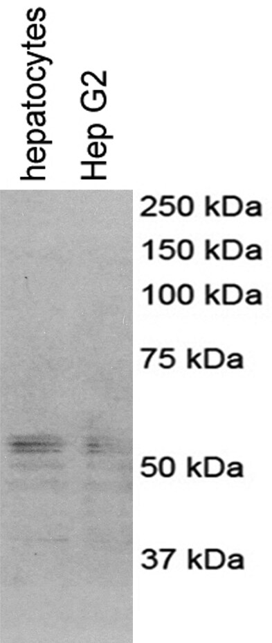
Application: Western BlotSample Tested: human primary hepatocytesSpecies: HumanVerified Customer | Posted 12/18/2017Review for anti-cGAS antibody (NBP1-86761)Name: Anti-cGAS antibody (NBP1-86761) Catalog #: Anti-cGAS antibody (NBP1-86761) Lot Number: Anti-cGAS antibody (NBP1-86761, Lot # G105753) PO/Order Number: Click here to enter text.. WB Image Description (Please provide labels for all lanes): lane 1: human primary hepatocytes; lane 2: Hep G2 Sample Information: Cell Line or Tissue: human primary hepatocytes, Hep G2 Species: human Treatment: No Lysate Preparation: Date of lysate preparation: December 7, 2017 Lysis buffer used: 1X lysis buffer from Cell Signaling by adding PMSF Reducing agent: beta-mercaptoethanol, DTT If boiled (temperature/time): Yes Controls: Positive Control: No Negative Control: No Loading Control (please attach additional images if applicable): No Protein Amount Loaded per lane: 20 ug Antibody Storage Conditions: -20℃ Electrophoresis: Gel Percentage: 10% Electrophoresis Conditions: Tris-Glycine-SDS at room temperature Voltage: 120V Time: 2 hours Membrane Transfer: Method (Submersion/Semi-dry): wet transfer Membrane Type (PVDF/Nitrocellulose): Nitrocellulose Time: 2 hours Voltage: 100V Blocking: Blocking Solution: 5% milk in 1X TBST Time: 1 hour at room temperature Primary Antibody: Dilution: 1/500 Diluent Buffer: 2.5% BSA Incubation Time: overnight Incubation Temperature: 4℃ Washing Conditions: Wash Solution: 1X TBST Time and Repetitions: 5 min each for 3 times Secondary Antibody Manufacturer and Catalog #: Promega, cat # W401B, Lot # 0000187662 Secondary description: goat anti-rabbit secondary antibody Dilution: 1/2000 Diluent Buffer: 3% milk Incubation Time: 1 hour Incubation Temperature: room temperature Detection Method: Detection: ECL (GE, cat # RPN2209, lot #n9838243) Procedure: Add equal volume of A and B, mix and apply on the membranes for 3-5 min before exposure Development Time: 3 min Molecular weight of band(s): ~55 kDa Experimental Concerns and Observations: Specific bands around 55 kDa were observed

There are no reviews that match your criteria.
Protocols
Find general support by application which include: protocols, troubleshooting, illustrated assays, videos and webinars.
- Cellular Response to Hypoxia Protocols
- R&D Systems Quality Control Western Blot Protocol
- Troubleshooting Guide: Western Blot Figures
- Western Blot Conditions
- Western Blot Protocol
- Western Blot Protocol for Cell Lysates
- Western Blot Troubleshooting
- Western Blot Troubleshooting Guide
- View all Protocols, Troubleshooting, Illustrated assays and Webinars
Loading...