CPD Antibody - BSA Free
Novus Biologicals | Catalog # NBP1-91447
![Western Blot: CPD Antibody [NBP1-91447]](https://resources.rndsystems.com/images/products/CPD-Antibody-Western-Blot-NBP1-91447-img0002.jpg "Western Blot: CPD Antibody [NBP1-91447]")
Loading...
Key Product Details
Species Reactivity
Validated:
Human
Cited:
Human
Applications
Validated:
Immunohistochemistry, Immunohistochemistry-Paraffin, Western Blot
Cited:
Western Blot
Label
Unconjugated
Antibody Source
Polyclonal Rabbit IgG
Format
BSA Free
Loading...
Product Specifications
Immunogen
Synthetic peptide directed towards the N terminal of human CPD. Peptide sequence SLNPDGFERAREGDCGFGDGGPSGASGRDNSRGRDLNRSFPDQFSTGEPP. The peptide sequence for this immunogen was taken from within the described region.
Clonality
Polyclonal
Host
Rabbit
Isotype
IgG
Description
The addition of 50% glycerol is optional for those storing this antibody at -20C and not aliquoting smaller units. However, please note that glycerol may interrupt some downstream antibody applications and should be added with caution.
Scientific Data Images for CPD Antibody - BSA Free
Western Blot: CPD Antibody [NBP1-91447]
Western Blot: CPD Antibody [NBP1-91447] - HeLa cell lysate, recommended antibody concentration 0.2 - 1 ug/mL.![Immunohistochemistry-Paraffin: CPD Antibody [NBP1-91447]](https://resources.rndsystems.com/images/products/CPD-Antibody-Immunohistochemistry-Paraffin-NBP1-91447-img0004.jpg "Immunohistochemistry-Paraffin: CPD Antibody [NBP1-91447]")
Immunohistochemistry-Paraffin: CPD Antibody [NBP1-91447]
Immunohistochemistry-Paraffin: CPD Antibody [NBP1-91447] - Human Pineal Tissue.Applications for CPD Antibody - BSA Free
Application
Recommended Usage
Immunohistochemistry
1:10 - 1:500
Immunohistochemistry-Paraffin
1:10-1:500
Western Blot
1.0 ug/ml
Reviewed Applications
Read 1 review rated 2 using NBP1-91447 in the following applications:
Formulation, Preparation, and Storage
Purification
Affinity purified
Formulation
PBS, 2% Sucrose
Format
BSA Free
Preservative
0.09% Sodium Azide
Concentration
0.5 mg/ml
Shipping
The product is shipped with polar packs. Upon receipt, store it immediately at the temperature recommended below.
Stability & Storage
Store at 4C short term. Aliquot and store at -20C long term. Avoid freeze-thaw cycles.
Background: CPD
Alternate Names
carboxypeptidase D, GP180, metallocarboxypeptidase D
Gene Symbol
CPD
UniProt
Additional CPD Products
Product Documents for CPD Antibody - BSA Free
Certificate of Analysis
To download a Certificate of Analysis, please enter a lot or batch number in the search box below.
Product Specific Notices for CPD Antibody - BSA Free
This product is for research use only and is not approved for use in humans or in clinical diagnosis. Primary Antibodies are guaranteed for 1 year from date of receipt.
Citations for CPD Antibody - BSA Free
 Powered by Bioz
Customer Reviews for CPD Antibody - BSA Free (1)
2 out of 5
1 Customer Rating
Have you used CPD Antibody - BSA Free?
Submit a review and receive an Amazon gift card!
$25/€18/£15/$25CAN/¥2500 Yen for a review with an image
$10/€7/£6/$10CAN/¥1110 Yen for a review without an image
Submit a review
Customer Images
Showing
1
-
1 of
1 review
Showing All
Filter By:
-
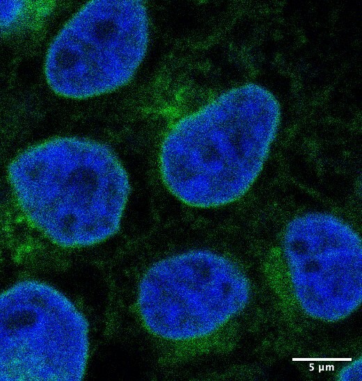
Application: ImmunocytochemistrySample Tested: Hek 293TSpecies: HEK 293T and HumanVerified Customer | Posted 01/06/2020DAPI in blue, anti-CPD in green. Stain in the nuclear membrane and in the cytoplasm. CPD has been reported at the Golgi
 Bio-Techne ResponseThis review was submitted through the legacy Novus Innovators Program, reflecting a new species or application tested on a primary antibody.
There are no reviews that match your criteria.
Protocols
Find general support by application which include: protocols, troubleshooting, illustrated assays, videos and webinars.
- Antigen Retrieval Protocol (PIER)
- Antigen Retrieval for Frozen Sections Protocol
- Appropriate Fixation of IHC/ICC Samples
- Cellular Response to Hypoxia Protocols
- Chromogenic IHC Staining of Formalin-Fixed Paraffin-Embedded (FFPE) Tissue Protocol
- Chromogenic Immunohistochemistry Staining of Frozen Tissue
- ClariTSA™ Fluorophore Kits
- Detection & Visualization of Antibody Binding
- Fluorescent IHC Staining of Frozen Tissue Protocol
- Graphic Protocol for Heat-induced Epitope Retrieval
- Graphic Protocol for the Preparation and Fluorescent IHC Staining of Frozen Tissue Sections
- Graphic Protocol for the Preparation and Fluorescent IHC Staining of Paraffin-embedded Tissue Sections
- Graphic Protocol for the Preparation of Gelatin-coated Slides for Histological Tissue Sections
- IHC Sample Preparation (Frozen sections vs Paraffin)
- Immunofluorescent IHC Staining of Formalin-Fixed Paraffin-Embedded (FFPE) Tissue Protocol
- Immunohistochemistry (IHC) and Immunocytochemistry (ICC) Protocols
- Immunohistochemistry Frozen Troubleshooting
- Immunohistochemistry Paraffin Troubleshooting
- Preparing Samples for IHC/ICC Experiments
- Preventing Non-Specific Staining (Non-Specific Binding)
- Primary Antibody Selection & Optimization
- Protocol for Heat-Induced Epitope Retrieval (HIER)
- Protocol for Making a 4% Formaldehyde Solution in PBS
- Protocol for VisUCyte™ HRP Polymer Detection Reagent
- Protocol for the Preparation & Fixation of Cells on Coverslips
- Protocol for the Preparation and Chromogenic IHC Staining of Frozen Tissue Sections
- Protocol for the Preparation and Chromogenic IHC Staining of Frozen Tissue Sections - Graphic
- Protocol for the Preparation and Chromogenic IHC Staining of Paraffin-embedded Tissue Sections
- Protocol for the Preparation and Chromogenic IHC Staining of Paraffin-embedded Tissue Sections - Graphic
- Protocol for the Preparation and Fluorescent IHC Staining of Frozen Tissue Sections
- Protocol for the Preparation and Fluorescent IHC Staining of Paraffin-embedded Tissue Sections
- Protocol for the Preparation of Gelatin-coated Slides for Histological Tissue Sections
- R&D Systems Quality Control Western Blot Protocol
- TUNEL and Active Caspase-3 Detection by IHC/ICC Protocol
- The Importance of IHC/ICC Controls
- Troubleshooting Guide: Immunohistochemistry
- Troubleshooting Guide: Western Blot Figures
- Western Blot Conditions
- Western Blot Protocol
- Western Blot Protocol for Cell Lysates
- Western Blot Troubleshooting
- Western Blot Troubleshooting Guide
- View all Protocols, Troubleshooting, Illustrated assays and Webinars
Loading...