Dectin-2/CLEC6A Antibody (3D1) - BSA Free
Novus Biologicals | Catalog # NBP2-27159
Key Product Details
Species Reactivity
Human, Primate
Applications
Immunohistochemistry, Immunohistochemistry-Paraffin, Western Blot, Flow Cytometry, Flow (Cell Surface), CyTOF-ready
Label
Unconjugated
Antibody Source
Monoclonal Mouse IgG3 Kappa Clone # 3D1
Format
BSA Free
Loading...
Product Specifications
Immunogen
A blend of two peptides made to amino acids 44-63 and 90-106 of human Dectin-2.
Reactivity Notes
Bovine, Sheep (83%), Mouse (65%), Rat (52%).
Clonality
Monoclonal
Host
Mouse
Isotype
IgG3 Kappa
Scientific Data Images for Dectin-2/CLEC6A Antibody (3D1) - BSA Free
![Western Blot: Dectin-2/CLEC6A Antibody (3D1)BSA Free [NBP2-27159]](https://resources.rndsystems.com/images/products/Dectin-2-CLEC6A-Antibody-3D1-Western-Blot-NBP2-27159-img0006.jpg "Western Blot: Dectin-2/CLEC6A Antibody (3D1)BSA Free [NBP2-27159]")
Western Blot: Dectin-2/CLEC6A Antibody (3D1)BSA Free [NBP2-27159]
Western Blot: Dectin-2/CLEC6A Antibody (3D1) [NBP2-27159] - Analysis of Dectin-2 in human heart lysate in the 1) absence and 2) presence of immunizing peptide, 3) mouse heart lysate, 4) rat heart lysate and 5) human Ramos cell lysate using Dectin-2 antibody at 2 ug/mL. Goat anti-mouse Ig HRP secondary antibody and PicoTect ECL substrate solution were used for this test.![Immunohistochemistry-Paraffin: Dectin-2/CLEC6A Antibody (3D1) - BSA Free [NBP2-27159]](https://resources.rndsystems.com/images/products/Dectin-2-CLEC6A-Antibody-3D1-Immunohistochemistry-Paraffin-NBP2-27159-img0005.jpg "Immunohistochemistry-Paraffin: Dectin-2/CLEC6A Antibody (3D1) - BSA Free [NBP2-27159]")
Immunohistochemistry-Paraffin: Dectin-2/CLEC6A Antibody (3D1) - BSA Free [NBP2-27159]
Immunohistochemistry-Paraffin: Dectin-2/CLEC6A Antibody (3D1) [NBP2-27159] - Normal human spleen tissue stained with Dectin-2 antibody (5 ug/mL), peroxidase-conjugate and DAB chromogen. Human tissue TMA was used for this test.![Flow Cytometry: Dectin-2/CLEC6A Antibody (3D1) - BSA Free [NBP2-27159]](https://resources.rndsystems.com/images/products/Dectin-2-CLEC6A-Antibody-3D1-Flow-Cytometry-NBP2-27159-img0007.jpg "Flow Cytometry: Dectin-2/CLEC6A Antibody (3D1) - BSA Free [NBP2-27159]")
Flow Cytometry: Dectin-2/CLEC6A Antibody (3D1) - BSA Free [NBP2-27159]
Flow Cytometry: Dectin-2/CLEC6A Antibody (3D1) [NBP2-27159] - An intracellular stain was performed on THP-1 cells with Dectin-2/CLEC6A (3D1) antibody NBP2-27159AF488 (blue) and a matched isotype control (orange). Cells were fixed with 4% PFA and then permeablized with 0.1% saponin. Cells were incubated in an antibody dilution of 5 ug/mL for 30 minutes at room temperature. Both antibodies were conjugated to Alexa Fluor 488.![Flow Cytometry: Dectin-2/CLEC6A Antibody (3D1) - BSA Free [NBP2-27159]](https://resources.rndsystems.com/images/products/Dectin-2-CLEC6A-Antibody-3D1-Flow-Cytometry-NBP2-27159-img0004.jpg "Flow Cytometry: Dectin-2/CLEC6A Antibody (3D1) - BSA Free [NBP2-27159]")
Flow Cytometry: Dectin-2/CLEC6A Antibody (3D1) - BSA Free [NBP2-27159]
Flow Cytometry: Dectin-2/CLEC6A Antibody (3D1) [NBP2-27159] - Surface staining of Dectin-2 on 5x10^5 human PBMCs using 0.5 ug of Dectin-2 antibody (red) and 0.5 ug of isotype control antibody (green). [IMGENEX: IMG-6682A] [NBP2-27159]")
Flow Cytometry: Mouse Monoclonal Dectin-2/CLEC6A Antibody (3D1) [IMGENEX: IMG-6682A] [NBP2-27159]
Flow Cytometry: Mouse Monoclonal Dectin-2/CLEC6A Antibody (3D1) [IMGENEX: IMG-6682A] [NBP2-27159] - Dectin-2/CLEC6A expression on human monocyte-derived macrophages. Macrophages were treated with 10 μg/ml of the Dectin-2/CLEC6A Antibody (3D1) [PE] (catalog # NBP2-27159PE) (green) or mouse IgG3 PE isotype control antibody (red) for 30 minutes on ice. Image from a verified customer review. [PE] [NBP2-27159PE] -")
Flow Cytometry: Mouse Monoclonal Dectin-2/CLEC6A Antibody (3D1) [PE] [NBP2-27159PE] -
Flow Cytometry: Mouse Monoclonal Dectin-2/CLEC6A Antibody (3D1) [PE] [NBP2-27159PE] - Dectin-2/CLEC6A expression on human monocyte-derived dendritic cells. DCs were treated with 10 ug/ml of the Dectin-2/CLEC6A antibody (Catalog# NBP2-27159PE) (blue) or mouse IgG3 PE isotype control antibody (red) for 30 minutes on ice. Image from a verified customer review.Applications for Dectin-2/CLEC6A Antibody (3D1) - BSA Free
Application
Recommended Usage
Flow Cytometry
0.5 ug/5x10^5 cells
Immunohistochemistry
1:200
Immunohistochemistry-Paraffin
1:200
Western Blot
2 - 5ug/mL
Reviewed Applications
Read 1 review rated 5 using NBP2-27159 in the following applications:
Flow Cytometry Panel Builder
Bio-Techne Knows Flow Cytometry
Save time and reduce costly mistakes by quickly finding compatible reagents using the Panel Builder Tool.
Advanced Features
- Spectra Viewer - Custom analysis of spectra from multiple fluorochromes
- Spillover Popups - Visualize the spectra of individual fluorochromes
- Antigen Density Selector - Match fluorochrome brightness with antigen density

Formulation, Preparation, and Storage
Purification
Protein G purified
Formulation
PBS
Format
BSA Free
Preservative
0.02% Sodium Azide
Concentration
1.0 mg/ml
Shipping
The product is shipped with polar packs. Upon receipt, store it immediately at the temperature recommended below.
Stability & Storage
Store at 4C short term. Aliquot and store at -20C long term. Avoid freeze-thaw cycles.
Background: Dectin-2/CLEC6A
Additional Dectin-2/CLEC6A Products
Product Documents for Dectin-2/CLEC6A Antibody (3D1) - BSA Free
Certificate of Analysis
To download a Certificate of Analysis, please enter a lot or batch number in the search box below.
Product Specific Notices for Dectin-2/CLEC6A Antibody (3D1) - BSA Free
This product is for research use only and is not approved for use in humans or in clinical diagnosis. Primary Antibodies are guaranteed for 1 year from date of receipt.
Related Research Areas
Customer Reviews for Dectin-2/CLEC6A Antibody (3D1) - BSA Free (1)
5 out of 5
1 Customer Rating
Have you used Dectin-2/CLEC6A Antibody (3D1) - BSA Free?
Submit a review and receive an Amazon gift card!
$25/€18/£15/$25CAN/¥2500 Yen for a review with an image
$10/€7/£6/$10CAN/¥1110 Yen for a review without an image
Submit a review
Customer Images
Showing
1
-
1 of
1 review
Showing All
Filter By:
-
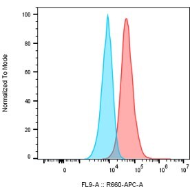
Application: Flow CytometrySample Tested: macrophagesSpecies: HumanVerified Customer | Posted 06/20/2026Detection of CLEC6A in M2c macrophages. The cells were incubated with 2 μl of the human CLEC6A antibody (clone 3D1) (Red) or isotype control (blue) for 30 minutes on ice, followed by a secondary a-mouse IgG APC.The antibody was used to detect human Clec6a expressed in monocyte-derived human macrophages by flow cytometry

There are no reviews that match your criteria.
Protocols
View specific protocols for Dectin-2/CLEC6A Antibody (3D1) - BSA Free (NBP2-27159):
Protocol for Flow Cytometry Cell Surface Staining
Sample Preparation.
1. Grow cells to 60-85% confluency. Flow cytometry requires between 2 x 10⁵ and 1 x 10⁶ cells for optimal performance.
2. If cells are adherent, harvest gently by washing once with staining buffer and then scraping. Avoid using trypsin as this can disrupt certain epitopes of interest. If enzymatic harvest is required, use Accutase, Collagenase, or TrypLE Express for a less damaging option.
3. Reserve 100 uL for counting, then transfer cell volume into a 15 mL conical tube and centrifuge for 4 minutes at 400 RCF.
a. Count cells using a hemocytometer and a 1:1 trypan blue exclusion stain to determine cell viability before starting the flow protocol. If cells appear blue, do not proceed.
4. Re-suspend cells to a concentration of 1 x 10⁶ cells/mL in staining buffer (NBP2-26247).
5. Aliquot out 100 uL samples in accordance with your experimental samples.
Tip: When cell surface and intracellular staining are required in the same sample, it is advisable that the cell surface staining be performed first since the fixation and permeablization steps might reduce the availability of surface antigens.
Cell surface staining
1. Recommended: Block non-specific interactions using 0.5-1 ug of a species specific Fc-blocking reagent such as an anti-mouse CD16/CD32 antibody (NBP1-27946).
2. Add appropriate amount of each antibody (eg. 1 test or 1 ug per sample, as experimentally determined) to 100 uL of staining buffer (NBP2-26247) per sample (eg. use 1 mL of staining buffer for 10 samples).
3. Mix well and incubate at room temperature in dark for 20 minutes.
4. Add 1-2 mL of staining buffer and centrifuge at 400 RCF for 1 minute and discard supernatant.
5. Wash twice by re-suspending cells in staining buffer (2 mL for tubes or 200 uL for wells) and centrifuging at 400 RCF for 5 minutes. Discard supernatant.
6. Add appropriate amount of secondary antibody (as experimentally determined) to each sample.
7. Incubate at room temperature in dark for 20 minutes.
8. Add 1-2 mL of staining buffer and centrifuge at 400 RCF for 1 minute and discard supernatant.
9. Wash twice by re-suspending cells in staining buffer (2 mL for tubes or 200 uL for wells) and centrifuging at 400 RCF for 5 minutes. Discard supernatant.
10. Resuspend in an appropriate volume of staining buffer (usually 500 uL per sample) and proceed with analysis on your flow cytometer.
Immunocytochemistry Protocol
Culture cells to appropriate density in 35 mm culture dishes or 6-well plates.
1. Remove culture medium and wash the cells briefly in PBS. Add 10% formalin to the dish and fix at room temperature for 10 minutes.
2. Remove the formalin and wash the cells in PBS.
3. Permeablize the cells with 0.1% Triton X100 or other suitable detergent for 10 min.
4. Remove the permeablization buffer and wash three times for 10 minutes each in PBS. Be sure to not let the specimen dry out.
5. To block nonspecific antibody binding, incubate in 10% normal goat serum from 1 hour to overnight at room temperature.
6. Add primary antibody at appropriate dilution and incubate overnight at 4C.
7. Remove primary antibody and replace with PBS. Wash three times for 10 minutes each.
8. Add secondary antibody at appropriate dilution. Incubate for 1 hour at room temperature.
9. Remove secondary antibody and replace with PBS. Wash three times for 10 minutes each.
10. Counter stain DNA with DAPi if required.
Western Blot Protocol
1. Perform SDS-PAGE on samples to be analyzed, loading 10-25 ug of total protein per lane.
2. Transfer proteins to PVDF membrane according to the instructions provided by the manufacturer of the membrane and transfer apparatus.
3. Stain the membrane with Ponceau S (or similar product) to assess transfer success, and mark molecular weight standards where appropriate.
4. Rinse the blot TBS -0.05% Tween 20 (TBST).
5. Block the membrane in 5% Non-fat milk in TBST (blocking buffer) for at least 1 hour.
6. Wash the membrane in TBST three times for 10 minutes each.
7. Dilute primary antibody in blocking buffer and incubate overnight at 4C with gentle rocking.
8. Wash the membrane in TBST three times for 10 minutes each.
9. Incubate the membrane in diluted HRP conjugated secondary antibody in blocking buffer (as per manufacturer's instructions) for 1 hour at room temperature.
10. Wash the blot in TBST three times for 10 minutes each (this step can be repeated as required to reduce background).
11. Apply the detection reagent of choice in accordance with the manufacturer's instructions.
Find general support by application which include: protocols, troubleshooting, illustrated assays, videos and webinars.
- 7-Amino Actinomycin D (7-AAD) Cell Viability Flow Cytometry Protocol
- Antigen Retrieval Protocol (PIER)
- Antigen Retrieval for Frozen Sections Protocol
- Appropriate Fixation of IHC/ICC Samples
- Cellular Response to Hypoxia Protocols
- Chromogenic IHC Staining of Formalin-Fixed Paraffin-Embedded (FFPE) Tissue Protocol
- Chromogenic Immunohistochemistry Staining of Frozen Tissue
- ClariTSA™ Fluorophore Kits
- Detection & Visualization of Antibody Binding
- Extracellular Membrane Flow Cytometry Protocol
- Flow Cytometry Protocol for Cell Surface Markers
- Flow Cytometry Protocol for Staining Membrane Associated Proteins
- Flow Cytometry Staining Protocols
- Flow Cytometry Troubleshooting Guide
- Fluorescent IHC Staining of Frozen Tissue Protocol
- Graphic Protocol for Heat-induced Epitope Retrieval
- Graphic Protocol for the Preparation and Fluorescent IHC Staining of Frozen Tissue Sections
- Graphic Protocol for the Preparation and Fluorescent IHC Staining of Paraffin-embedded Tissue Sections
- Graphic Protocol for the Preparation of Gelatin-coated Slides for Histological Tissue Sections
- IHC Sample Preparation (Frozen sections vs Paraffin)
- Immunofluorescent IHC Staining of Formalin-Fixed Paraffin-Embedded (FFPE) Tissue Protocol
- Immunohistochemistry (IHC) and Immunocytochemistry (ICC) Protocols
- Immunohistochemistry Frozen Troubleshooting
- Immunohistochemistry Paraffin Troubleshooting
- Intracellular Flow Cytometry Protocol Using Alcohol (Methanol)
- Intracellular Flow Cytometry Protocol Using Detergents
- Intracellular Nuclear Staining Flow Cytometry Protocol Using Detergents
- Intracellular Staining Flow Cytometry Protocol Using Alcohol Permeabilization
- Intracellular Staining Flow Cytometry Protocol Using Detergents to Permeabilize Cells
- Preparing Samples for IHC/ICC Experiments
- Preventing Non-Specific Staining (Non-Specific Binding)
- Primary Antibody Selection & Optimization
- Propidium Iodide Cell Viability Flow Cytometry Protocol
- Protocol for Heat-Induced Epitope Retrieval (HIER)
- Protocol for Liperfluo
- Protocol for Making a 4% Formaldehyde Solution in PBS
- Protocol for VisUCyte™ HRP Polymer Detection Reagent
- Protocol for the Characterization of Human Th22 Cells
- Protocol for the Characterization of Human Th9 Cells
- Protocol for the Preparation & Fixation of Cells on Coverslips
- Protocol for the Preparation and Chromogenic IHC Staining of Frozen Tissue Sections
- Protocol for the Preparation and Chromogenic IHC Staining of Frozen Tissue Sections - Graphic
- Protocol for the Preparation and Chromogenic IHC Staining of Paraffin-embedded Tissue Sections
- Protocol for the Preparation and Chromogenic IHC Staining of Paraffin-embedded Tissue Sections - Graphic
- Protocol for the Preparation and Fluorescent IHC Staining of Frozen Tissue Sections
- Protocol for the Preparation and Fluorescent IHC Staining of Paraffin-embedded Tissue Sections
- Protocol for the Preparation of Gelatin-coated Slides for Histological Tissue Sections
- Protocol: Annexin V and PI Staining by Flow Cytometry
- Protocol: Annexin V and PI Staining for Apoptosis by Flow Cytometry
- R&D Systems Quality Control Western Blot Protocol
- TUNEL and Active Caspase-3 Detection by IHC/ICC Protocol
- The Importance of IHC/ICC Controls
- Troubleshooting Guide: Fluorokine Flow Cytometry Kits
- Troubleshooting Guide: Immunohistochemistry
- Troubleshooting Guide: Western Blot Figures
- Western Blot Conditions
- Western Blot Protocol
- Western Blot Protocol for Cell Lysates
- Western Blot Troubleshooting
- Western Blot Troubleshooting Guide
- View all Protocols, Troubleshooting, Illustrated assays and Webinars
Loading...
Associated Pathways
