
EIF3C Antibody - BSA Free
Novus Biologicals | Catalog # NB100-511

![Western Blot: EIF3C Antibody [NB100-511]](https://resources.rndsystems.com/images/products/EIF3C-Antibody-Western-Blot-NB100-511-img0005.jpg "Western Blot: EIF3C Antibody [NB100-511]")
Scientific images may be resized, cropped, or adjusted for brightness or contrast for presentation purposes. These updates are limited to presentation and do not affect the underlying scientific interpretation of the data. Where available, additional source data may be provided upon request.
Key Product Details
Validated by
Species Reactivity
Validated:
Cited:
Predicted:
Applications
Validated:
Cited:
Label
Antibody Source
Format
Product Specifications
Immunogen
Reactivity Notes
Clonality
Host
Isotype
Theoretical MW
Disclaimer note: The observed molecular weight of the protein may vary from the listed predicted molecular weight due to post translational modifications, post translation cleavages, relative charges, and other experimental factors.
Scientific Data Images for EIF3C Antibody - BSA Free
Western Blot: EIF3C Antibody [NB100-511]
Western Blot: EIF3C Antibody [NB100-511] - eIF3C expression in naive and activated (overnight) murine B cells. Image submitted by a verified customer review.![Western Blot: EIF3C Antibody [NB100-511]](https://resources.rndsystems.com/images/products/EIF3C-Antibody-Western-Blot-NB100-511-img0004.jpg "Western Blot: EIF3C Antibody [NB100-511]")
Western Blot: EIF3C Antibody [NB100-511]
Western Blot: EIF3C Antibody [NB100-511] - Detection of Human eIF3C/eIF3S8 by Western Blot. Samples: Whole cell lysate (50 ug) from HeLa, 293T, and Jurkat cells prepared using NETN lysis buffer. Antibodies: Affinity purified rabbit anti-eIF3C/eIF3S8 antibody NB100-511 used for WB at 0.1 ug/ml. Detection: Chemiluminescence with an exposure time of 3 minutes.![Immunoprecipitation: EIF3C Antibody [NB100-511]](https://resources.rndsystems.com/images/products/EIF3C-Antibody-Immunoprecipitation-NB100-511-img0006.jpg "Immunoprecipitation: EIF3C Antibody [NB100-511]")
Immunoprecipitation: EIF3C Antibody [NB100-511]
Immunoprecipitation: EIF3C Antibody [NB100-511] - Detection of human eIF3C/eIF3S8 by western blot of immunoprecipitates. Samples: Whole cell lysate (0.5 or 1.0 mg per IP reaction; 20% of IP loaded) from HEK293T cells prepared using NETN lysis buffer. Antibodies: Affinity purified rabbit anti-eIF3C/eIF3S8 antibody NB100-511 used for IP at 6 ug per reaction. eIF3C/eIF3S8 was inefficiently immunoprecipitated by rabbit anti-eIF3C/eIF3S8 antibody from Company B. For blotting immunoprecipitated eIF3C/eIF3S8, NB100-511 was used at 1 ug/ml. Detection: Chemiluminescence with an exposure time of 3 minutes.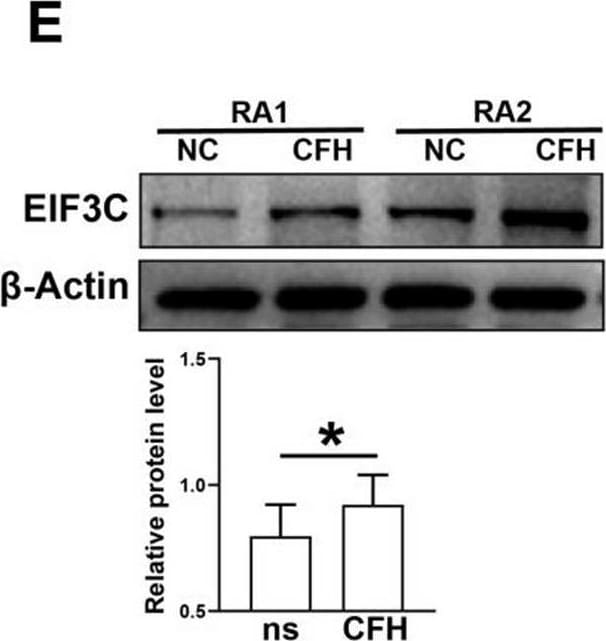
Western Blot: EIF3C Antibody - BSA Free [NB100-511] -
EIF3C is a potential target for CFH to play a role in inhibiting FLSs and monocyte function. Monocytes and FLS from RA patients were selected respectively, and cells from each patient were divided into two groups. One group was left untreated, and one group was treated with 5 μg/ml CFH for 6 h. A Volcano plot indicated up-regulated (red dots) and down-regulated (blue dots) genes (P value < 0.05 and |log2FC|> 0.5) by RNA sequencing in CFH-treated versus untreated RA monocytes. EIF3C is indicated. B Volcano plot indicated up-regulated (red dots) and down-regulated (blue dots) genes (P value < 0.05 and |log2FC|> 0.5) by RNA sequencing in CFH-treated versus untreated RA monocytes. EIF3C and EIF3CL are indicated. C We selected the genes upregulated in the CFH-treated monocytes and the genes upregulated in the CFH-treated FLS identified by RNA-sequencing and determined the intersecting proteins. We obtained one gene, namely EIF3C. DEGs: Differentially Expressed Genes. D The boxplot shows the relative expression of EIF3C and EIF3CL. E CFH upregulated the protein expression of EIF3C by using Western blot. F CFH combined with TNF-alpha upregulated protein expression of EIF3C in both RA FLSs and monocytes by using Western blot. Data are expressed as mean +/- SEM (n = 6). *p < 0.05 Image collected and cropped by CiteAb from the following open publication (https://pubmed.ncbi.nlm.nih.gov/37996918), licensed under a CC-BY license. Not internally tested by Novus Biologicals.
Western Blot: EIF3C Antibody - BSA Free [NB100-511] -
EIF3C is a potential target for CFH to play a role in inhibiting FLSs and monocyte function. Monocytes and FLS from RA patients were selected respectively, and cells from each patient were divided into two groups. One group was left untreated, and one group was treated with 5 μg/ml CFH for 6 h. A Volcano plot indicated up-regulated (red dots) and down-regulated (blue dots) genes (P value < 0.05 and |log2FC|> 0.5) by RNA sequencing in CFH-treated versus untreated RA monocytes. EIF3C is indicated. B Volcano plot indicated up-regulated (red dots) and down-regulated (blue dots) genes (P value < 0.05 and |log2FC|> 0.5) by RNA sequencing in CFH-treated versus untreated RA monocytes. EIF3C and EIF3CL are indicated. C We selected the genes upregulated in the CFH-treated monocytes and the genes upregulated in the CFH-treated FLS identified by RNA-sequencing and determined the intersecting proteins. We obtained one gene, namely EIF3C. DEGs: Differentially Expressed Genes. D The boxplot shows the relative expression of EIF3C and EIF3CL. E CFH upregulated the protein expression of EIF3C by using Western blot. F CFH combined with TNF-alpha upregulated protein expression of EIF3C in both RA FLSs and monocytes by using Western blot. Data are expressed as mean +/- SEM (n = 6). *p < 0.05 Image collected and cropped by CiteAb from the following open publication (https://pubmed.ncbi.nlm.nih.gov/37996918), licensed under a CC-BY license. Not internally tested by Novus Biologicals.Applications for EIF3C Antibody - BSA Free
Immunoprecipitation
Western Blot
Reviewed Applications
Read 1 review rated 5 using NB100-511 in the following applications:
Formulation, Preparation, and Storage
Purification
Formulation
Format
Preservative
Concentration
Shipping
Stability & Storage
Background: EIF3C
Alternate Names
Gene Symbol
UniProt
Additional EIF3C Products
Product Documents for EIF3C Antibody - BSA Free
Certificate of Analysis
To download a Certificate of Analysis, please enter a lot or batch number in the search box below.
Product Specific Notices for EIF3C Antibody - BSA Free
This product is for research use only and is not approved for use in humans or in clinical diagnosis. Primary Antibodies are guaranteed for 1 year from date of receipt.
Citations for EIF3C Antibody - BSA Free
 Powered by Bioz
Customer Reviews for EIF3C Antibody - BSA Free (1)
Have you used EIF3C Antibody - BSA Free?
Submit a review and receive an Amazon gift card!
$25/€18/£15/$25CAN/¥2500 for a review with an image
$10/€7/£6/$10CAN/¥1110 for a review without an image
Submit a review
Customer Images
-
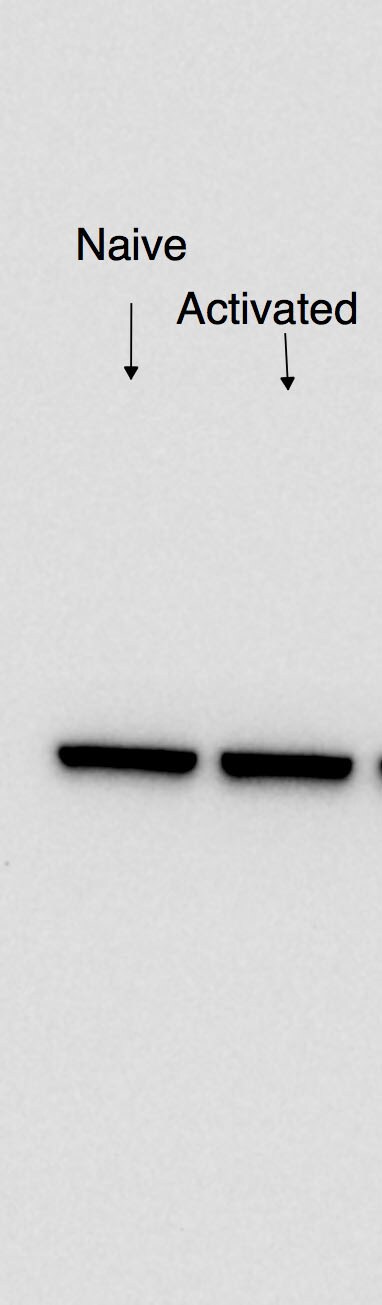
Application: Western BlotSample Tested: B cellsSpecies: MouseVerified Customer | Posted 02/28/2017eIF3C expression in naive and activated (overnight) murine B cells.

There are no reviews that match your criteria.
Protocols
Find general support by application which include: protocols, troubleshooting, illustrated assays, videos and webinars.
- Cellular Response to Hypoxia Protocols
- Immunoprecipitation Protocol
- R&D Systems Quality Control Western Blot Protocol
- Troubleshooting Guide: Western Blot Figures
- Western Blot Conditions
- Western Blot Protocol
- Western Blot Protocol for Cell Lysates
- Western Blot Troubleshooting
- Western Blot Troubleshooting Guide
- View all Protocols, Troubleshooting, Illustrated assays and Webinars