
FGF activity is mediated by a family of type I transmembrane tyrosine kinases, which undergo dimerization and autophosphorylation after ligand binding. Five distinct genes encode closely related FGF receptors, FGFR1 through 5. FGFRs contain three Ig-like domains and a stretch of acidic residues between the first and second Ig-like domains. FGFR1, 2, 3, and -4 have a cytoplasmic split tyrosine-kinase domain, but FGFR5 does not. Multiple forms of FGFR1, 2, and 3 are generated by alternative splicing.
FGFR5/FGFRL1 Antibody (019) - Azide and BSA Free
Novus Biologicals | Catalog # NBP2-90467
Recombinant Monoclonal Antibody

Scientific images may be resized, cropped, or adjusted for brightness or contrast for presentation purposes. These updates are limited to presentation and do not affect the underlying scientific interpretation of the data. Where available, additional source data may be provided upon request.
Key Product Details
Species Reactivity
Mouse
Applications
Western Blot, Immunoprecipitation
Label
Unconjugated
Antibody Source
Recombinant Monoclonal Rabbit IgG Clone # 019 expressed in HEK293
Format
Azide and BSA Free
Loading...
Product Specifications
Immunogen
This antibody was obtained from a rabbit immunized with purified, recombinant Mouse FGFR5/FGFRL1 (Accession#: NP_473412.1; Met1-Pro374).
Clonality
Monoclonal
Host
Rabbit
Isotype
IgG
Description
This antibody can be stored at 2C-8C for one month without detectable loss of activity. Antibody products are stable for twelve months from date of receipt when stored at -20C to -80C. Avoid repeated freeze-thaw cycles.
Scientific Data Images for FGFR5/FGFRL1 Antibody (019) - Azide and BSA Free
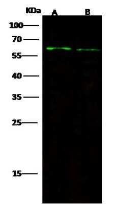
Western Blot: FGFR5/FGFRL1 Antibody (019) [NBP2-90467]
Western Blot: FGFR5/FGFRL1 Antibody (019) [NBP2-90467] - Anti-FGFR5/FGFRL1 rabbit monoclonal antibody at 1:500 dilution. Lane A: 293T Whole Cell Lysate. Lane B: HepG2 Whole Cell Lysate. Lysates/proteins at 30 ug per lane. Secondary Goat Anti-Rabbit IgG H&L (Dylight 800) at 1/10000 dilution. Developed using the Odyssey technique. Performed under reducing conditions. Predicted band size: 55 kDa. Observed band size: 62 kDa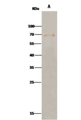
Immunoprecipitation: FGFR5/FGFRL1 Antibody (019) [NBP2-90467]
Immunoprecipitation: FGFR5/FGFRL1 Antibody (019) [NBP2-90467] - Mouse FGFR5/FGFRL1 was immunoprecipitated using: Lane A: 0.5 mg Hela Whole Cell Lysate. 2 ul anti-Mouse FGFR5/FGFRL1 rabbit monoclonal antibody and 15 ul of 50 % Protein G agarose. Primary antibody: Anti-Mouse FGFR5/FGFRL1 rabbit monoclonal antibody, at 1:100 dilution. Secondary antibody: Clean-Bloto IP Detection Reagent (HRP) at 1:1000 dilution. Developed using the DAB staining technique. Performed under reducing conditions. Predicted band size: 55 kDa. Observed band size: 70 kDaApplications for FGFR5/FGFRL1 Antibody (019) - Azide and BSA Free
Application
Recommended Usage
Immunoprecipitation
4-6 uL/mg of lysate
Western Blot
1:500-1:2000
Formulation, Preparation, and Storage
Purification
Protein A purified
Formulation
0.2 um filtered solution in PBS
Format
Azide and BSA Free
Preservative
No Preservative
Concentration
Please see the vial label for concentration. If unlisted please contact technical services.
Shipping
The product is shipped with polar packs. Upon receipt, store it immediately at the temperature recommended below.
Stability & Storage
Store at 4C short term. Aliquot and store at -20C long term. Avoid freeze-thaw cycles.
Background: FGFR5/FGFRL1
Long Name
Fibroblast Growth Factor Receptor-like 1
Alternate Names
FGF R5, FGFRL1
Gene Symbol
FGFRL1
Additional FGFR5/FGFRL1 Products
Product Documents for FGFR5/FGFRL1 Antibody (019) - Azide and BSA Free
Certificate of Analysis
To download a Certificate of Analysis, please enter a lot or batch number in the search box below.
Product Specific Notices for FGFR5/FGFRL1 Antibody (019) - Azide and BSA Free
This product is for research use only and is not approved for use in humans or in clinical diagnosis. Primary Antibodies are guaranteed for 1 year from date of receipt.
Related Research Areas
Customer Reviews for FGFR5/FGFRL1 Antibody (019) - Azide and BSA Free
There are currently no reviews for this product. Be the first to review FGFR5/FGFRL1 Antibody (019) - Azide and BSA Free and earn rewards!
Have you used FGFR5/FGFRL1 Antibody (019) - Azide and BSA Free?
Submit a review and receive an Amazon gift card!
$25/€18/£15/$25CAN/¥2500 for a review with an image
$10/€7/£6/$10CAN/¥1110 for a review without an image
Submit a review
Protocols
Find general support by application which include: protocols, troubleshooting, illustrated assays, videos and webinars.
- Cellular Response to Hypoxia Protocols
- Immunoprecipitation Protocol
- R&D Systems Quality Control Western Blot Protocol
- Troubleshooting Guide: Western Blot Figures
- Western Blot Conditions
- Western Blot Protocol
- Western Blot Protocol for Cell Lysates
- Western Blot Troubleshooting
- Western Blot Troubleshooting Guide
- View all Protocols, Troubleshooting, Illustrated assays and Webinars
Loading...
Associated Pathways

