Meet MitoBrilliant™ Fluorescent Mitochondria Stain
Next-generation fluorescent stains for the localization and labeling of mitochondria in both live and fixed cells.
Properties and Applications of MitoBrilliant Dyes
MitoBrilliant probes have been developed to overcome some common limitations encountered with standard mitochondrial trackers. The range includes 2 types of dyes, MitoBrilliant Live dyes, suitable for live-cell imaging; and a ‘universal’ dye, suitable for both live-cell and fixed-cell imaging.
Excellent Brightness and Photostability
MitoBrilliant Live 646 (Cat. No. 7417) produced a more stable, longer-lasting fluorescent signal than a leading competitor dye in live-cell time lapse imaging.
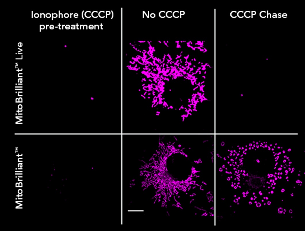
Δψm-Dependent and Δψm-Independent Mitochondrial Staining
- MitoBrilliant Live dyes (Cat. No. 7417 and 7693) accumulate in the mitochondria of live cells in a Δψm dependent manner.
- MitoBrilliant 646 (Cat. No. 7700) can be retained in mitochondria when Δψm is disrupted.
Improved Mitochondrial Staining Fidelity Post-Fixation
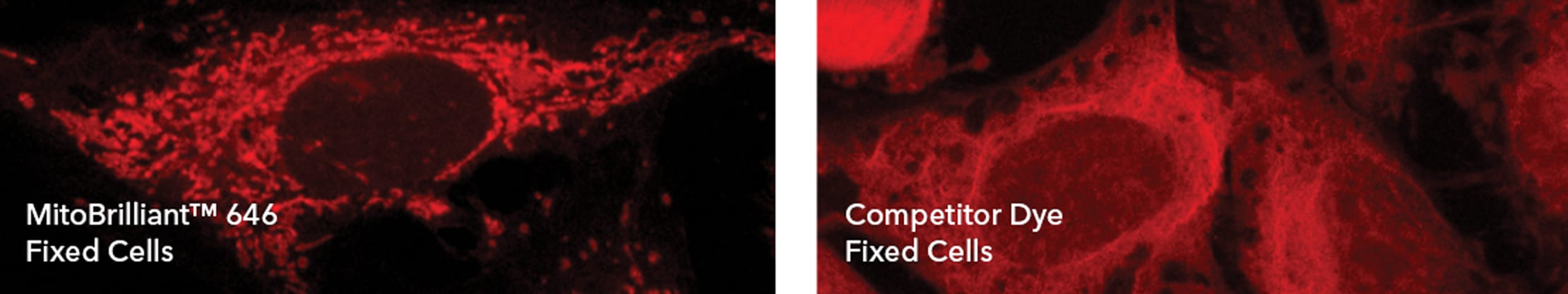
MitoBrilliant 646 (Cat. No. 7700) staining is retained after fixation with PFA and provides significantly improved staining resolution than a leading competitor dye.
MitoBrilliant Dyes Support a Wide Range of Applications
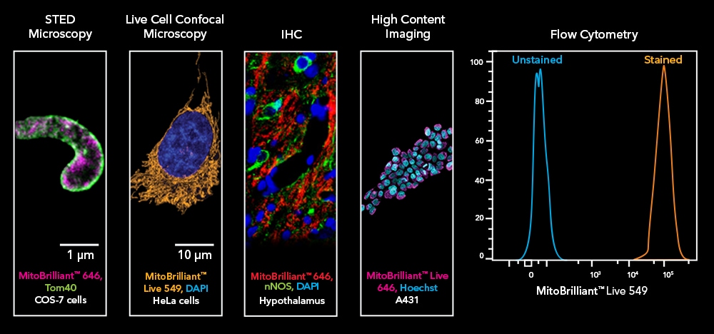
MitoBrilliant dyes enable highly accurate, sensitive, specific and persistent labeling of mitochondria in a wide variety of applications.
MitoBrilliant Technology - Key Features
| Product Name | Abs/Em (nm) | Δψm Dependent | Live/Fixed Cell Use? | Image Without Wash Step? | Demonstrated Applications |
|---|---|---|---|---|---|
| 655/668 | No | Suitable for live and fixed cell work | Yes, but replacing media recommended | Fixed-cell imaging, Live-cell imaging Flow cytometry, IHC/ICC Super-resolution microscopy (STED), High-content screening | |
| 648/662 | Yes | Live-cell work only | Yes, but replacing media recommended | Live-cell imaging, Flow cytometry, High-content screening | |
| 550/568 | Yes | Live-cell work only | Yes, but replacing media recommended | Live-cell imaging, Flow cytometry, High-content screening |

Customer Success Stories
Dr. Jessica Cale
Researcher at CMMIT, Murdoch University, Australia
"MitoBrilliant™ 646 produced excellent results as both a live and fixed cell stain. As a live cell stain MitoBrilliant™ 646 produced a stable signal within 20 minutes. For use as fixed-cell stain optimal conditions were found to be a 1-hour incubation in live cells followed by fixation with ice-cold acetone-methanol."
MitoBrilliant Product Guide
Our guide provides protocols and data for use in different research applications including flow cytometry, immunohistochemistry, live-cell and fixed-cell imaging, and super-resolution microscopy. Please download for more information.