G3BP2 Antibody - BSA Free
Novus Biologicals | Catalog # NBP1-82976
![Immunohistochemistry-Paraffin: G3BP2 Antibody [NBP1-82976]](https://resources.rndsystems.com/images/products/G3BP2-Antibody-Immunohistochemistry-Paraffin-NBP1-82976-img0025.jpg "Immunohistochemistry-Paraffin: G3BP2 Antibody [NBP1-82976]")
Loading...
Key Product Details
Validated by
Orthogonal Validation, Independent Antibodies
Species Reactivity
Validated:
Human
Cited:
Human, Mouse
Predicted:
Rat (92%). Backed by our 100% Guarantee.
Applications
Validated:
Immunohistochemistry, Immunohistochemistry-Paraffin
Cited:
Immunohistochemistry, Western Blot, Immunocytochemistry/ Immunofluorescence, Immunoprecipitation, Proximity Ligation Assay, IF/IHC
Label
Unconjugated
Antibody Source
Polyclonal Rabbit IgG
Format
BSA Free
Loading...
Product Specifications
Immunogen
This antibody was developed against Recombinant Protein corresponding to amino acids: KNLEELEEKSTTPPPAEPVSLPQEPPKPRVEAKPEVQSQPPRVREQRPRERPGFPPRGPRPGRGDMEQNDS
Reactivity Notes
Mouse reactivity reported in scientific literature (PMID: 25893917).
Clonality
Polyclonal
Host
Rabbit
Isotype
IgG
Scientific Data Images for G3BP2 Antibody - BSA Free
![Immunohistochemistry-Paraffin: G3BP2 Antibody [NBP1-82976]](https://resources.rndsystems.com/images/products/G3BP2-Antibody-Immunohistochemistry-Paraffin-NBP1-82976-img0024.jpg "Immunohistochemistry-Paraffin: G3BP2 Antibody [NBP1-82976]")
Immunohistochemistry-Paraffin: G3BP2 Antibody [NBP1-82976]
Immunohistochemistry-Paraffin: G3BP2 Antibody [NBP1-82976] - Staining of human cerebellum, cerebral cortex, skin and testis using Anti-G3BP2 antibody NBP1-82976 (A) shows similar protein distribution across tissues to independent antibody NBP1-82977 (B).![Immunohistochemistry-Paraffin: G3BP2 Antibody [NBP1-82976]](https://resources.rndsystems.com/images/products/G3BP2-Antibody-Immunohistochemistry-Paraffin-NBP1-82976-img0028.jpg "Immunohistochemistry-Paraffin: G3BP2 Antibody [NBP1-82976]")
Immunohistochemistry-Paraffin: G3BP2 Antibody [NBP1-82976]
Immunohistochemistry-Paraffin: G3BP2 Antibody [NBP1-82976] - Staining of human cerebral cortex shows strong cytoplasmic positivity in neuronal cells.![Immunohistochemistry-Paraffin: G3BP2 Antibody [NBP1-82976]](https://resources.rndsystems.com/images/products/G3BP2-Antibody-Immunohistochemistry-Paraffin-NBP1-82976-img0017.jpg "Immunohistochemistry-Paraffin: G3BP2 Antibody [NBP1-82976]")
Immunohistochemistry-Paraffin: G3BP2 Antibody [NBP1-82976]
Immunohistochemistry-Paraffin: G3BP2 Antibody [NBP1-82976] - Staining of human testis shows moderate cytoplasmic positivity in cells in seminiferous ducts and Leydig cells.![Immunohistochemistry-Paraffin: G3BP2 Antibody [NBP1-82976]](https://resources.rndsystems.com/images/products/G3BP2-Antibody-Immunohistochemistry-Paraffin-NBP1-82976-img0026.jpg "Immunohistochemistry-Paraffin: G3BP2 Antibody [NBP1-82976]")
Immunohistochemistry-Paraffin: G3BP2 Antibody [NBP1-82976]
Immunohistochemistry-Paraffin: G3BP2 Antibody [NBP1-82976] - Staining of human skin shows very weak cytoplasmic positivity in epidermal cells.![Immunohistochemistry-Paraffin: G3BP2 Antibody [NBP1-82976]](https://resources.rndsystems.com/images/products/G3BP2-Antibody-Immunohistochemistry-Paraffin-NBP1-82976-img0027.jpg "Immunohistochemistry-Paraffin: G3BP2 Antibody [NBP1-82976]")
Immunohistochemistry-Paraffin: G3BP2 Antibody [NBP1-82976]
Immunohistochemistry-Paraffin: G3BP2 Antibody [NBP1-82976] - Staining of human cerebellum shows strong cytoplasmic positivity in Purkinje cells and in molecular layer.
Western Blot: G3BP2 Antibody - BSA Free [NBP1-82976] -
NCAP interacts with SG proteins. (A) Interaction of NCAP with G3BP1, G3BP2, YTHDF3, USP10 and PKR. A549 cells transfected with GFP‐NCAP were unstressed, treated with arsenite or heat shock and subjected to co‐IP using anti‐His antibodies. The pulldown samples and total cell lysates were subjected to western blotting with indicated antibodies. (B) NCAP reduces global protein synthesis in A549 cells as measured by the Click chemistry‐AHA method (see Methods for more details). Ponceau staining and actin were used as loading controls. (C) NCAP does not affect the phosphorylation of eIF2 alpha. A549 cells transfected with GFP‐NCAP were untreated, treated with arsenite or heat shock and subjected to western blotting with antibodies against p‐eIF2 alpha, eIF2 alpha, NCAP and ACTIN. Image collected and cropped by CiteAb from the following open publication (https://pubmed.ncbi.nlm.nih.gov/34780058), licensed under a CC-BY license. Not internally tested by Novus Biologicals.Applications for G3BP2 Antibody - BSA Free
Application
Recommended Usage
Immunohistochemistry
1:50 - 1:200
Immunohistochemistry-Paraffin
1:50 - 1:200
Application Notes
For IHC-Paraffin, HIER pH 6 retrieval is recommended.
Reviewed Applications
Read 1 review rated 5 using NBP1-82976 in the following applications:
Formulation, Preparation, and Storage
Purification
Affinity purified
Formulation
PBS (pH 7.2) and 40% Glycerol
Format
BSA Free
Preservative
0.02% Sodium Azide
Concentration
Concentrations vary lot to lot. See vial label for concentration. If unlisted please contact technical services.
Shipping
The product is shipped with polar packs. Upon receipt, store it immediately at the temperature recommended below.
Stability & Storage
Store at 4C short term. Aliquot and store at -20C long term. Avoid freeze-thaw cycles.
Background: G3BP2
Alternate Names
G3BP-2, GAP SH3 domain-binding protein 2, GTPase activating protein (SH3 domain) binding protein 2, KIAA0660, ras GTPase-activating protein-binding protein 2, Ras-GTPase activating protein SH3 domain-binding protein 2
Gene Symbol
G3BP2
Additional G3BP2 Products
Product Documents for G3BP2 Antibody - BSA Free
Certificate of Analysis
To download a Certificate of Analysis, please enter a lot or batch number in the search box below.
Product Specific Notices for G3BP2 Antibody - BSA Free
This product is for research use only and is not approved for use in humans or in clinical diagnosis. Primary Antibodies are guaranteed for 1 year from date of receipt.
Citations for G3BP2 Antibody - BSA Free
 Powered by Bioz
Customer Reviews for G3BP2 Antibody - BSA Free (1)
5 out of 5
1 Customer Rating
Have you used G3BP2 Antibody - BSA Free?
Submit a review and receive an Amazon gift card!
$25/€18/£15/$25CAN/¥2500 Yen for a review with an image
$10/€7/£6/$10CAN/¥1110 Yen for a review without an image
Submit a review
Customer Images
Showing
1
-
1 of
1 review
Showing All
Filter By:
-
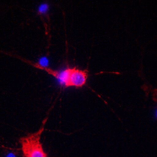
Application: ImmunofluorescenceSample Tested: Cortical neuronsSpecies: MouseVerified Customer | Posted 09/01/2017G3BP2 staning is detected in neurites and cell bodies in mouse cortical neuros (dilution: 1:500 [red], DAPI: blue).

There are no reviews that match your criteria.
Protocols
Find general support by application which include: protocols, troubleshooting, illustrated assays, videos and webinars.
- Antigen Retrieval Protocol (PIER)
- Antigen Retrieval for Frozen Sections Protocol
- Appropriate Fixation of IHC/ICC Samples
- Cellular Response to Hypoxia Protocols
- Chromogenic IHC Staining of Formalin-Fixed Paraffin-Embedded (FFPE) Tissue Protocol
- Chromogenic Immunohistochemistry Staining of Frozen Tissue
- ClariTSA™ Fluorophore Kits
- Detection & Visualization of Antibody Binding
- Fluorescent IHC Staining of Frozen Tissue Protocol
- Graphic Protocol for Heat-induced Epitope Retrieval
- Graphic Protocol for the Preparation and Fluorescent IHC Staining of Frozen Tissue Sections
- Graphic Protocol for the Preparation and Fluorescent IHC Staining of Paraffin-embedded Tissue Sections
- Graphic Protocol for the Preparation of Gelatin-coated Slides for Histological Tissue Sections
- IHC Sample Preparation (Frozen sections vs Paraffin)
- Immunofluorescent IHC Staining of Formalin-Fixed Paraffin-Embedded (FFPE) Tissue Protocol
- Immunohistochemistry (IHC) and Immunocytochemistry (ICC) Protocols
- Immunohistochemistry Frozen Troubleshooting
- Immunohistochemistry Paraffin Troubleshooting
- Preparing Samples for IHC/ICC Experiments
- Preventing Non-Specific Staining (Non-Specific Binding)
- Primary Antibody Selection & Optimization
- Protocol for Heat-Induced Epitope Retrieval (HIER)
- Protocol for Making a 4% Formaldehyde Solution in PBS
- Protocol for VisUCyte™ HRP Polymer Detection Reagent
- Protocol for the Preparation & Fixation of Cells on Coverslips
- Protocol for the Preparation and Chromogenic IHC Staining of Frozen Tissue Sections
- Protocol for the Preparation and Chromogenic IHC Staining of Frozen Tissue Sections - Graphic
- Protocol for the Preparation and Chromogenic IHC Staining of Paraffin-embedded Tissue Sections
- Protocol for the Preparation and Chromogenic IHC Staining of Paraffin-embedded Tissue Sections - Graphic
- Protocol for the Preparation and Fluorescent IHC Staining of Frozen Tissue Sections
- Protocol for the Preparation and Fluorescent IHC Staining of Paraffin-embedded Tissue Sections
- Protocol for the Preparation of Gelatin-coated Slides for Histological Tissue Sections
- TUNEL and Active Caspase-3 Detection by IHC/ICC Protocol
- The Importance of IHC/ICC Controls
- Troubleshooting Guide: Immunohistochemistry
- View all Protocols, Troubleshooting, Illustrated assays and Webinars
Loading...