GLI-2 Antibody - BSA Free
Novus Biologicals | Catalog # NBP2-23602
![Immunocytochemistry/ Immunofluorescence: GLI-2 Antibody - BSA Free [NBP2-23602]](https://resources.rndsystems.com/images/products/GLI-2-Antibody-Immunocytochemistry-Immunofluorescence-NBP2-23602-img0007.jpg "Immunocytochemistry/ Immunofluorescence: GLI-2 Antibody - BSA Free [NBP2-23602]")
Key Product Details
Species Reactivity
Validated:
Human, Mouse, Rat
Cited:
Mouse, Rat
Predicted:
Hamster (92%). Backed by our 100% Guarantee.
Applications
Validated:
Immunohistochemistry, Immunohistochemistry-Paraffin, Immunohistochemistry-Frozen, Immunocytochemistry/ Immunofluorescence
Cited:
Immunohistochemistry-Frozen, IF/IHC
Label
Unconjugated
Antibody Source
Polyclonal Rabbit IgG
Format
BSA Free
Loading...
Product Specifications
Immunogen
A synthetic peptide made to an internal portion of the human GLI-2 protein (between residues 300-400) [UniProt# P10070]
Reactivity Notes
Rat reactivity reported in scientific literature (PMID: 26210874).
Localization
Nuclear and cytoplasm
Clonality
Polyclonal
Host
Rabbit
Isotype
IgG
Scientific Data Images for GLI-2 Antibody - BSA Free
Immunocytochemistry/ Immunofluorescence: GLI-2 Antibody - BSA Free [NBP2-23602]
Immunocytochemistry/Immunofluorescence: GLI-2 Antibody [NBP2-23602] - HeLa cells were fixed in 4% paraformaldehyde for 10 minutes and permeabilized in 0.5% Triton X-100 in PBS for 5 minutes. The cells were incubated with GLI-2 Antibody (NBP2-23602) at 1 ug/ml overnight at 4C and detected with an anti-rabbit Dylight 488 (Green) at a 1:1000 dilution for 60 minutes. Nuclei were counterstained with DAPI (Blue). Cells were imaged using a 100X objective and digitally deconvolved.![Immunohistochemistry-Paraffin: GLI-2 Antibody - BSA Free [NBP2-23602]](https://resources.rndsystems.com/images/products/GLI-2-Antibody-Immunohistochemistry-Paraffin-NBP2-23602-img0006.jpg "Immunohistochemistry-Paraffin: GLI-2 Antibody - BSA Free [NBP2-23602]")
Immunohistochemistry-Paraffin: GLI-2 Antibody - BSA Free [NBP2-23602]
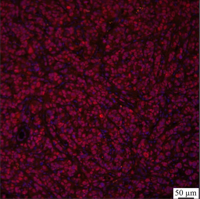
Immunohistochemistry-Paraffin: GLI-2 Antibody [NBP2-23602] - GLI-2 (red) and DAPI (blue) in mouse BT-474 primary tumors (breast cancer tissue). IHC-P image submitted by a verified customer review.![Immunocytochemistry/ Immunofluorescence: GLI-2 Antibody - BSA Free [NBP2-23602]](https://resources.rndsystems.com/images/products/GLI-2-Antibody-Immunocytochemistry-Immunofluorescence-NBP2-23602-img0004.jpg "Immunocytochemistry/ Immunofluorescence: GLI-2 Antibody - BSA Free [NBP2-23602]")
Immunocytochemistry/ Immunofluorescence: GLI-2 Antibody - BSA Free [NBP2-23602]
Immunocytochemistry/Immunofluorescence: GLI-2 Antibody [NBP2-23602] - GLI-2 antibody was tested in HeLa cells with DyLight 488 (green). Nuclei and alpha-tubulin were counterstained with DAPI (blue) and DyLight 550 (red). An antibody dilution of 1:50 was used. Image objective 40x.![Immunohistochemistry: GLI-2 Antibody - BSA Free [NBP2-23602]](https://resources.rndsystems.com/images/products/GLI-2-Antibody-Immunohistochemistry-NBP2-23602-img0005.jpg "Immunohistochemistry: GLI-2 Antibody - BSA Free [NBP2-23602]")
Immunohistochemistry: GLI-2 Antibody - BSA Free [NBP2-23602]
Immunohistochemistry: GLI-2 Antibody [NBP2-23602] - Analysis of GLI-2 in mouse heart.Applications for GLI-2 Antibody - BSA Free
Application
Recommended Usage
Immunocytochemistry/ Immunofluorescence
1:50 - 1:200
Immunohistochemistry
1:200 - 1:300
Immunohistochemistry-Frozen
reported in scientific literature (PMID 26210874)
Immunohistochemistry-Paraffin
1:200 - 1:300
Application Notes
In Immunocytochemistry/Immunofluorescence, cytoplasmic and nuclear staining was observed in HeLa cells. Prior to immunostaining paraffin tissues, antigen retrieval with sodium citrate buffer (pH 6.0) is recommended.
Reviewed Applications
Read 1 review rated 4 using NBP2-23602 in the following applications:
Formulation, Preparation, and Storage
Purification
Immunogen affinity purified
Formulation
PBS
Format
BSA Free
Preservative
0.02% Sodium Azide
Concentration
1.0 mg/ml
Shipping
The product is shipped with polar packs. Upon receipt, store it immediately at the temperature recommended below.
Stability & Storage
Store at 4C short term. Aliquot and store at -20C long term. Avoid freeze-thaw cycles.
Background: GLI-2
Long Name
GLI-Kruppel family member GLI2
Alternate Names
GLI2, THP2
Gene Symbol
GLI2
UniProt
Additional GLI-2 Products
Product Documents for GLI-2 Antibody - BSA Free
Certificate of Analysis
To download a Certificate of Analysis, please enter a lot or batch number in the search box below.
Product Specific Notices for GLI-2 Antibody - BSA Free
This product is for research use only and is not approved for use in humans or in clinical diagnosis. Primary Antibodies are guaranteed for 1 year from date of receipt.
Related Research Areas
Citations for GLI-2 Antibody - BSA Free
 Powered by Bioz
Customer Reviews for GLI-2 Antibody - BSA Free (1)
4 out of 5
1 Customer Rating
Have you used GLI-2 Antibody - BSA Free?
Submit a review and receive an Amazon gift card!
$25/€18/£15/$25CAN/¥2500 Yen for a review with an image
$10/€7/£6/$10CAN/¥1110 Yen for a review without an image
Submit a review
Customer Images
Showing
1
-
1 of
1 review
Showing All
Filter By:
-
Application: Immunohistochemistry-ParaffinSample Tested: Breast cancer tissueSpecies: MouseVerified Customer | Posted 08/17/2021Gli-2 (red) and DAPI (blue) in BT-474 primary tumorsCitrate retrieval buffer

There are no reviews that match your criteria.
Protocols
View specific protocols for GLI-2 Antibody - BSA Free (NBP2-23602):
GLI-2 Antibody:
Immunocytochemistry Protocol
Culture cells to appropriate density on suitable glass coverslips in 35 mm culture dishes or 6-well plates.
1. Remove culture medium and add 10% formalin to the dish. Fix at room temperature for 5-10 minutes.
2. Remove the formalin and add 0.5% Triton-X 100 in TBS to permeabilize the cells. Incubate for 5-10 minutes.
3. Remove the permeabilization buffer and add wash buffer (i.e. PBS or PBS with 0.1% Tween-20). Be sure to not let the specimen dry out. Gently wash three times for 10 minutes.
4. Alternatively, cells can be fixed with -20C methanol for 10 min at room temperature. Remove the methanol and rehydrate in PBS for 10 min before proceeding.
5. To block nonspecific antibody binding incubate in 10% normal goat serum for 1 hour at room temperature.
6. Add primary antibody at appropriate dilution and incubate at room temperature for 1 hour or at 4 degrees C overnight.
7. Remove primary antibody and replace with wash buffer. Gently wash three times for 10 minutes.
8. Add secondary antibody at the appropriate dilution. Incubate for 1 hour at room temperature.
9. Remove antibody and replace with wash buffer. Gently wash three times for 10 minutes.
10. Nuclei can be staining with 4',6' diamino phenylindole (DAPI) at 0.1 ug/ml, or coverslips can be directly mounted in media containing DAPI.
11. Cells can now be viewed with a fluorescence microscope.
*The above information is only intended as a guide. The researcher should determine what protocol best meets their needs. Please follow proper laboratory procedures for the disposal of formalin.
GLI-2 Antibody:
Immunohistochemistry-Paraffin Embedded Sections
Antigen Unmasking:
Bring slides to a boil in 10 mM sodium citrate buffer (pH 6.0) then maintain at a sub-boiling temperature for 10 minutes. Cool slides on bench-top for 30 minutes.
Staining:
1. Wash sections in deionized water three times for 5 minutes each.
2. Wash sections in wash buffer for 5 minutes.
3. Block each section with 100-400 ul blocking solution for 1 hour at room temperature.
4. Remove blocking solution and add 100-400 ul diluted primary antibody. Incubate overnight at 4 degrees C.
5. Remove antibody solution and wash sections in wash buffer three times for 5 minutes each.
6. Add 100-400 ul biotinylated diluted secondary antibody. Incubate 30 minutes at room temperature.
7. Remove secondary antibody solution and wash sections three times with wash buffer for 5 minutes each.
8. Add 100-400 ul Streptavidin-HRP reagent to each section and incubate for 30 minutes at room temperature.
9. Wash sections three times in wash buffer for 5 minutes each.
10. Add 100-400 ul DAB substrate to each section and monitor staining closely.
11. As soon as the sections develop, immerse slides in deionized water.
12. Counterstain sections in hematoxylin.
13. Wash sections in deionized water two times for 5 minutes each.
14. Dehydrate sections.
15. Mount coverslips.
Find general support by application which include: protocols, troubleshooting, illustrated assays, videos and webinars.
- Antigen Retrieval Protocol (PIER)
- Antigen Retrieval for Frozen Sections Protocol
- Appropriate Fixation of IHC/ICC Samples
- Cellular Response to Hypoxia Protocols
- Chromogenic IHC Staining of Formalin-Fixed Paraffin-Embedded (FFPE) Tissue Protocol
- Chromogenic Immunohistochemistry Staining of Frozen Tissue
- ClariTSA™ Fluorophore Kits
- Detection & Visualization of Antibody Binding
- Fluorescent IHC Staining of Frozen Tissue Protocol
- Graphic Protocol for Heat-induced Epitope Retrieval
- Graphic Protocol for the Preparation and Fluorescent IHC Staining of Frozen Tissue Sections
- Graphic Protocol for the Preparation and Fluorescent IHC Staining of Paraffin-embedded Tissue Sections
- Graphic Protocol for the Preparation of Gelatin-coated Slides for Histological Tissue Sections
- ICC Cell Smear Protocol for Suspension Cells
- ICC Immunocytochemistry Protocol Videos
- ICC for Adherent Cells
- IHC Sample Preparation (Frozen sections vs Paraffin)
- Immunocytochemistry (ICC) Protocol
- Immunocytochemistry Troubleshooting
- Immunofluorescence of Organoids Embedded in Cultrex Basement Membrane Extract
- Immunofluorescent IHC Staining of Formalin-Fixed Paraffin-Embedded (FFPE) Tissue Protocol
- Immunohistochemistry (IHC) and Immunocytochemistry (ICC) Protocols
- Immunohistochemistry Frozen Troubleshooting
- Immunohistochemistry Paraffin Troubleshooting
- Preparing Samples for IHC/ICC Experiments
- Preventing Non-Specific Staining (Non-Specific Binding)
- Primary Antibody Selection & Optimization
- Protocol for Heat-Induced Epitope Retrieval (HIER)
- Protocol for Making a 4% Formaldehyde Solution in PBS
- Protocol for VisUCyte™ HRP Polymer Detection Reagent
- Protocol for the Fluorescent ICC Staining of Cell Smears - Graphic
- Protocol for the Fluorescent ICC Staining of Cultured Cells on Coverslips - Graphic
- Protocol for the Preparation & Fixation of Cells on Coverslips
- Protocol for the Preparation and Chromogenic IHC Staining of Frozen Tissue Sections
- Protocol for the Preparation and Chromogenic IHC Staining of Frozen Tissue Sections - Graphic
- Protocol for the Preparation and Chromogenic IHC Staining of Paraffin-embedded Tissue Sections
- Protocol for the Preparation and Chromogenic IHC Staining of Paraffin-embedded Tissue Sections - Graphic
- Protocol for the Preparation and Fluorescent ICC Staining of Cells on Coverslips
- Protocol for the Preparation and Fluorescent ICC Staining of Non-adherent Cells
- Protocol for the Preparation and Fluorescent ICC Staining of Stem Cells on Coverslips
- Protocol for the Preparation and Fluorescent IHC Staining of Frozen Tissue Sections
- Protocol for the Preparation and Fluorescent IHC Staining of Paraffin-embedded Tissue Sections
- Protocol for the Preparation of Gelatin-coated Slides for Histological Tissue Sections
- Protocol for the Preparation of a Cell Smear for Non-adherent Cell ICC - Graphic
- TUNEL and Active Caspase-3 Detection by IHC/ICC Protocol
- The Importance of IHC/ICC Controls
- Troubleshooting Guide: Immunohistochemistry
- View all Protocols, Troubleshooting, Illustrated assays and Webinars
Loading...