HIST1H3A [p Ser10] Antibody (rPHH3/6824)
Novus Biologicals | Catalog # NBP3-13802
Recombinant Monoclonal Antibody
Key Product Details
Species Reactivity
Human
Applications
Immunohistochemistry-Paraffin
Label
Unconjugated
Antibody Source
Recombinant Monoclonal Mouse IgG1 kappa Clone # rPHH3/6824
Loading...
Product Specifications
Immunogen
A synthetic peptide corresponding to (ARK-pS-TGGKAPRKQLc) of Phosphohistone H3 (phospho S10) (Uniprot: P68431)
Modification
p Ser 10
Localization
Nucleus, Chromosome
Marker
Nuclear Marker
Specificity
Phosphohistone H3 (PHH3) is a marker specific for cells undergoing mitosis. Serine 10 of Histone H3 is phosphorylated in association with mitotic chromatin condensation in late G2 and M phase of the cell cycle and thus, PHH3 can distinguish mitosis from apoptotic nuclei. The range of percentage PHH3 positive tumor nuclei was from 0.0 to 6.6% (median value 0.8%). Increased expression of PHH3was significantly associated with tumor thickness (p = 0.031), presence of tumor ulceration (p =0.041) and tumor necrosis (p =0.027), but not with Clarks level of invasion. High levels of PHH3 was associated with increased mitotic count (p = 0.003) and high Ki-67 expression (p = 0.002). For central nervous system tumors, melanoma, soft tissue tumors, GIST, etc., PHH3 mAb is helpful for tumor pathological classification and prognosis.
Clonality
Monoclonal
Host
Mouse
Isotype
IgG1 kappa
Theoretical MW
15 kDa.
Disclaimer note: The observed molecular weight of the protein may vary from the listed predicted molecular weight due to post translational modifications, post translation cleavages, relative charges, and other experimental factors.
Disclaimer note: The observed molecular weight of the protein may vary from the listed predicted molecular weight due to post translational modifications, post translation cleavages, relative charges, and other experimental factors.
Description
200ug/ml of antibody purified from Bioreactor Concentrate by Protein A or G. Prepared in 1 mM PBS with 0.05% BSA & 0.05% azide. Also available WITHOUT BSA & azide at 1.0 mg/ml. (NBP3-14043)
Antibody with azide - store at 2 to 8C. Antibody without azide - store at -20 to -80 C.
Antibody with azide - store at 2 to 8C. Antibody without azide - store at -20 to -80 C.
Scientific Data Images for HIST1H3A [p Ser10] Antibody (rPHH3/6824)
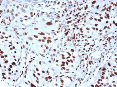
Immunohistochemistry-Paraffin: HIST1H3A [p Ser10] Antibody (rPHH3/6824) [NBP3-13802] - Formalin-fixed, paraffin-embedded human bladder stained with HIST1H3A [p Ser10] antibody (rPHH3/6824).
![HIST1H3A [p Ser10] Antibody (rPHH3/6824) Western Blot: HIST1H3A [p Ser10] Antibody (rPHH3/6824) [NBP3-13802] -](https://resources.rndsystems.com/images/products/nbp3-13802_mouse-hist1h3a-p-ser-10-mab-rphh3-6824-26202521345241.jpg "Western Blot: HIST1H3A [p Ser10] Antibody (rPHH3/6824) [NBP3-13802] -")
Western Blot: HIST1H3A [p Ser10] Antibody (rPHH3/6824) [NBP3-13802] -
Western Blot Analysis of Human MCF-7 lysate using HIST1H3A [p Ser10] Antibody (rPHH3/6824).Applications for HIST1H3A [p Ser10] Antibody (rPHH3/6824)
Application
Recommended Usage
Immunohistochemistry-Paraffin
1-2 ug/ml
Application Notes
Immunohistochemistry (Formalin-fixed): 1-2ug/ml for 30 minutes at RT. Staining of formalin-fixed tissues requires heating tissue sections in 10mM Tris with 1mM EDTA, pH 9.0, for 45 min at 95C followed by cooling at RT for 20 minutes
Formulation, Preparation, and Storage
Purification
Protein A or G purified
Formulation
1mM PBS with 0.05% BSA
Preservative
0.05% Sodium Azide
Concentration
0.2 mg/ml
Shipping
The product is shipped with polar packs. Upon receipt, store it immediately at the temperature recommended below.
Stability & Storage
Store at 4C.
Background: HIST1H3A
Alternate Names
H3 histone family, member A, H3/A, H3FAHIST1H3E, H3FB, H3FC, H3FD, H3FF, H3FH, H3FI, H3FJ, H3FK, H3FL, HIST1H3B, HIST1H3C, HIST1H3D, HIST1H3F, HIST1H3G, HIST1H3H, HIST1H3I, HIST1H3J, histone 1, H3a, histone cluster 1, H3a, histone H3.1, Histone H3/a, Histone H3/b, Histone H3/c, Histone H3/d, Histone H3/f, Histone H3/h, Histone H3/i, Histone H3/j, Histone H3/k, Histone H3/l
Gene Symbol
H3C1
UniProt
Additional HIST1H3A Products
Product Documents for HIST1H3A [p Ser10] Antibody (rPHH3/6824)
Certificate of Analysis
To download a Certificate of Analysis, please enter a lot or batch number in the search box below.
Product Specific Notices for HIST1H3A [p Ser10] Antibody (rPHH3/6824)
This product is for research use only and is not approved for use in humans or in clinical diagnosis. Primary Antibodies are guaranteed for 1 year from date of receipt.
Customer Reviews for HIST1H3A [p Ser10] Antibody (rPHH3/6824)
There are currently no reviews for this product. Be the first to review HIST1H3A [p Ser10] Antibody (rPHH3/6824) and earn rewards!
Have you used HIST1H3A [p Ser10] Antibody (rPHH3/6824)?
Submit a review and receive an Amazon gift card!
$25/€18/£15/$25CAN/¥2500 Yen for a review with an image
$10/€7/£6/$10CAN/¥1110 Yen for a review without an image
Submit a review
Protocols
Find general support by application which include: protocols, troubleshooting, illustrated assays, videos and webinars.
- Antigen Retrieval Protocol (PIER)
- Antigen Retrieval for Frozen Sections Protocol
- Appropriate Fixation of IHC/ICC Samples
- Cellular Response to Hypoxia Protocols
- Chromogenic IHC Staining of Formalin-Fixed Paraffin-Embedded (FFPE) Tissue Protocol
- Chromogenic Immunohistochemistry Staining of Frozen Tissue
- ClariTSA™ Fluorophore Kits
- Detection & Visualization of Antibody Binding
- Fluorescent IHC Staining of Frozen Tissue Protocol
- Graphic Protocol for Heat-induced Epitope Retrieval
- Graphic Protocol for the Preparation and Fluorescent IHC Staining of Frozen Tissue Sections
- Graphic Protocol for the Preparation and Fluorescent IHC Staining of Paraffin-embedded Tissue Sections
- Graphic Protocol for the Preparation of Gelatin-coated Slides for Histological Tissue Sections
- IHC Sample Preparation (Frozen sections vs Paraffin)
- Immunofluorescent IHC Staining of Formalin-Fixed Paraffin-Embedded (FFPE) Tissue Protocol
- Immunohistochemistry (IHC) and Immunocytochemistry (ICC) Protocols
- Immunohistochemistry Frozen Troubleshooting
- Immunohistochemistry Paraffin Troubleshooting
- Preparing Samples for IHC/ICC Experiments
- Preventing Non-Specific Staining (Non-Specific Binding)
- Primary Antibody Selection & Optimization
- Protocol for Heat-Induced Epitope Retrieval (HIER)
- Protocol for Making a 4% Formaldehyde Solution in PBS
- Protocol for VisUCyte™ HRP Polymer Detection Reagent
- Protocol for the Preparation & Fixation of Cells on Coverslips
- Protocol for the Preparation and Chromogenic IHC Staining of Frozen Tissue Sections
- Protocol for the Preparation and Chromogenic IHC Staining of Frozen Tissue Sections - Graphic
- Protocol for the Preparation and Chromogenic IHC Staining of Paraffin-embedded Tissue Sections
- Protocol for the Preparation and Chromogenic IHC Staining of Paraffin-embedded Tissue Sections - Graphic
- Protocol for the Preparation and Fluorescent IHC Staining of Frozen Tissue Sections
- Protocol for the Preparation and Fluorescent IHC Staining of Paraffin-embedded Tissue Sections
- Protocol for the Preparation of Gelatin-coated Slides for Histological Tissue Sections
- TUNEL and Active Caspase-3 Detection by IHC/ICC Protocol
- The Importance of IHC/ICC Controls
- Troubleshooting Guide: Immunohistochemistry
- View all Protocols, Troubleshooting, Illustrated assays and Webinars
Loading...