![Western Blot: hnRNP U Antibody [NB100-2134]](https://resources.rndsystems.com/images/products/hnRNP-U-Antibody-Western-Blot-NB100-2134-img0007.jpg "Western Blot: hnRNP U Antibody [NB100-2134]")
Loading...
Key Product Details
Species Reactivity
Human, Mouse
Applications
Immunohistochemistry, Immunohistochemistry-Paraffin, Western Blot, Simple Western, Immunoprecipitation, Immunoprecipitation (Negative)
Label
Unconjugated
Antibody Source
Polyclonal Rabbit IgG
Loading...
Product Specifications
Immunogen
The immunogen recognized by this antibody maps to a region between residue 450 and 500 of human Heterogeneous Nuclear Ribonucleoprotein U using the numbering given in entry NP_114032.1 (GeneID 3192).
Clonality
Polyclonal
Host
Rabbit
Isotype
IgG
Scientific Data Images for hnRNP U Antibody
Western Blot: hnRNP U Antibody [NB100-2134]
Western Blot: hnRNP U Antibody [NB100-2134] - Whole cell lysate from HeLa (5, 15 and 50 ug), HEK293T (T; 50 ug), and mouse NIH 3T3 (M; 50 ug) cells. Antibodies: Affinity purified rabbit anti-hnRNP-U antibody used for WB at 0.04 ug/ml. Detection: Chemiluminescence with exposure time of 10seconds.![Immunohistochemistry-Paraffin: hnRNP U Antibody [NB100-2134]](https://resources.rndsystems.com/images/products/hnRNP-U-Antibody-Immunohistochemistry-Paraffin-NB100-2134-img0004.jpg "Immunohistochemistry-Paraffin: hnRNP U Antibody [NB100-2134]")
Immunohistochemistry-Paraffin: hnRNP U Antibody [NB100-2134]
Immunohistochemistry-Paraffin: hnRNP U Antibody [NB100-2134] - Human breast carcinoma (left) and mouse squamous cell carcinoma (right). Antibody: Affinity purified rabbit anti-hnRNP-U used at a dilution of 1:200 (1 ug/mL). Detection: DAB![Western Blot: hnRNP U Antibody [NB100-2134]](https://resources.rndsystems.com/images/products/hnRNP-U-Antibody-Immunoprecipitation-NB100-2134-img0006.jpg "Western Blot: hnRNP U Antibody [NB100-2134]")
Western Blot: hnRNP U Antibody [NB100-2134]
Western Blot: hnRNP U Antibody [NB100-2134] - Detection of human hnRNP-U by western blot of immunoprecipitates. Samples: Whole cell lysate (1 mg for IP, 20% of IP loaded) from HeLa cells. Antibodies: Affinity purified rabbit anti-hnRNP-U antibody NB100-2134 used for WB at 1 ug/ml. hnRNP-U was immunoprecipitated using a rabbit anti-hnRNP-U antibody at 3 ug/mg lysate. Detection: Chemiluminescence with exposure time of 30 seconds.![Simple Western: hnRNP U Antibody [NB100-2134]](https://resources.rndsystems.com/images/products/hnRNP-U-Antibody-Simple-Western-NB100-2134-img0005.jpg "Simple Western: hnRNP U Antibody [NB100-2134]")
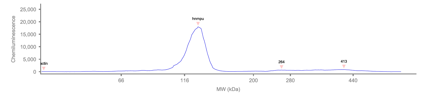
Simple Western: hnRNP U Antibody [NB100-2134]
Simple Western: hnRNP U Antibody [NB100-2134] - Electropherogram from Simple Western (WES) of HNRNPU (NB100-2134). Antibody at 1:100 on protein sample from Neural Epithelial Stem Cells. Simple Western image submitted by a verified customer review.Applications for hnRNP U Antibody
Application
Recommended Usage
Immunohistochemistry
1:100 - 1:500
Immunohistochemistry-Paraffin
1:100 - 1:500
Simple Western
1:100
Western Blot
1:2000 - 1:10000
Application Notes
Immunoprecipitation: Not Recommended, use rabbit anti-hnRNP-U antibody NB100-2135. Epitope retrieval with citrate buffer pH 6.0 is recommended for FFPE tissue sections. This hnRNP U antibody is validated for Simple Western from a verified customer review.
See Simple Western Antibody Database for Simple Western validation: Tested in Neural Epithelial Stem Cells, separated by Size, antibody dilution of 1:100
See Simple Western Antibody Database for Simple Western validation: Tested in Neural Epithelial Stem Cells, separated by Size, antibody dilution of 1:100
Reviewed Applications
Read 1 review rated 4 using NB100-2134 in the following applications:
Formulation, Preparation, and Storage
Purification
Immunogen affinity purified
Formulation
TBS, 0.1% BSA
Preservative
0.09% Sodium Azide
Concentration
0.2 mg/ml
Shipping
The product is shipped with polar packs. Upon receipt, store it immediately at the temperature recommended below.
Stability & Storage
Store at 4C. Do not freeze.
Background: hnRNP U
Alternate Names
heterogeneous nuclear ribonucleoprotein U, heterogeneous nuclear ribonucleoprotein U (scaffold attachment factor A), hnRNP U, HNRPU, p120, p120 nuclear protein, pp120, SAFA, SAF-AhnRNPU, Scaffold attachment factor A, U21.1
Gene Symbol
HNRNPU
UniProt
Additional hnRNP U Products
Product Documents for hnRNP U Antibody
Certificate of Analysis
To download a Certificate of Analysis, please enter a lot or batch number in the search box below.
Product Specific Notices for hnRNP U Antibody
This product is for research use only and is not approved for use in humans or in clinical diagnosis. Primary Antibodies are guaranteed for 1 year from date of receipt.
Citations for hnRNP U Antibody
 Powered by Bioz
Customer Reviews for hnRNP U Antibody (1)
4 out of 5
1 Customer Rating
Have you used hnRNP U Antibody?
Submit a review and receive an Amazon gift card!
$25/€18/£15/$25CAN/¥2500 Yen for a review with an image
$10/€7/£6/$10CAN/¥1110 Yen for a review without an image
Submit a review
Customer Images
Showing
1
-
1 of
1 review
Showing All
Filter By:
-
Application: Simple WesternSample Tested: Human fibroblast and neural stem cellsSpecies: HumanVerified Customer | Posted 11/28/2019Elecropherogram from Simple Western (WES) of HNRNPU (NB100-2134) 1:100 on protein sample from Neural Epithelial Stem CellsDilution 1:100

There are no reviews that match your criteria.
Protocols
Find general support by application which include: protocols, troubleshooting, illustrated assays, videos and webinars.
- Antigen Retrieval Protocol (PIER)
- Antigen Retrieval for Frozen Sections Protocol
- Appropriate Fixation of IHC/ICC Samples
- Cellular Response to Hypoxia Protocols
- Chromogenic IHC Staining of Formalin-Fixed Paraffin-Embedded (FFPE) Tissue Protocol
- Chromogenic Immunohistochemistry Staining of Frozen Tissue
- ClariTSA™ Fluorophore Kits
- Detection & Visualization of Antibody Binding
- Fluorescent IHC Staining of Frozen Tissue Protocol
- Graphic Protocol for Heat-induced Epitope Retrieval
- Graphic Protocol for the Preparation and Fluorescent IHC Staining of Frozen Tissue Sections
- Graphic Protocol for the Preparation and Fluorescent IHC Staining of Paraffin-embedded Tissue Sections
- Graphic Protocol for the Preparation of Gelatin-coated Slides for Histological Tissue Sections
- IHC Sample Preparation (Frozen sections vs Paraffin)
- Immunofluorescent IHC Staining of Formalin-Fixed Paraffin-Embedded (FFPE) Tissue Protocol
- Immunohistochemistry (IHC) and Immunocytochemistry (ICC) Protocols
- Immunohistochemistry Frozen Troubleshooting
- Immunohistochemistry Paraffin Troubleshooting
- Immunoprecipitation Protocol
- Preparing Samples for IHC/ICC Experiments
- Preventing Non-Specific Staining (Non-Specific Binding)
- Primary Antibody Selection & Optimization
- Protocol for Heat-Induced Epitope Retrieval (HIER)
- Protocol for Making a 4% Formaldehyde Solution in PBS
- Protocol for VisUCyte™ HRP Polymer Detection Reagent
- Protocol for the Preparation & Fixation of Cells on Coverslips
- Protocol for the Preparation and Chromogenic IHC Staining of Frozen Tissue Sections
- Protocol for the Preparation and Chromogenic IHC Staining of Frozen Tissue Sections - Graphic
- Protocol for the Preparation and Chromogenic IHC Staining of Paraffin-embedded Tissue Sections
- Protocol for the Preparation and Chromogenic IHC Staining of Paraffin-embedded Tissue Sections - Graphic
- Protocol for the Preparation and Fluorescent IHC Staining of Frozen Tissue Sections
- Protocol for the Preparation and Fluorescent IHC Staining of Paraffin-embedded Tissue Sections
- Protocol for the Preparation of Gelatin-coated Slides for Histological Tissue Sections
- R&D Systems Quality Control Western Blot Protocol
- TUNEL and Active Caspase-3 Detection by IHC/ICC Protocol
- The Importance of IHC/ICC Controls
- Troubleshooting Guide: Immunohistochemistry
- Troubleshooting Guide: Western Blot Figures
- Western Blot Conditions
- Western Blot Protocol
- Western Blot Protocol for Cell Lysates
- Western Blot Troubleshooting
- Western Blot Troubleshooting Guide
- View all Protocols, Troubleshooting, Illustrated assays and Webinars
Loading...