Arginase 1 (ARG1) is a 35‑40 kDa member of the arginase family of enzymes. It is expressed in multiple cell types, including erythrocytes, hepatocytes, neutrophils, smooth muscle and macrophages. ARG1 demonstrates two distinct functions: in the hepatocyte cytoplasm, it catalyzes the conversion of arginine to ornithine and urea, while in multiple cells, it degrades arginine, thus indirectly downregulating NO synthase (NOS) activity by depriving this enzyme of its substrate. Human ARG1 is 322 amino acids (aa) in length. Its enzyme region comprises aa 9‑309 and contains two Mn atoms. ARG1 is moderately active as a monomer, but highly active as a 105 kDa homotrimer. Trimerization is promoted by nitrosylation of Cys303, creating a regulatory feedback loop with NOS. There are two isoform variants, one that shows an eight aa insertion after Gln43, and another that shows a deletion of aa 204‑289. Full-length human ARG1 shares 87% aa identity with mouse and rat ARG1.
Human Arginase 1/ARG1 PE‑conjugated Antibody
R&D Systems | Catalog # IC8026P

Key Product Details
Species Reactivity
Validated:
Cited:
Applications
Validated:
Cited:
Label
Antibody Source
Product Specifications
Immunogen
Met1-Lys322
Accession # P05089
Specificity
Clonality
Host
Isotype
Scientific Data Images for Human Arginase 1/ARG1 PE‑conjugated Antibody
Detection of Arginase 1/ARG1 in HepG2 Human Cell Line by Flow Cytometry.
HepG2 human hepatocellular carcinoma cell line was stained with Mouse Anti-Human Arginase 1/ARG1 PE-conjugated Mono-clonal Antibody (Catalog # IC8026P, filled histogram) or isotype control antibody (Catalog # IC0041P, open histogram). To facilitate intracellular staining, cells were fixed with Flow Cytometry Fixation Buffer (Catalog # FC004) and permeabilized with Flow Cytometry Permeabilization/Wash Buffer I (Catalog # FC005). View our protocol for Staining Intracellular Molecules.
Detection of Arginase 1/ARG1 in Human PBMCs by Flow Cytometry.
Human peripheral blood mononuclear cells (PBMCs) were stained with Mouse Anti-Human Integrin aM/CD11b APC-conjugated Monoclonal Antibody (Catalog # FAB1699A) and either (A) Mouse Anti-Human Arginase 1/ARG1 PE-conjugated Monoclonal Antibody (Catalog # IC8026P) or (B) Mouse IgG2BPhycoerythrin Isotype Control (Catalog # IC0041P). To facilitate intracellular staining, cells were fixed with Flow Cytometry Fixation Buffer (Catalog # FC004) and permeabilized with Flow Cytometry Permeabilization/Wash Buffer I (Catalog # FC005). View our protocol for Staining Intracellular Molecules.Applications for Human Arginase 1/ARG1 PE‑conjugated Antibody
Intracellular Staining by Flow Cytometry
Sample: HepG2 human hepatocellular carcinoma cell line fixed with Flow Cytometry Fixation Buffer (Catalog # FC004) and permeabilized with Flow Cytometry Permeabilization/Wash Buffer I (Catalog # FC005)
Reviewed Applications
Read 1 review rated 5 using IC8026P in the following applications:
Spectra Viewer
Plan Your Experiments
Use our spectra viewer to interactively plan your experiments, assessing multiplexing options. View the excitation and emission spectra for our fluorescent dye range and other commonly used dyes.
Spectra Viewer
Flow Cytometry Panel Builder
Bio-Techne Knows Flow Cytometry
Save time and reduce costly mistakes by quickly finding compatible reagents using the Panel Builder Tool.
Advanced Features
- Spectra Viewer - Custom analysis of spectra from multiple fluorochromes
- Spillover Popups - Visualize the spectra of individual fluorochromes
- Antigen Density Selector - Match fluorochrome brightness with antigen density

Formulation, Preparation, and Storage
Purification
Formulation
Shipping
Stability & Storage
- 12 months from date of receipt, 2 to 8 °C as supplied.
Background: Arginase 1/ARG1
Long Name
Alternate Names
Gene Symbol
UniProt
Additional Arginase 1/ARG1 Products
Product Documents for Human Arginase 1/ARG1 PE‑conjugated Antibody
Certificate of Analysis
To download a Certificate of Analysis, please enter a lot or batch number in the search box below.
Note: Certificate of Analysis not available for kit components.
Product Specific Notices for Human Arginase 1/ARG1 PE‑conjugated Antibody
For research use only
Related Research Areas
Citations for Human Arginase 1/ARG1 PE‑conjugated Antibody
 Powered by Bioz
Customer Reviews for Human Arginase 1/ARG1 PE‑conjugated Antibody (1)
Have you used Human Arginase 1/ARG1 PE‑conjugated Antibody?
Submit a review and receive an Amazon gift card!
$25/€18/£15/$25CAN/¥2500 Yen for a review with an image
$10/€7/£6/$10CAN/¥1110 Yen for a review without an image
Submit a review
Customer Images
-
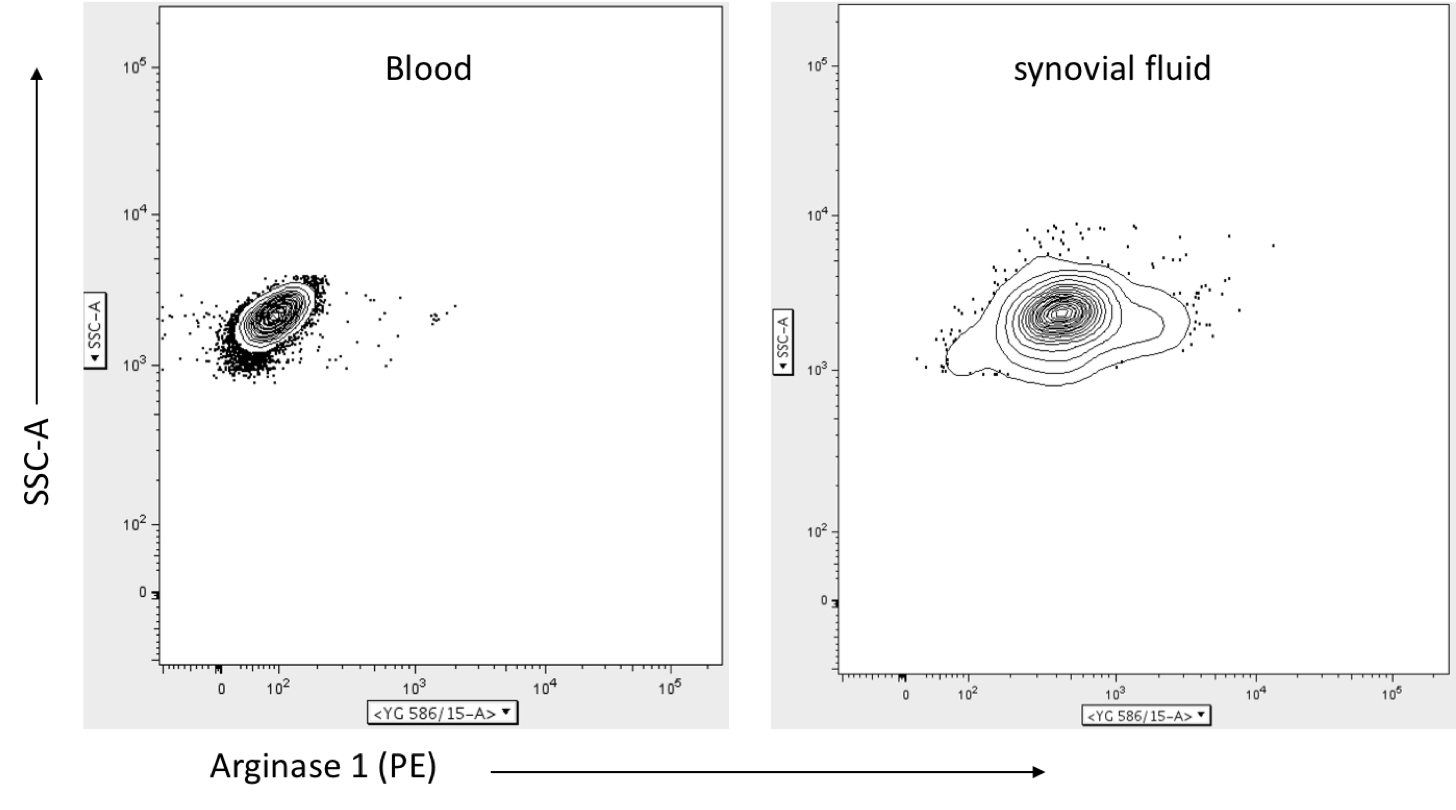
Application: Flow CytometrySample Tested: Peripheral blood neutrophils and synovial fluid neutrophilsSpecies: HumanVerified Customer | Posted 05/15/2020I used this antibody to stain human neutrophils from blood and synovial fluid of arthritis patients. Blood neutrophils had low surface expression of arginase 1, while neutrophils isolated from the synovial fluid of the inflamed knee were found to retain this enzyme on the cell surface. This antibody may be used to stain other cell types as well including macrophages. Cells were stained on ice to minimize internalization of the antibody, but I did not assess cross-reactivity with the mitochondria-association isoform arginase-2.

There are no reviews that match your criteria.
Protocols
Find general support by application which include: protocols, troubleshooting, illustrated assays, videos and webinars.
- 7-Amino Actinomycin D (7-AAD) Cell Viability Flow Cytometry Protocol
- Extracellular Membrane Flow Cytometry Protocol
- Flow Cytometry Protocol for Cell Surface Markers
- Flow Cytometry Protocol for Staining Membrane Associated Proteins
- Flow Cytometry Staining Protocols
- Flow Cytometry Troubleshooting Guide
- Intracellular Flow Cytometry Protocol Using Alcohol (Methanol)
- Intracellular Flow Cytometry Protocol Using Detergents
- Intracellular Nuclear Staining Flow Cytometry Protocol Using Detergents
- Intracellular Staining Flow Cytometry Protocol Using Alcohol Permeabilization
- Intracellular Staining Flow Cytometry Protocol Using Detergents to Permeabilize Cells
- Propidium Iodide Cell Viability Flow Cytometry Protocol
- Protocol for Liperfluo
- Protocol for the Characterization of Human Th22 Cells
- Protocol for the Characterization of Human Th9 Cells
- Protocol: Annexin V and PI Staining by Flow Cytometry
- Protocol: Annexin V and PI Staining for Apoptosis by Flow Cytometry
- Troubleshooting Guide: Fluorokine Flow Cytometry Kits
- View all Protocols, Troubleshooting, Illustrated assays and Webinars