
The cadherin superfamily is a large family of membrane-associated glycoproteins that engage in homotypic, calcium‑dependent, cell-to-cell adhesion events. The superfamily can be divided into at least four subfamilies based on its member’s extracellular (EC) regions and cytoplasmic domains (1, 2). These include classical cadherins, desmosomal cadherins, protocadherins, and cadherin-like molecules that contain a variable number of EC and transmembrane (TM) domains (1). Cadherin‑6, also known as KCAD or K-cadherin, is a classical cadherin of 110‑120 kD that has at least one full length and two alternate splice forms ranging in size from 105‑120 kDa (3). Human Cadherin-6 is synthesized as a 790 amino acid (aa) type I transmembrane glycoprotein that contains a 18 aa signal peptide, a 35 aa prosequence, a 562 aa extracellular region, a 21 aa transmembrane segment, and a 154 aa cytoplasmic domain (4, 5). There are five EC cadherin domains that are approximately 110 aa in length. This pattern is consistent with classical cadherin family molecules that are modular in their extracellular region and mediate calcium‑dependent cell-to-cell adhesion through their Ca++-binding repeats (2). Due to the absence of a His-Ala-Val motif in its most N-terminal cadherin repeat, Cadherin-6 can be further classified as a type II classical cadherin (4). One Cadherin‑6 splice variant (termed 6/2) shows a 9 aa substitution for the 94 aa that spans residues 283 to 376 of the full-length extracellular region (3). A second splice variant shows a 36 aa substitution for the C-terminal 163 aa of the transmembrane and cytoplasmic region (6). The extracellular region of human Cadherin‑6 is 98% aa identical to rat Cadherin‑6 extracellular region, plus 60% and 58% aa identical to the extracellular regions of human Cadherin-8 and -11, respectively. Cadherin-6 has high expression in kidney, brain, and cerebellum, and low expression in lung, pancreas, gastric mucosa, and cytotrophoblasts (4, 5, 7, 8, 9). Cadherin-6 is also found in renal, lung, and ovarian carcinoma (7, 10). As a classic cadherin, Cadherin‑6 will form homodimers and promote intercellular adhesion with itself and possibly, Cadherin-9 and -14 (4, 11).
Human Cadherin-6/KCAD Antibody (427909) [PE]
R&D Systems | Catalog # FAB2715P

Key Product Details
Species Reactivity
Validated:
Cited:
Applications
Validated:
Cited:
Label
Antibody Source
Product Specifications
Immunogen
Ser54-Ala615
Accession # P55285
Specificity
Clonality
Host
Isotype
Scientific Data Images for Human Cadherin-6/KCAD Antibody (427909) [PE]
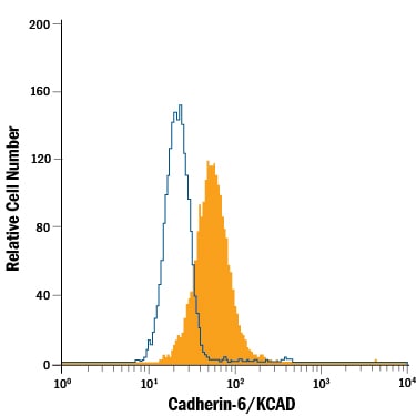
Detection of Cadherin‑6/KCAD in MG‑63 Human Cell Line by Flow Cytometry.
MG-63 human osteosarcoma cell line was stained with Mouse Anti-Human Cadherin-6/KCAD PE-conjugated Monoclonal Antibody (Catalog # FAB2715P, filled histogram) or isotype control antibody (Catalog # IC002P, open histogram). Cells were stained in a buffer containing Ca2+and Mg2+. View our protocol for Staining Membrane-associated Proteins.Applications for Human Cadherin-6/KCAD Antibody (427909) [PE]
Flow Cytometry
Sample: MG‑63 human osteosarcoma cell line stained in a buffer containing Ca2+ and Mg2+
Spectra Viewer
Plan Your Experiments
Use our spectra viewer to interactively plan your experiments, assessing multiplexing options. View the excitation and emission spectra for our fluorescent dye range and other commonly used dyes.
Spectra Viewer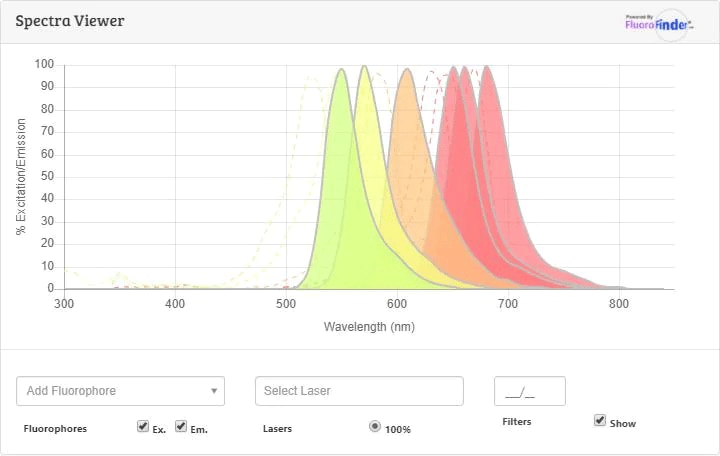
Flow Cytometry Panel Builder
Bio-Techne Knows Flow Cytometry
Save time and reduce costly mistakes by quickly finding compatible reagents using the Panel Builder Tool.
Advanced Features
- Spectra Viewer - Custom analysis of spectra from multiple fluorochromes
- Spillover Popups - Visualize the spectra of individual fluorochromes
- Antigen Density Selector - Match fluorochrome brightness with antigen density

Formulation, Preparation, and Storage
Purification
Formulation
Shipping
Stability & Storage
- 12 months from date of receipt, 2 to 8 °C as supplied.
Background: Cadherin-6/KCAD
References
- Koch, A.W. et al. (2004) Cell. Mol. Life Sci. 61:1884.
- Angst, B.D. et al. (2001) J. Cell Sci. 114:629.
- Mbalaviele, G. et al. (1998) J. Cell Biol. 141:1467.
- Shimoyama, Y. et al. (2000) Biochem. J. 349:159.
- Shimoyama, Y. et al. (1995) Cancer Res. 55:2206.
- GenBank Accession # P55285.
- Xiang Y.Y. et al. (1994) Cancer Res. 54:3034.
- Marthiens V. et al. (2002) Mol. Cell Neurosci. 20:458.
- MacCalman C.D. et al. (1998) Am J Reprod. Immunol. 39:96.
- Sella, G.C. et al. (2001) Cancer Res. 61:6977.
- Shimoyama, Y. et al. (1999) J. Biol. Chem. 274:11987.
Alternate Names
Entrez Gene IDs
Gene Symbol
UniProt
Additional Cadherin-6/KCAD Products
Product Documents for Human Cadherin-6/KCAD Antibody (427909) [PE]
Certificate of Analysis
To download a Certificate of Analysis, please enter a lot or batch number in the search box below.
Note: Certificate of Analysis not available for kit components.
Product Specific Notices for Human Cadherin-6/KCAD Antibody (427909) [PE]
For research use only
Related Research Areas
Citations for Human Cadherin-6/KCAD Antibody (427909) [PE]
 Powered by Bioz
Customer Reviews for Human Cadherin-6/KCAD Antibody (427909) [PE]
There are currently no reviews for this product. Be the first to review Human Cadherin-6/KCAD Antibody (427909) [PE] and earn rewards!
Have you used Human Cadherin-6/KCAD Antibody (427909) [PE]?
Submit a review and receive an Amazon gift card!
$25/€18/£15/$25CAN/¥2500 for a review with an image
$10/€7/£6/$10CAN/¥1110 for a review without an image
Submit a review
Protocols
Find general support by application which include: protocols, troubleshooting, illustrated assays, videos and webinars.
- 7-Amino Actinomycin D (7-AAD) Cell Viability Flow Cytometry Protocol
- Extracellular Membrane Flow Cytometry Protocol
- Flow Cytometry Protocol for Cell Surface Markers
- Flow Cytometry Protocol for Staining Membrane Associated Proteins
- Flow Cytometry Staining Protocols
- Flow Cytometry Troubleshooting Guide
- Intracellular Flow Cytometry Protocol Using Alcohol (Methanol)
- Intracellular Flow Cytometry Protocol Using Detergents
- Intracellular Nuclear Staining Flow Cytometry Protocol Using Detergents
- Intracellular Staining Flow Cytometry Protocol Using Alcohol Permeabilization
- Intracellular Staining Flow Cytometry Protocol Using Detergents to Permeabilize Cells
- Propidium Iodide Cell Viability Flow Cytometry Protocol
- Protocol for Liperfluo
- Protocol for the Characterization of Human Th22 Cells
- Protocol for the Characterization of Human Th9 Cells
- Protocol: Annexin V and PI Staining by Flow Cytometry
- Protocol: Annexin V and PI Staining for Apoptosis by Flow Cytometry
- Troubleshooting Guide: Fluorokine Flow Cytometry Kits
- View all Protocols, Troubleshooting, Illustrated assays and Webinars
FAQs for Human Cadherin-6/KCAD Antibody (427909) [PE]
-
Q: Why does the staining protocol with this Cadherin antibody use buffers containing Ca2+ and Mg2+?
A: The staining protocol with this and other Cadherin antibodies uses buffer containing Ca2+ and Mg2+ because Cadherin function is Calcium-dependent.