
Discontinued Product
MAB1038 has been discontinued.
View all STRO-1 products.
Key Product Details
Species Reactivity
Validated:
Human
Cited:
Human, Mouse, Rat, Porcine, Canine, Deer, Feline
Applications
Validated:
Immunocytochemistry
Cited:
Immunohistochemistry, Immunohistochemistry-Paraffin, Immunohistochemistry-Frozen, Western Blot, Flow Cytometry, Immunocytochemistry, Immunoprecipitation, Cell Selection, MACS
Label
Unconjugated
Antibody Source
Monoclonal Mouse IgM Clone # STRO-1
Loading...
Product Specifications
Immunogen
Human CD34+ bone marrow cells
Specificity
Detects human STRO-1.
Clonality
Monoclonal
Host
Mouse
Isotype
IgM
Scientific Data Images for Human STRO-1 Antibody (STRO-1)
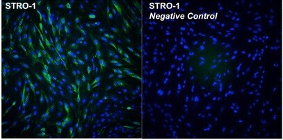
STRO‑1 in Human MG‑63 Cell Line.
STRO-1 was detected in immersion fixed MG-63 human osteosarcoma cell line using Human STRO-1 Monoclonal Antibody (Catalog # MAB1038) at 10 µg/mL for 3 hours at room temperature. Cells were stained using the NorthernLights™ 557-conjugated Anti-Mouse IgM Secondary Antibody (red; NL019), and counterstained with DAPI (blue). View our protocol for Fluorescent ICC Staining of Cells on Coverslips.Applications for Human STRO-1 Antibody (STRO-1)
Application
Recommended Usage
Immunocytochemistry
8-25 µg/mL
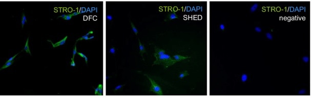
Sample: Immersion fixed human long term bone marrow culture and immersion fixed embryonic mouse parietal endoderm cells (E10.5)
Sample: Immersion fixed human long term bone marrow culture and immersion fixed embryonic mouse parietal endoderm cells (E10.5)
Reviewed Applications
Read 9 reviews rated 4.2 using MAB1038 in the following applications:
Formulation, Preparation, and Storage
Purification
IgM-specific Affinity-purified from hybridoma culture supernatant
Reconstitution
Reconstitute at 0.5 mg/mL in sterile PBS. For liquid material, refer to CoA for concentration.
Formulation
Lyophilized from a 0.2 μm filtered solution in PBS with Trehalose. *Small pack size (SP) is supplied either lyophilized or as a 0.2 µm filtered solution in PBS.
Shipping
Lyophilized product is shipped at ambient temperature. Liquid small pack size (-SP) is shipped with polar packs. Upon receipt, store immediately at the temperature recommended below.
Stability & Storage
Use a manual defrost freezer and avoid repeated freeze-thaw cycles.
- 12 months from date of receipt, -20 to -70 °C as supplied.
- 1 month, 2 to 8 °C under sterile conditions after reconstitution.
- 6 months, -20 to -70 °C under sterile conditions after reconstitution.
Calculators
Background: STRO-1
References
- Simmons, P.J. and B. Torok-Storb (1991) Blood 78:55.
- Dennis, J.E. et al. (2002) Cells Tissues Organs 170:73.
Long Name
A Stromal Cell Precursor Surface Antigen
Alternate Names
STRO1
Additional STRO-1 Products
Product Documents for Human STRO-1 Antibody (STRO-1)
Certificate of Analysis
To download a Certificate of Analysis, please enter a lot or batch number in the search box below.
Note: Certificate of Analysis not available for kit components.
Product Specific Notices for Human STRO-1 Antibody (STRO-1)
For research use only
Related Research Areas
Citations for Human STRO-1 Antibody (STRO-1)
 Powered by Bioz
Customer Reviews for Human STRO-1 Antibody (STRO-1) (9)
4.2 out of 5
9 Customer Ratings
Have you used Human STRO-1 Antibody (STRO-1)?
Submit a review and receive an Amazon gift card!
$25/€18/£15/$25CAN/¥2500 Yen for a review with an image
$10/€7/£6/$10CAN/¥1110 Yen for a review without an image
Submit a review
Customer Images
Showing
1
-
5 of
9 reviews
Showing All
Filter By:
-
Application: Immunocytochemistry/ImmunofluorescenceSample Tested: Human dental pulp cellsSpecies: HumanVerified Customer | Posted 07/20/2021

-
Application: Immunocytochemistry/ImmunofluorescenceSample Tested: Stem cellsSpecies: HumanVerified Customer | Posted 07/02/2021
-
Application: ImmunofluorescenceSample Tested: See PMID 23603337Species: HumanVerified Customer | Posted 02/03/2015
-
Application: Western BlotSample Tested: See PMID 23312853Species: HumanVerified Customer | Posted 02/03/2015
-
Application: ImmunofluorescenceSample Tested: See PMID 23072705Species: HumanVerified Customer | Posted 02/03/2015
-
Application: Flow CytometrySample Tested: See PMID 23067388Species: HumanVerified Customer | Posted 02/03/2015
-
Application: Flow CytometrySample Tested: See PMID 22226689Species: HumanVerified Customer | Posted 02/03/2015
-
Application: Flow CytometrySample Tested: See PMID 21550429Species: HumanVerified Customer | Posted 02/03/2015
-
Application: Flow CytometrySample Tested: See PMID 20682625Species: HumanVerified Customer | Posted 02/03/2015
There are no reviews that match your criteria.
Protocols
Find general support by application which include: protocols, troubleshooting, illustrated assays, videos and webinars.
- Appropriate Fixation of IHC/ICC Samples
- Cellular Response to Hypoxia Protocols
- ClariTSA™ Fluorophore Kits
- Detection & Visualization of Antibody Binding
- ICC Cell Smear Protocol for Suspension Cells
- ICC Immunocytochemistry Protocol Videos
- ICC for Adherent Cells
- Immunocytochemistry (ICC) Protocol
- Immunocytochemistry Troubleshooting
- Immunofluorescence of Organoids Embedded in Cultrex Basement Membrane Extract
- Immunohistochemistry (IHC) and Immunocytochemistry (ICC) Protocols
- Preparing Samples for IHC/ICC Experiments
- Preventing Non-Specific Staining (Non-Specific Binding)
- Primary Antibody Selection & Optimization
- Protocol for VisUCyte™ HRP Polymer Detection Reagent
- Protocol for the Fluorescent ICC Staining of Cell Smears - Graphic
- Protocol for the Fluorescent ICC Staining of Cultured Cells on Coverslips - Graphic
- Protocol for the Preparation and Fluorescent ICC Staining of Cells on Coverslips
- Protocol for the Preparation and Fluorescent ICC Staining of Non-adherent Cells
- Protocol for the Preparation and Fluorescent ICC Staining of Stem Cells on Coverslips
- Protocol for the Preparation of a Cell Smear for Non-adherent Cell ICC - Graphic
- TUNEL and Active Caspase-3 Detection by IHC/ICC Protocol
- The Importance of IHC/ICC Controls
- View all Protocols, Troubleshooting, Illustrated assays and Webinars
Loading...