![Western Blot: IP3R1 Antibody [NB120-5908]](https://resources.rndsystems.com/images/products/IP3R1-Antibody-Western-Blot-NB120-5908-img0001.jpg "Western Blot: IP3R1 Antibody [NB120-5908]")
Loading...
Key Product Details
Species Reactivity
Validated:
Human, Mouse, Rat
Cited:
Mouse
Applications
Validated:
Western Blot, Immunocytochemistry/ Immunofluorescence
Cited:
Western Blot, Immunocytochemistry/ Immunofluorescence
Label
Unconjugated
Antibody Source
Polyclonal Rabbit IgG
Loading...
Product Specifications
Immunogen
Synthetic peptide corresponding to residues N(1829) K K K D D E V D R D A P S R K K A K E(1848) of human IP3 Receptor Type I.
Reactivity Notes
Human reactivity reported by a customer review. Mouse reactivity reported in scientific literature (PMID: 19382908). Rat reactivity reported in scientific literature (PMID: 12767046).
Localization
Endoplasmic reticulum - Integral membrane protein.
Specificity
IP3R1
Clonality
Polyclonal
Host
Rabbit
Isotype
IgG
Scientific Data Images for IP3R1 Antibody
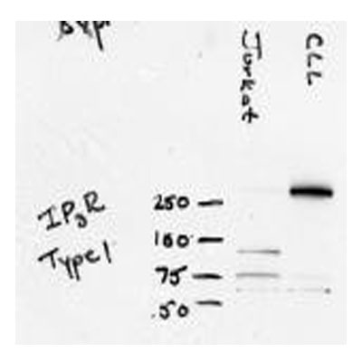
Western Blot: IP3R1 Antibody [NB120-5908]
IP3R1-Antibody-Western-Blot-NB120-5908-img0001.jpgApplications for IP3R1 Antibody
Application
Recommended Usage
Immunocytochemistry/ Immunofluorescence
1:10 - 1:500
Western Blot
1:100 - 1:2000
Application Notes
WB: Detects an approx. 240 - 260 kDa protein representing IP3R-I from rat and mouse brain extract.
Reviewed Applications
Read 1 review rated 5 using NB120-5908 in the following applications:
Formulation, Preparation, and Storage
Purification
Unpurified
Formulation
Whole serum diluted in PBS
Preservative
0.05% Sodium Azide
Concentration
This product is unpurified. The exact concentration of antibody is not quantifiable.
Shipping
The product is shipped with polar packs. Upon receipt, store it immediately at the temperature recommended below.
Stability & Storage
Store at -20C. Avoid freeze-thaw cycles.
Background: IP3R1
Long Name
Inositol 1,4,5-trisphosphate receptor type 1
Alternate Names
InsP3R1, IP3R 1, ITPR1
Gene Symbol
ITPR1
UniProt
Additional IP3R1 Products
Product Documents for IP3R1 Antibody
Certificate of Analysis
To download a Certificate of Analysis, please enter a lot or batch number in the search box below.
Product Specific Notices for IP3R1 Antibody
This product is for research use only and is not approved for use in humans or in clinical diagnosis. Primary Antibodies are guaranteed for 1 year from date of receipt.
Citations for IP3R1 Antibody
 Powered by Bioz
Customer Reviews for IP3R1 Antibody (1)
5 out of 5
1 Customer Rating
Have you used IP3R1 Antibody?
Submit a review and receive an Amazon gift card!
$25/€18/£15/$25CAN/¥2500 Yen for a review with an image
$10/€7/£6/$10CAN/¥1110 Yen for a review without an image
Submit a review
Customer Images
Showing
1
-
1 of
1 review
Showing All
Filter By:
-
Application: Western BlotSample Tested: Jurkat whole cell lysate, primary CLL cells from human blood, Sample Amount: 80ugSpecies: HumanVerified Customer | Posted 11/17/2010

There are no reviews that match your criteria.
Protocols
Find general support by application which include: protocols, troubleshooting, illustrated assays, videos and webinars.
- Appropriate Fixation of IHC/ICC Samples
- Cellular Response to Hypoxia Protocols
- ClariTSA™ Fluorophore Kits
- Detection & Visualization of Antibody Binding
- ICC Cell Smear Protocol for Suspension Cells
- ICC Immunocytochemistry Protocol Videos
- ICC for Adherent Cells
- Immunocytochemistry (ICC) Protocol
- Immunocytochemistry Troubleshooting
- Immunofluorescence of Organoids Embedded in Cultrex Basement Membrane Extract
- Immunohistochemistry (IHC) and Immunocytochemistry (ICC) Protocols
- Preparing Samples for IHC/ICC Experiments
- Preventing Non-Specific Staining (Non-Specific Binding)
- Primary Antibody Selection & Optimization
- Protocol for VisUCyte™ HRP Polymer Detection Reagent
- Protocol for the Fluorescent ICC Staining of Cell Smears - Graphic
- Protocol for the Fluorescent ICC Staining of Cultured Cells on Coverslips - Graphic
- Protocol for the Preparation and Fluorescent ICC Staining of Cells on Coverslips
- Protocol for the Preparation and Fluorescent ICC Staining of Non-adherent Cells
- Protocol for the Preparation and Fluorescent ICC Staining of Stem Cells on Coverslips
- Protocol for the Preparation of a Cell Smear for Non-adherent Cell ICC - Graphic
- R&D Systems Quality Control Western Blot Protocol
- TUNEL and Active Caspase-3 Detection by IHC/ICC Protocol
- The Importance of IHC/ICC Controls
- Troubleshooting Guide: Western Blot Figures
- Western Blot Conditions
- Western Blot Protocol
- Western Blot Protocol for Cell Lysates
- Western Blot Troubleshooting
- Western Blot Troubleshooting Guide
- View all Protocols, Troubleshooting, Illustrated assays and Webinars
Loading...