MAT2B Antibody - BSA Free
Novus Biologicals | Catalog # NBP1-82797
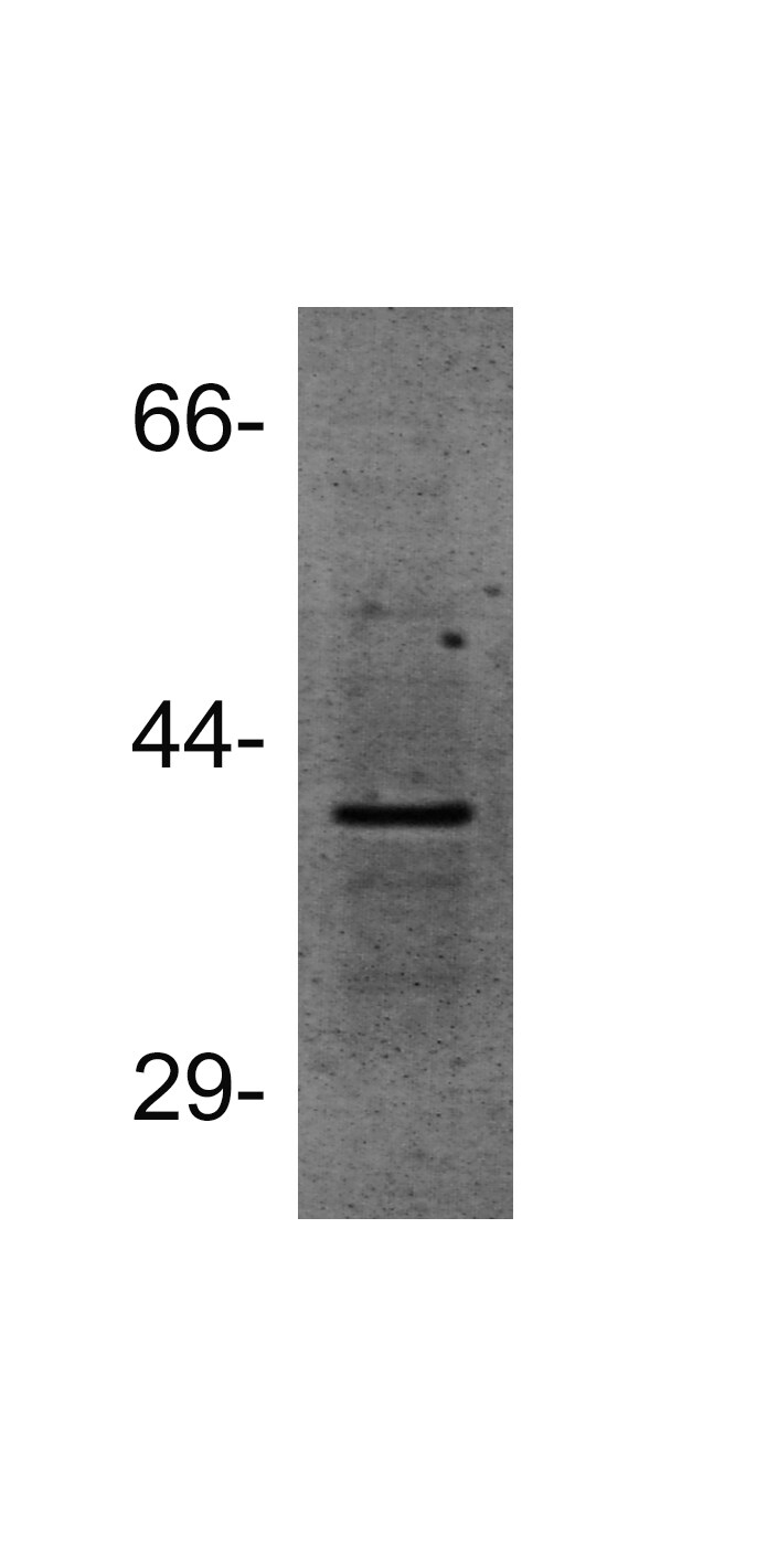
![Western Blot: MAT2B Antibody [NBP1-82797]](https://resources.rndsystems.com/images/products/MAT2B-Antibody-Western-Blot-NBP1-82797-img0004.jpg "Western Blot: MAT2B Antibody [NBP1-82797]")
Loading...
Key Product Details
Species Reactivity
Validated:
Human
Cited:
Human, Rat
Predicted:
Mouse (99%). Backed by our 100% Guarantee.
Applications
Validated:
Immunohistochemistry, Immunohistochemistry-Paraffin, Western Blot
Cited:
Western Blot
Label
Unconjugated
Antibody Source
Polyclonal Rabbit IgG
Format
BSA Free
Loading...
Product Specifications
Immunogen
This antibody was developed against Recombinant Protein corresponding to amino acids: MFDKVQFSNKSANMDHWQQRFPTHVKDVATVCRQLAEKRMLDPSIKGTFHWSGNEQMTKYEMACAIADAFN
Reactivity Notes
Rat reactivity reported in the scientific literature (PMID: 20043323).
Clonality
Polyclonal
Host
Rabbit
Isotype
IgG
Theoretical MW
38 kDa.
Disclaimer note: The observed molecular weight of the protein may vary from the listed predicted molecular weight due to post translational modifications, post translation cleavages, relative charges, and other experimental factors.
Disclaimer note: The observed molecular weight of the protein may vary from the listed predicted molecular weight due to post translational modifications, post translation cleavages, relative charges, and other experimental factors.
Scientific Data Images for MAT2B Antibody - BSA Free
Western Blot: MAT2B Antibody [NBP1-82797]
Western Blot: MAT2B Antibody [NBP1-82797] - Lane 1: Marker [kDa] 230, 130, 95, 72, 56, 36, 28, 17, 11. Lane 2: Human cell line RT-4. Lane 3: Human cell line U-251MG sp![Immunohistochemistry-Paraffin: MAT2B Antibody [NBP1-82797]](https://resources.rndsystems.com/images/products/MAT2B-Antibody-Immunohistochemistry-Paraffin-NBP1-82797-img0009.jpg "Immunohistochemistry-Paraffin: MAT2B Antibody [NBP1-82797]")
Immunohistochemistry-Paraffin: MAT2B Antibody [NBP1-82797]
Immunohistochemistry-Paraffin: MAT2B Antibody [NBP1-82797] - Staining of human skin shows no positivity in keratinocytes as expected.![Immunohistochemistry-Paraffin: MAT2B Antibody [NBP1-82797]](https://resources.rndsystems.com/images/products/MAT2B-Antibody-Immunohistochemistry-Paraffin-NBP1-82797-img0005.jpg "Immunohistochemistry-Paraffin: MAT2B Antibody [NBP1-82797]")
Immunohistochemistry-Paraffin: MAT2B Antibody [NBP1-82797]
Immunohistochemistry-Paraffin: MAT2B Antibody [NBP1-82797] - Staining of human kidney shows moderate cytoplasmic and nuclear positivity in cells in tubules.![Immunohistochemistry-Paraffin: MAT2B Antibody [NBP1-82797]](https://resources.rndsystems.com/images/products/MAT2B-Antibody-Immunohistochemistry-Paraffin-NBP1-82797-img0006.jpg "Immunohistochemistry-Paraffin: MAT2B Antibody [NBP1-82797]")
Immunohistochemistry-Paraffin: MAT2B Antibody [NBP1-82797]
Immunohistochemistry-Paraffin: MAT2B Antibody [NBP1-82797] - Staining of human duodenum shows moderate positivity in luminal membrane in glandular cells.![Immunohistochemistry-Paraffin: MAT2B Antibody [NBP1-82797]](https://resources.rndsystems.com/images/products/MAT2B-Antibody-Immunohistochemistry-Paraffin-NBP1-82797-img0007.jpg "Immunohistochemistry-Paraffin: MAT2B Antibody [NBP1-82797]")
Immunohistochemistry-Paraffin: MAT2B Antibody [NBP1-82797]
Immunohistochemistry-Paraffin: MAT2B Antibody [NBP1-82797] - Staining of human liver shows moderate positivity in bile canaliculi in hepatocytes.![Immunohistochemistry-Paraffin: MAT2B Antibody [NBP1-82797]](https://resources.rndsystems.com/images/products/MAT2B-Antibody-Immunohistochemistry-Paraffin-NBP1-82797-img0008.jpg "Immunohistochemistry-Paraffin: MAT2B Antibody [NBP1-82797]")
Immunohistochemistry-Paraffin: MAT2B Antibody [NBP1-82797]
Immunohistochemistry-Paraffin: MAT2B Antibody [NBP1-82797] - Staining of human cerebral cortex shows moderate positivity in neuronal processes in astrocytes.
Western Blot: MAT2B Antibody - BSA Free [NBP1-82797] -
MAT1A, MAT2A, MAT2B and miR-203 expression in human HCC with different prognosis(A) MAT1A, MAT2A, MAT2B, Ki67 and MDK gene expression. The results are expressed as N-fold differences in target gene expression relative to the RNR-18 expression, named N Target (NT). NT = 2– delta CT, delta CT of each sample was calculated by subtracting the Ct of the target gene from the Ct of the RNR-18 gene. (B) Representative Western Blot of MAT alpha 1, MAT alpha 2, and MAT beta 2, in HCCs with different prognosis and correspondent surroundings. Chemiluminescence analysis: optical densities were normalized to beta -actin levels and expressed in arbitrary units. (C) miR-203 expression (NT = 2– delta CT) and Spearman’s correlation analysis of miR-203. A total of 26 cases (13 HCCB and 13 HCCP) were used for the correlation analysis. Data are means +/- standard deviation (SD) of six experiments. Mann-Whitney test: Point, different from NL for P < 0.001. Asterisk, different from SL for at least P < 0.01. Dagger, HCCP/SLP different from HCCB/SLB for P < 0.001. Abbreviations: HCCB, HCC with better prognosis (survival > 3 years) and correspondent surrounding (SLB); HCCP, HCC with poorer prognosis (survival < 3 years) and corresponding surrounding (SLP). Image collected and cropped by CiteAb from the following open publication (https://pubmed.ncbi.nlm.nih.gov/31073374), licensed under a CC-BY license. Not internally tested by Novus Biologicals.Applications for MAT2B Antibody - BSA Free
Application
Recommended Usage
Immunohistochemistry
1:50 - 1:200
Immunohistochemistry-Paraffin
1:50 - 1:200
Western Blot
0.04-0.4 ug/ml
Application Notes
For IHC-Paraffin, HIER pH 6 retrieval is recommended.
Reviewed Applications
Read 1 review rated 4 using NBP1-82797 in the following applications:
Formulation, Preparation, and Storage
Purification
Affinity purified
Formulation
PBS (pH 7.2) and 40% Glycerol
Format
BSA Free
Preservative
0.02% Sodium Azide
Concentration
Concentrations vary lot to lot. See vial label for concentration. If unlisted please contact technical services.
Shipping
The product is shipped with polar packs. Upon receipt, store it immediately at the temperature recommended below.
Stability & Storage
Store at 4C short term. Aliquot and store at -20C long term. Avoid freeze-thaw cycles.
Background: MAT2B
Alternate Names
beta regulatory subunit of methionine adenosyltransferase, DTDP-4-keto-6-deoxy-D-glucose 4-reductase, MAT II beta, MAT-II, MATIIbeta, methionine adenosyltransferase 2 subunit beta, Methionine adenosyltransferase II beta, methionine adenosyltransferase II, beta, MGC12237, Nbla02999, putative protein product of Nbla02999, SDR23E1, short chain dehydrogenase/reductase family 23E, member 1, TGR
Entrez Gene IDs
27430 (Human)
Gene Symbol
MAT2B
UniProt
Additional MAT2B Products
Product Documents for MAT2B Antibody - BSA Free
Certificate of Analysis
To download a Certificate of Analysis, please enter a lot or batch number in the search box below.
Product Specific Notices for MAT2B Antibody - BSA Free
This product is for research use only and is not approved for use in humans or in clinical diagnosis. Primary Antibodies are guaranteed for 1 year from date of receipt.
Citations for MAT2B Antibody - BSA Free
 Powered by Bioz
Customer Reviews for MAT2B Antibody - BSA Free (1)
4 out of 5
1 Customer Rating
Have you used MAT2B Antibody - BSA Free?
Submit a review and receive an Amazon gift card!
$25/€18/£15/$25CAN/¥2500 Yen for a review with an image
$10/€7/£6/$10CAN/¥1110 Yen for a review without an image
Submit a review
Customer Images
Showing
1
-
1 of
1 review
Showing All
Filter By:
-
Application: Western BlotSample Tested: Whole cell lysate prepared from RT-4 cellsSpecies: HumanVerified Customer | Posted 12/14/2014Western blot for MAT2B in RT-4 cells

There are no reviews that match your criteria.
Protocols
Find general support by application which include: protocols, troubleshooting, illustrated assays, videos and webinars.
- Antigen Retrieval Protocol (PIER)
- Antigen Retrieval for Frozen Sections Protocol
- Appropriate Fixation of IHC/ICC Samples
- Cellular Response to Hypoxia Protocols
- Chromogenic IHC Staining of Formalin-Fixed Paraffin-Embedded (FFPE) Tissue Protocol
- Chromogenic Immunohistochemistry Staining of Frozen Tissue
- ClariTSA™ Fluorophore Kits
- Detection & Visualization of Antibody Binding
- Fluorescent IHC Staining of Frozen Tissue Protocol
- Graphic Protocol for Heat-induced Epitope Retrieval
- Graphic Protocol for the Preparation and Fluorescent IHC Staining of Frozen Tissue Sections
- Graphic Protocol for the Preparation and Fluorescent IHC Staining of Paraffin-embedded Tissue Sections
- Graphic Protocol for the Preparation of Gelatin-coated Slides for Histological Tissue Sections
- IHC Sample Preparation (Frozen sections vs Paraffin)
- Immunofluorescent IHC Staining of Formalin-Fixed Paraffin-Embedded (FFPE) Tissue Protocol
- Immunohistochemistry (IHC) and Immunocytochemistry (ICC) Protocols
- Immunohistochemistry Frozen Troubleshooting
- Immunohistochemistry Paraffin Troubleshooting
- Preparing Samples for IHC/ICC Experiments
- Preventing Non-Specific Staining (Non-Specific Binding)
- Primary Antibody Selection & Optimization
- Protocol for Heat-Induced Epitope Retrieval (HIER)
- Protocol for Making a 4% Formaldehyde Solution in PBS
- Protocol for VisUCyte™ HRP Polymer Detection Reagent
- Protocol for the Preparation & Fixation of Cells on Coverslips
- Protocol for the Preparation and Chromogenic IHC Staining of Frozen Tissue Sections
- Protocol for the Preparation and Chromogenic IHC Staining of Frozen Tissue Sections - Graphic
- Protocol for the Preparation and Chromogenic IHC Staining of Paraffin-embedded Tissue Sections
- Protocol for the Preparation and Chromogenic IHC Staining of Paraffin-embedded Tissue Sections - Graphic
- Protocol for the Preparation and Fluorescent IHC Staining of Frozen Tissue Sections
- Protocol for the Preparation and Fluorescent IHC Staining of Paraffin-embedded Tissue Sections
- Protocol for the Preparation of Gelatin-coated Slides for Histological Tissue Sections
- R&D Systems Quality Control Western Blot Protocol
- TUNEL and Active Caspase-3 Detection by IHC/ICC Protocol
- The Importance of IHC/ICC Controls
- Troubleshooting Guide: Immunohistochemistry
- Troubleshooting Guide: Western Blot Figures
- Western Blot Conditions
- Western Blot Protocol
- Western Blot Protocol for Cell Lysates
- Western Blot Troubleshooting
- Western Blot Troubleshooting Guide
- View all Protocols, Troubleshooting, Illustrated assays and Webinars
Loading...