Melanocortin-1 R/MC1R Antibody - BSA Free
Novus Biologicals | Catalog # NLS1040
![Immunohistochemistry-Paraffin: Melanocortin-1 R/MC1R Antibody - BSA Free [NLS1040]](https://resources.rndsystems.com/images/products/Melanocortin-1-R-MC1R-Antibody-Immunohistochemistry-Paraffin-NLS1040-img0002.jpg "Immunohistochemistry-Paraffin: Melanocortin-1 R/MC1R Antibody - BSA Free [NLS1040]")
Loading...
Key Product Details
Species Reactivity
Validated:
Human, Monkey
Predicted:
Chimpanzee (100%), Orangutan (100%), Primate (100%). Backed by our 100% Guarantee.
Applications
Immunohistochemistry, Immunohistochemistry-Paraffin
Label
Unconjugated
Antibody Source
Polyclonal Rabbit IgG
Format
BSA Free
Loading...
Product Specifications
Immunogen
Synthetic 16 amino acid peptide from 3rd cytoplasmic domain of human Melanocortin-1 R/MC1R.
Epitope
3rd cytoplasmic domain of human
Reactivity Notes
Predicted cross-reactivity based on sequence identity: Gorilla (100%), Gibbon (100%).
Specificity
Human Melanocortin-1 R/MC1R. BLAST analysis of the peptide immunogen showed no homology with other human proteins.
Clonality
Polyclonal
Host
Rabbit
Isotype
IgG
Description
Product can be stored undiluted at 4C for up to 1 month.
Scientific Data Images for Melanocortin-1 R/MC1R Antibody - BSA Free
Immunohistochemistry-Paraffin: Melanocortin-1 R/MC1R Antibody - BSA Free [NLS1040]
Immunohistochemistry-Paraffin: Melanocortin-1 R/MC1R Antibody [NLS1040] - Human Adrenal: Formalin-Fixed, Paraffin-Embedded (FFPE)![Immunohistochemistry-Paraffin: Melanocortin-1 R/MC1R Antibody - BSA Free [NLS1040]](https://resources.rndsystems.com/images/products/Melanocortin-1-R-MC1R-Antibody-Immunohistochemistry-Paraffin-NLS1040-img0001.jpg "Immunohistochemistry-Paraffin: Melanocortin-1 R/MC1R Antibody - BSA Free [NLS1040]")
Immunohistochemistry-Paraffin: Melanocortin-1 R/MC1R Antibody - BSA Free [NLS1040]
Immunohistochemistry-Paraffin: Melanocortin-1 R/MC1R Antibody [NLS1040] - Human Kidney: Formalin-Fixed, Paraffin-Embedded (FFPE)Applications for Melanocortin-1 R/MC1R Antibody - BSA Free
Application
Recommended Usage
Immunohistochemistry-Paraffin
2.5 - 5 ug/ml
Application Notes
.
Reviewed Applications
Read 1 review rated 5 using NLS1040 in the following applications:
Formulation, Preparation, and Storage
Purification
Immunogen affinity purified
Formulation
PBS
Format
BSA Free
Preservative
0.1% Sodium Azide
Concentration
1.0 mg/ml
Shipping
The product is shipped with polar packs. Upon receipt, store it immediately at the temperature recommended below.
Stability & Storage
Keep as concentrated solution. Aliquot and store at -20C or below. Avoid multiple freeze-thaw cycles.
Background: Melanocortin-1 R/MC1R
Long Name
Melanocortin 1 Receptor
Alternate Names
CMM5, MC1R, Melanocortin-1R, Melanocortin1R, MSH-R, Mshra, SHEP2, Tob
Entrez Gene IDs
4157 (Human)
Gene Symbol
MC1R
Additional Melanocortin-1 R/MC1R Products
Product Documents for Melanocortin-1 R/MC1R Antibody - BSA Free
Certificate of Analysis
To download a Certificate of Analysis, please enter a lot or batch number in the search box below.
Product Specific Notices for Melanocortin-1 R/MC1R Antibody - BSA Free
This product is for research use only and is not approved for use in humans or in clinical diagnosis. Primary Antibodies are guaranteed for 1 year from date of receipt.
Customer Reviews for Melanocortin-1 R/MC1R Antibody - BSA Free (1)
5 out of 5
1 Customer Rating
Have you used Melanocortin-1 R/MC1R Antibody - BSA Free?
Submit a review and receive an Amazon gift card!
$25/€18/£15/$25CAN/¥2500 Yen for a review with an image
$10/€7/£6/$10CAN/¥1110 Yen for a review without an image
Submit a review
Customer Images
Showing
1
-
1 of
1 review
Showing All
Filter By:
-
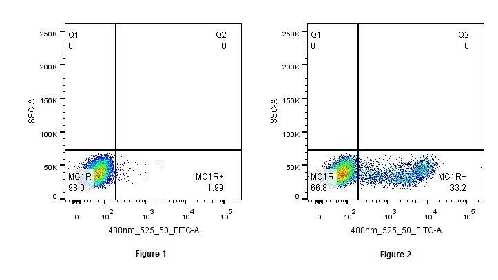
Application: Flow CytometrySample Tested: Blood mononuclear cells (PBMCs)Species: HumanVerified Customer | Posted 02/05/2019Figure 1 shows Lymphocytes stained with Alexa Fluor 488 Donkey anti-rabbit IgG alone, and Figure 2 shows Lymphocytes stained with human MC1R and Alexa Fluor 488 Donkey anti-rabbit IgG.Blocking: Natural Goat Serum Primary antibody: 2 uL (1 ug) Secondary antibody: Alexa Fluor 488 Donkey anti-rabbit IgG (2.5 uL / 1 ug) Details: Human lymphocytes were isolated from PBMCs. Lymphocytes were blocked with Natural Goat Serum for 15 minutes, and then stained and subsequently incubated at -20 C with anti-human MC1R primary antibody for 30 minutes. After washing, cells were stained with Alexa Fluor 488 Donkey anti-rabbit IgG and incubated at -20 C for 40 minutes. After washing, cells were detected using a Fortessa 4-15 flow cytometer.

There are no reviews that match your criteria.
Protocols
View specific protocols for Melanocortin-1 R/MC1R Antibody - BSA Free (NLS1040):
Immunohistochemistry Protocol for MC1 Receptor Antibody (NLS1040):
Immunohistochemistry
1. Prepare tissue with formalin fixation and by embedding it in paraffin wax.
2. Make 4-um sections and place on pre-cleaned and charged microscope slides.
3. Heat in a tissue-drying oven for 45 minutes at 60 degrees Celcius.
4. Deparaffinize the tissues by wash drying the slides in 3 changes of xylene approximately 5 minutes each @ RT.
5. Rehydrate the tissues by washing the slides in 3 changes of 100% alcohol approximately 3 minutes each @ RT.
6. Wash the slides in 2 changes of 95% alcohol approximately 3 minutes each @ RT.
7. Wash the slides in 1 change of 80% alcohol approximately 3 minutes @ RT.
8. Rinse the slides in gentle running distilled water approximately 5 minutes @ RT.
9. Perform antigen retrieval by steaming the slides in 0.01M sodium citrate buffer (pH 6.0) @ 99-100 degrees Celcius for 20 minutes.
10. Remove the slides from the heat and let stand in buffer @ RT for 20 minutes.
11. Rinse the slides in 1X TBS-T for 1 minute @ RT.
**Do not allow the tissues to dry at any time during the staining procedure**
12. Begin the immunostaining by applying a universal protein block approximately 20 minutes @ RT.
13. Drain protein block from the slides and apply the diluted primary antibody approximately 45 minutes @ RT.
14. Rinse the slide in 1X TBS-T approximately 1 minute @ RT.
15. Apply a biotinylated anti-rabbit IgG (H+L) secondary approximately 30 minutes @ RT.
16. Rinse the slide in 1X TBS-T approximately 1 minute at RT.
17. Apply an alkaline phosphatase steptavidin approximately 30 minutes at RT.
18. Rinse the slide in 1X TBS-T approximately 1 minute at RT.
19. Apply an alkaline phosphatase chromagen substrate approximately 30 minutes at RT.
20. Rinse the slide in distilled water approximately 1 minute @ RT.
**This method should only be used if the chromagen substrate is alcohol insoluble (ie: Vector Red, DAB)**
21. Dehydrate the tissue by washing the slides in 2 changes of 80% alcohol approximately 1 minute each @ RT.
22. Wash the slides in 2 changes of 95% alcohol approximately 1 minute each @ RT.
23. Wash the slides in 3 changes of 100% alcohol approximately 1 minute each @ RT.
24. Wash the slides in 3 changes of xyleneapproximately 1 minute each @ RT.
25. Apply cover slip.
Find general support by application which include: protocols, troubleshooting, illustrated assays, videos and webinars.
- Antigen Retrieval Protocol (PIER)
- Antigen Retrieval for Frozen Sections Protocol
- Appropriate Fixation of IHC/ICC Samples
- Cellular Response to Hypoxia Protocols
- Chromogenic IHC Staining of Formalin-Fixed Paraffin-Embedded (FFPE) Tissue Protocol
- Chromogenic Immunohistochemistry Staining of Frozen Tissue
- ClariTSA™ Fluorophore Kits
- Detection & Visualization of Antibody Binding
- Fluorescent IHC Staining of Frozen Tissue Protocol
- Graphic Protocol for Heat-induced Epitope Retrieval
- Graphic Protocol for the Preparation and Fluorescent IHC Staining of Frozen Tissue Sections
- Graphic Protocol for the Preparation and Fluorescent IHC Staining of Paraffin-embedded Tissue Sections
- Graphic Protocol for the Preparation of Gelatin-coated Slides for Histological Tissue Sections
- IHC Sample Preparation (Frozen sections vs Paraffin)
- Immunofluorescent IHC Staining of Formalin-Fixed Paraffin-Embedded (FFPE) Tissue Protocol
- Immunohistochemistry (IHC) and Immunocytochemistry (ICC) Protocols
- Immunohistochemistry Frozen Troubleshooting
- Immunohistochemistry Paraffin Troubleshooting
- Preparing Samples for IHC/ICC Experiments
- Preventing Non-Specific Staining (Non-Specific Binding)
- Primary Antibody Selection & Optimization
- Protocol for Heat-Induced Epitope Retrieval (HIER)
- Protocol for Making a 4% Formaldehyde Solution in PBS
- Protocol for VisUCyte™ HRP Polymer Detection Reagent
- Protocol for the Preparation & Fixation of Cells on Coverslips
- Protocol for the Preparation and Chromogenic IHC Staining of Frozen Tissue Sections
- Protocol for the Preparation and Chromogenic IHC Staining of Frozen Tissue Sections - Graphic
- Protocol for the Preparation and Chromogenic IHC Staining of Paraffin-embedded Tissue Sections
- Protocol for the Preparation and Chromogenic IHC Staining of Paraffin-embedded Tissue Sections - Graphic
- Protocol for the Preparation and Fluorescent IHC Staining of Frozen Tissue Sections
- Protocol for the Preparation and Fluorescent IHC Staining of Paraffin-embedded Tissue Sections
- Protocol for the Preparation of Gelatin-coated Slides for Histological Tissue Sections
- TUNEL and Active Caspase-3 Detection by IHC/ICC Protocol
- The Importance of IHC/ICC Controls
- Troubleshooting Guide: Immunohistochemistry
- View all Protocols, Troubleshooting, Illustrated assays and Webinars
Loading...