


Mouse VEGFR1/Flt-1 PE‑conjugated Antibody

R&D Systems | Catalog # FAB4711P

R&D Systems
Discontinued Product
FAB4711P has been discontinued. View all VEGFR1/Flt-1 products.

Key Product Details

Validated by

Biological Validation

Species Reactivity

Validated:

Mouse

Cited:

Mouse, Xenograft

Applications

Validated:

Flow Cytometry

Cited:

Flow Cytometry

Label

Phycoerythrin (Excitation = 488 nm, Emission = 565-605 nm)

Antibody Source

Monoclonal Rat IgG2B Clone # 141522
Loading...

Product Specifications

Immunogen

Mouse myeloma cell line NS0-derived recombinant mouse VEGF R1/Flt‑1
Ser27-Glu759
Accession # P35969

Specificity

Detects mouse VEGF R1/Flt‑1 in direct ELISAs and Western blots. In direct ELISAs and Western blots, no cross-reactivity with recombinant human VEGF R1, recombinant mouse (rm) VEGF R2, or rmVEGF R3 is observed.

Clonality

Monoclonal

Host

Rat

Isotype

IgG2B

Scientific Data Images for Mouse VEGFR1/Flt-1 PE‑conjugated Antibody

Detection of VEGF R1/Flt-1 antibody in bEnd.3 Mouse Cell Line antibody by Flow Cytometry.

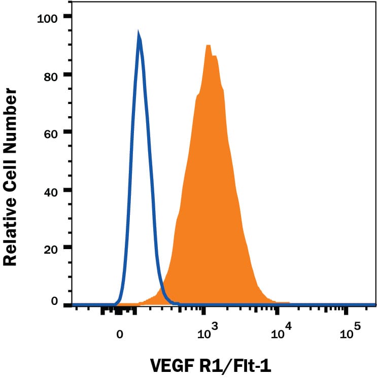
Detection of VEGF R1/Flt‑1 in bEnd.3 Mouse Cell Line by Flow Cytometry.

bEnd.3 mouse endothelioma cell line was stained with Rat Anti-Mouse VEGF R1/Flt-1 PE-conjugated Monoclonal Antibody (Catalog # FAB4711P, filled histogram) or isotype control antibody (Catalog # IC013P, open histogram). View our protocol for Staining Membrane-associated Proteins.
Detection of Mouse VEGFR1/Flt-1 by Flow Cytometry

Detection of Mouse VEGFR1/Flt-1 by Flow Cytometry

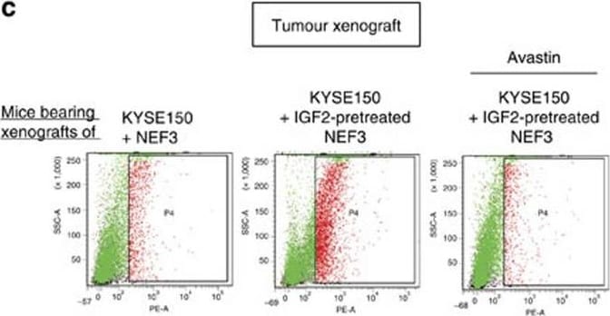
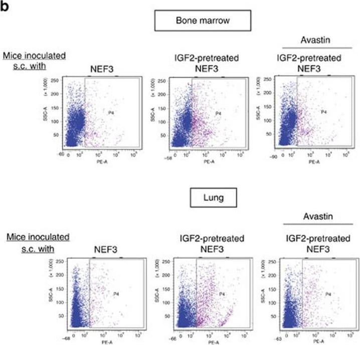
Fibroblast-derived VEGF enriches VEGFR1+ cells bone marrow and pre-metastatic sites.(a) Immunohistochemical expression of mouse VEGF in the tumour xenografts established with KYSE150-Id1, KYSE150-Id1-shIGF2 or vector control cells (scale bar, 20 μm). (b) Flow cytometry data showing the expression of VEGFR1+ cells in bone marrow (upper panel) and lungs (lower panel) of mice with subcutaneous implantation of indicated fibroblasts and treatment (female 6–8-week-old nude mice, n=3 per group). (c,d) Flow cytometry analysis of VEGFR1+ cells (c) and comparison of tumour volume (d) of subcutaneous tumour xenografts established from co-implantation of KYSE150 cells and indicated NEFs in the presence or absence of Avastin treatment. Photographs show representative tumours of the three groups (female 6–8-week-old nude mice, n=6 per group; scale bar, 1 cm). Bars, s.d.; *P<0.05; **P<0.01; ***P<0.001 by Student's t-test. Image collected and cropped by CiteAb from the following publication (https://www.nature.com/articles/ncomms14399), licensed under a CC-BY license. Not internally tested by R&D Systems.
Detection of Mouse VEGFR1/Flt-1 by Flow Cytometry

Detection of Mouse VEGFR1/Flt-1 by Flow Cytometry

Fibroblast-derived VEGF enriches VEGFR1+ cells bone marrow and pre-metastatic sites.(a) Immunohistochemical expression of mouse VEGF in the tumour xenografts established with KYSE150-Id1, KYSE150-Id1-shIGF2 or vector control cells (scale bar, 20 μm). (b) Flow cytometry data showing the expression of VEGFR1+ cells in bone marrow (upper panel) and lungs (lower panel) of mice with subcutaneous implantation of indicated fibroblasts and treatment (female 6–8-week-old nude mice, n=3 per group). (c,d) Flow cytometry analysis of VEGFR1+ cells (c) and comparison of tumour volume (d) of subcutaneous tumour xenografts established from co-implantation of KYSE150 cells and indicated NEFs in the presence or absence of Avastin treatment. Photographs show representative tumours of the three groups (female 6–8-week-old nude mice, n=6 per group; scale bar, 1 cm). Bars, s.d.; *P<0.05; **P<0.01; ***P<0.001 by Student's t-test. Image collected and cropped by CiteAb from the following publication (https://www.nature.com/articles/ncomms14399), licensed under a CC-BY license. Not internally tested by R&D Systems.

Applications for Mouse VEGFR1/Flt-1 PE‑conjugated Antibody

Application
Recommended Usage

Flow Cytometry

10 µL/106 cells
Sample: bEnd.3 mouse endothelioma cell line

Reviewed Applications

Read 2 reviews rated 4.5 using FAB4711P in the following applications:

Spectra Viewer

Plan Your Experiments

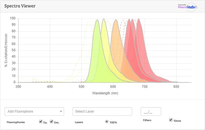
Use our spectra viewer to interactively plan your experiments, assessing multiplexing options. View the excitation and emission spectra for our fluorescent dye range and other commonly used dyes.

Spectra Viewer

Flow Cytometry Panel Builder

Bio-Techne Knows Flow Cytometry

Save time and reduce costly mistakes by quickly finding compatible reagents using the Panel Builder Tool.

Advanced Features

  • Spectra Viewer - Custom analysis of spectra from multiple fluorochromes
  • Spillover Popups - Visualize the spectra of individual fluorochromes
  • Antigen Density Selector - Match fluorochrome brightness with antigen density
Build Your Panel Now

Formulation, Preparation, and Storage

Purification

Protein A or G purified from hybridoma culture supernatant

Formulation

Supplied in a saline solution containing BSA and Sodium Azide.

Shipping

The product is shipped with polar packs. Upon receipt, store it immediately at the temperature recommended below.

Stability & Storage

Protect from light. Do not freeze.
  • 12 months from date of receipt, 2 to 8 °C as supplied.

Background: VEGFR1/Flt-1

VEGF R1 is one of the five receptor tyrosine kinases (RTKs) (VEGF R1, KDR/Flk-1, Flt-4, Tie-1, and Tek/Tie-2) whose expression is almost exclusively restricted to the endothelial cells. Tie-1 and tek/tie-2 define a new class of RTKs containing two immunoglobulin-like domains, three EGF homology domains and three fibronectin type III domains in their extracellular regions. VEGF R1/Flt-1, VEGF R2/KDR/Flk-1, VEGF R3/Flt-4 are members of the class III subfamily of RTKs containing seven immunoglobulin-like repeats in their extracellular domains. All five RTKs are likely to play central roles in vasculogenesis and angiogenesis.

Full length mouse VEGF R1 mRNA encodes a 1333 amino acid (aa) residue precursor with a predicted 22 aa residue signal peptide. Mature VEGF R1 is composed of a 737 aa residue extracellular domain, a 22 aa residue transmembrane domain and a 552 aa residue cytoplasmic domain. As a result of alternative splicing of the mRNA, a cDNA encoding a truncated form of VEGF R1, lacking the seventh immunoglobulin-like domain, the transmembrane and intracellular domains, has been cloned. The recombinant soluble VEGF R1/Fc chimera binds VEGF and PlGF with high affinity and is a potent VEGF antagonist.

References

  1. He, Y. et al. (1999) Molecular Endocrinology 13:537.
  2. Ferrara, N. and T. Davis-Smyth (1997) Endocrine Reviews 8:4.

Long Name

Vascular Endothelial Growth Factor Receptor 1

Alternate Names

Flt-1, FLT1, FRT, VEGF R1, VEGFR-1

Entrez Gene IDs

2321 (Human); 14254 (Mouse)

Gene Symbol

FLT1

UniProt

Additional VEGFR1/Flt-1 Products

Product Documents for Mouse VEGFR1/Flt-1 PE‑conjugated Antibody

Certificate of Analysis

To download a Certificate of Analysis, please enter a lot or batch number in the search box below.

Note: Certificate of Analysis not available for kit components.

Product Specific Notices for Mouse VEGFR1/Flt-1 PE‑conjugated Antibody

For research use only

Citations for Mouse VEGFR1/Flt-1 PE‑conjugated Antibody

Customer Reviews for Mouse VEGFR1/Flt-1 PE‑conjugated Antibody (2)

4.5 out of 5
2 Customer Ratings
5 Stars
50%
4 Stars
50%
3 Stars
0%
2 Stars
0%
1 Stars
0%

Have you used Mouse VEGFR1/Flt-1 PE‑conjugated Antibody?

Submit a review and receive an Amazon gift card!

$25/€18/£15/$25CAN/¥2500 Yen for a review with an image

$10/€7/£6/$10CAN/¥1110 Yen for a review without an image

Submit a review
Amazon Gift Card
Showing  1 - 2 of 2 reviews Showing All
Filter By:
  • Name: Anonymous
    Application: Flow Cytometry
    Sample Tested: See PMID: 21447646
    Species: Rat
    Verified Customer | Posted 01/26/2015
  • Name: Anonymous
    Application: Flow Cytometry
    Sample Tested: See PMID: 24449499
    Species: Mouse
    Verified Customer | Posted 01/26/2015

There are no reviews that match your criteria.

Showing  1 - 2 of 2 reviews Showing All

FAQs

No product specific FAQs exist for this product.

View all FAQs for Antibodies
