MX2 Antibody - BSA Free
Novus Biologicals | Catalog # NBP1-81018
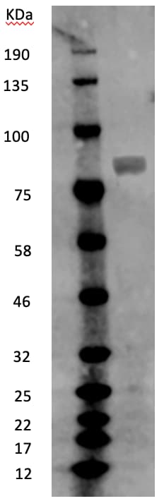
![Western Blot: MX2 Antibody [NBP1-81018]](https://resources.rndsystems.com/images/products/MX2-Antibody-Western-Blot-NBP1-81018-img0008.jpg "Western Blot: MX2 Antibody [NBP1-81018]")
Loading...
Key Product Details
Validated by
Biological Validation
Species Reactivity
Validated:
Human
Cited:
Human
Applications
Validated:
Immunohistochemistry, Immunohistochemistry-Paraffin
Cited:
Western Blot, Immunocytochemistry/ Immunofluorescence
Label
Unconjugated
Antibody Source
Polyclonal Rabbit IgG
Format
BSA Free
Loading...
Product Specifications
Immunogen
This antibody was developed against Recombinant Protein corresponding to amino acids: PYRRRSQFSSRKYLKKEMNSFQQQPPPFGTVPPQMMFPPNWQGAEKDAAFLAKDFNFLTLNNQPPPGNRSQPRAMG
Clonality
Polyclonal
Host
Rabbit
Isotype
IgG
Theoretical MW
82 kDa.
Disclaimer note: The observed molecular weight of the protein may vary from the listed predicted molecular weight due to post translational modifications, post translation cleavages, relative charges, and other experimental factors.
Disclaimer note: The observed molecular weight of the protein may vary from the listed predicted molecular weight due to post translational modifications, post translation cleavages, relative charges, and other experimental factors.
Scientific Data Images for MX2 Antibody - BSA Free
![Immunohistochemistry-Paraffin: MX2 Antibody [NBP1-81018]](https://resources.rndsystems.com/images/products/MX2-Antibody-Immunohistochemistry-Paraffin-NBP1-81018-img0005.jpg "Immunohistochemistry-Paraffin: MX2 Antibody [NBP1-81018]")
Immunohistochemistry-Paraffin: MX2 Antibody [NBP1-81018]
Immunohistochemistry-Paraffin: MX2 Antibody [NBP1-81018] - Staining of human spleen shows strong positivity in nuclear membrane in cells in red pulp.![Western Blot: MX2 Antibody [NBP1-81018]](https://resources.rndsystems.com/images/products/MX2-Antibody-Western-Blot-NBP1-81018-img0006.jpg "Western Blot: MX2 Antibody [NBP1-81018]")
Western Blot: MX2 Antibody [NBP1-81018]
Western Blot: MX2 Antibody [NBP1-81018] - Human glioma U87 cell lysate. WB image submitted by a verified customer review.![Immunohistochemistry-Paraffin: MX2 Antibody [NBP1-81018]](https://resources.rndsystems.com/images/products/MX2-Antibody-Immunohistochemistry-Paraffin-NBP1-81018-img0001.jpg "Immunohistochemistry-Paraffin: MX2 Antibody [NBP1-81018]")
Immunohistochemistry-Paraffin: MX2 Antibody [NBP1-81018]
Immunohistochemistry-Paraffin: MX2 Antibody [NBP1-81018] - Staining of human liver shows no postivity in hepatocytes as expected.![Immunohistochemistry-Paraffin: MX2 Antibody [NBP1-81018]](https://resources.rndsystems.com/images/products/MX2-Antibody-Immunohistochemistry-Paraffin-NBP1-81018-img0002.jpg "Immunohistochemistry-Paraffin: MX2 Antibody [NBP1-81018]")
Immunohistochemistry-Paraffin: MX2 Antibody [NBP1-81018]
Immunohistochemistry-Paraffin: MX2 Antibody [NBP1-81018] - Staining of human colon shows moderate postivity in nuclear membrane in lymphoid cells in the lamina propria.![Immunohistochemistry-Paraffin: MX2 Antibody [NBP1-81018]](https://resources.rndsystems.com/images/products/MX2-Antibody-Immunohistochemistry-Paraffin-NBP1-81018-img0003.jpg "Immunohistochemistry-Paraffin: MX2 Antibody [NBP1-81018]")
Immunohistochemistry-Paraffin: MX2 Antibody [NBP1-81018]
Immunohistochemistry-Paraffin: MX2 Antibody [NBP1-81018] - Staining of human skeletal muscle shows no postivity in myocytes as expected.![Immunohistochemistry-Paraffin: MX2 Antibody [NBP1-81018]](https://resources.rndsystems.com/images/products/MX2-Antibody-Immunohistochemistry-Paraffin-NBP1-81018-img0004.jpg "Immunohistochemistry-Paraffin: MX2 Antibody [NBP1-81018]")
Immunohistochemistry-Paraffin: MX2 Antibody [NBP1-81018]
Immunohistochemistry-Paraffin: MX2 Antibody [NBP1-81018] - Staining of human lymph node shows moderate positivity in nuclear membrane in non-germinal center cells.
Western Blot: MX2 Antibody [NBP1-81018] -
Western Blot: MX2 Antibody [NBP1-81018] - MxB is localized to mitochondria in cultured cells.Fluorescence images of Hep3B cells or HeLa cells, stained with a polyclonal antibody (IJ-GP) to MxB (a, c), or expressing exogenous human MxB-GFP (b, d). All cells were stained for the inner mitochondrial membrane marker CoxIV. MxB is visualized as small or large puncta that align closely with mitochondrial membranes. Boxed regions provide a higher magnification for color overlays showing the alignment of MxB (green) with mitochondria (red) in both cell types. Scale bars, 10 μm. e Western blot analysis of different human tissues (Br = brain, Lng = lung, Liv = liver, LN = lymph node, SP = spleen, Tst = testis, Tns = tonsil) for endogenous MxB that appears enriched in the liver, lymph node, testis, tonsil, & pig liver. f Western blotting comparing MxB expression in HeLa cells, three different hepatocyte cell lines, & a human monocyte cell line (THP-1), as well as primary human & pig hepatocytes. Expression levels are exceptionally high in primary hepatocytes, the HepG2 & Huh7 cell lines (H3B = Hep3B, HG2 = HepG2, Hu7 = Huh7, THP = THP-1, Hu = human hepatocytes). g Western blotting of four distinct cell lines comparing endogenous MxB expression under control conditions vs. treatment with 1,000 IU/ ml-1 of IFN-alpha −2Α. HeLa, Hep3B & HepG2 cells induced MxB expression by IFN treatment, but not Huh7 cells. Image collected & cropped by CiteAb from the following publication (https://pubmed.ncbi.nlm.nih.gov/32102993), licensed under a CC-BY license. Not internally tested by Novus Biologicals.
Western Blot: MX2 Antibody [NBP1-81018] -
Western Blot: MX2 Antibody [NBP1-81018] - MxB is localized to mitochondria in cultured cells.Fluorescence images of Hep3B cells or HeLa cells, stained with a polyclonal antibody (IJ-GP) to MxB (a, c), or expressing exogenous human MxB-GFP (b, d). All cells were stained for the inner mitochondrial membrane marker CoxIV. MxB is visualized as small or large puncta that align closely with mitochondrial membranes. Boxed regions provide a higher magnification for color overlays showing the alignment of MxB (green) with mitochondria (red) in both cell types. Scale bars, 10 μm. e Western blot analysis of different human tissues (Br = brain, Lng = lung, Liv = liver, LN = lymph node, SP = spleen, Tst = testis, Tns = tonsil) for endogenous MxB that appears enriched in the liver, lymph node, testis, tonsil, & pig liver. f Western blotting comparing MxB expression in HeLa cells, three different hepatocyte cell lines, & a human monocyte cell line (THP-1), as well as primary human & pig hepatocytes. Expression levels are exceptionally high in primary hepatocytes, the HepG2 & Huh7 cell lines (H3B = Hep3B, HG2 = HepG2, Hu7 = Huh7, THP = THP-1, Hu = human hepatocytes). g Western blotting of four distinct cell lines comparing endogenous MxB expression under control conditions vs. treatment with 1,000 IU/ ml-1 of IFN-alpha −2Α. HeLa, Hep3B & HepG2 cells induced MxB expression by IFN treatment, but not Huh7 cells. Image collected & cropped by CiteAb from the following publication (https://pubmed.ncbi.nlm.nih.gov/32102993), licensed under a CC-BY license. Not internally tested by Novus Biologicals.Applications for MX2 Antibody - BSA Free
Application
Recommended Usage
Immunohistochemistry
1:200 - 1:500
Immunohistochemistry-Paraffin
1:200 - 1:500
Application Notes
For IHC-Paraffin, HIER pH 6 retrieval is recommended.
Reviewed Applications
Read 1 review rated 4 using NBP1-81018 in the following applications:
Formulation, Preparation, and Storage
Purification
Affinity purified
Formulation
PBS (pH 7.2) and 40% Glycerol
Format
BSA Free
Preservative
0.02% Sodium Azide
Concentration
Concentrations vary lot to lot. See vial label for concentration. If unlisted please contact technical services.
Shipping
The product is shipped with polar packs. Upon receipt, store it immediately at the temperature recommended below.
Stability & Storage
Store at 4C short term. Aliquot and store at -20C long term. Avoid freeze-thaw cycles.
Background: MX2
Alternate Names
human interferon-regulated resistance GTP-binding protein MXB10Interferon-regulated resistance GTP-binding protein MxB, interferon-induced GTP-binding protein Mx2, MXB, myxovirus (influenza virus) resistance 2 (mouse), myxovirus (influenza) resistance 2, homolog of murine, Myxovirus resistance protein 2, p78-related protein, second interferon-induced protein p78
Entrez Gene IDs
4600 (Human)
Gene Symbol
MX2
UniProt
Additional MX2 Products
Product Documents for MX2 Antibody - BSA Free
Certificate of Analysis
To download a Certificate of Analysis, please enter a lot or batch number in the search box below.
Product Specific Notices for MX2 Antibody - BSA Free
This product is for research use only and is not approved for use in humans or in clinical diagnosis. Primary Antibodies are guaranteed for 1 year from date of receipt.
Citations for MX2 Antibody - BSA Free
 Powered by Bioz
Customer Reviews for MX2 Antibody - BSA Free (1)
4 out of 5
1 Customer Rating
Have you used MX2 Antibody - BSA Free?
Submit a review and receive an Amazon gift card!
$25/€18/£15/$25CAN/¥2500 Yen for a review with an image
$10/€7/£6/$10CAN/¥1110 Yen for a review without an image
Submit a review
Customer Images
Showing
1
-
1 of
1 review
Showing All
Filter By:
-
Application: Western BlotSample Tested: Human glioma U87 CellSpecies: HumanVerified Customer | Posted 01/31/2020

There are no reviews that match your criteria.
Protocols
Find general support by application which include: protocols, troubleshooting, illustrated assays, videos and webinars.
- Antigen Retrieval Protocol (PIER)
- Antigen Retrieval for Frozen Sections Protocol
- Appropriate Fixation of IHC/ICC Samples
- Cellular Response to Hypoxia Protocols
- Chromogenic IHC Staining of Formalin-Fixed Paraffin-Embedded (FFPE) Tissue Protocol
- Chromogenic Immunohistochemistry Staining of Frozen Tissue
- ClariTSA™ Fluorophore Kits
- Detection & Visualization of Antibody Binding
- Fluorescent IHC Staining of Frozen Tissue Protocol
- Graphic Protocol for Heat-induced Epitope Retrieval
- Graphic Protocol for the Preparation and Fluorescent IHC Staining of Frozen Tissue Sections
- Graphic Protocol for the Preparation and Fluorescent IHC Staining of Paraffin-embedded Tissue Sections
- Graphic Protocol for the Preparation of Gelatin-coated Slides for Histological Tissue Sections
- IHC Sample Preparation (Frozen sections vs Paraffin)
- Immunofluorescent IHC Staining of Formalin-Fixed Paraffin-Embedded (FFPE) Tissue Protocol
- Immunohistochemistry (IHC) and Immunocytochemistry (ICC) Protocols
- Immunohistochemistry Frozen Troubleshooting
- Immunohistochemistry Paraffin Troubleshooting
- Preparing Samples for IHC/ICC Experiments
- Preventing Non-Specific Staining (Non-Specific Binding)
- Primary Antibody Selection & Optimization
- Protocol for Heat-Induced Epitope Retrieval (HIER)
- Protocol for Making a 4% Formaldehyde Solution in PBS
- Protocol for VisUCyte™ HRP Polymer Detection Reagent
- Protocol for the Preparation & Fixation of Cells on Coverslips
- Protocol for the Preparation and Chromogenic IHC Staining of Frozen Tissue Sections
- Protocol for the Preparation and Chromogenic IHC Staining of Frozen Tissue Sections - Graphic
- Protocol for the Preparation and Chromogenic IHC Staining of Paraffin-embedded Tissue Sections
- Protocol for the Preparation and Chromogenic IHC Staining of Paraffin-embedded Tissue Sections - Graphic
- Protocol for the Preparation and Fluorescent IHC Staining of Frozen Tissue Sections
- Protocol for the Preparation and Fluorescent IHC Staining of Paraffin-embedded Tissue Sections
- Protocol for the Preparation of Gelatin-coated Slides for Histological Tissue Sections
- TUNEL and Active Caspase-3 Detection by IHC/ICC Protocol
- The Importance of IHC/ICC Controls
- Troubleshooting Guide: Immunohistochemistry
- View all Protocols, Troubleshooting, Illustrated assays and Webinars
FAQs for MX2 Antibody - BSA Free
Showing
1
-
1 of
1 FAQ
Showing All
-
Q: What is the concentration of this product?
A: The concentration of our product NBP1-81018 Lot A83265 is 0.2mg/ml.
Loading...