P2Y12/P2RY12 Antibody - BSA Free
Novus Biologicals | Catalog # NBP2-33870

Loading...
Key Product Details
Validated by
Orthogonal Validation, Independent Antibodies
Species Reactivity
Validated:
Human
Cited:
Human, Mouse, Primate - Macaca mulatta (Rhesus Macaque)
Applications
Validated:
Multiplex Immunofluorescence, Immunohistochemistry, Immunohistochemistry-Paraffin, COMET
Cited:
Immunohistochemistry, Western Blot, IF/IHC
Label
Unconjugated
Antibody Source
Polyclonal Rabbit IgG
Format
BSA Free
Loading...
Product Specifications
Immunogen
This antibody was developed against a recombinant protein corresponding to amino acids: KSFRNSLISMLKCPNSATSLSQDNRKKEQDGGDPNEETPM
Clonality
Polyclonal
Host
Rabbit
Isotype
IgG
Scientific Data Images for P2Y12/P2RY12 Antibody - BSA Free
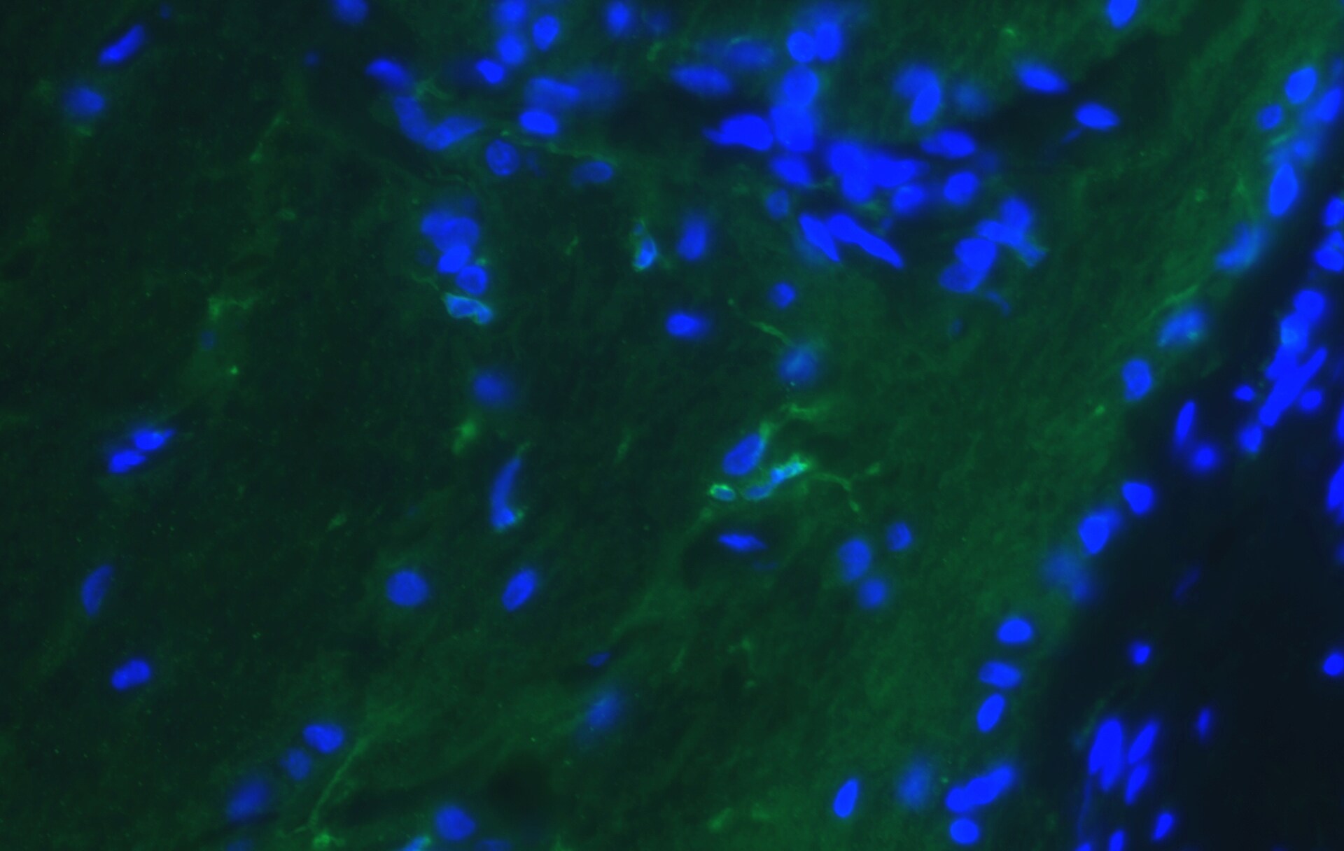
Detection of P2Y12/P2RY12 in human Brain Cortex via seqIF™ staining on COMET™
P2Y12/P2RY12 was detected in immersion fixed paraffin-embedded sections of human Cortex using Rabbit Anti-human P2Y12/P2RY12, Polyclonal Antibody (Catalog # NBP2-33870) at 1:100 at 37°Celsius for 4 minutes. Before incubation with the primary antibody, tissue underwent an all-in-one dewaxing and antigen retrieval preprocessing using PreTreatment Module (PT Module) and Dewax and HIER Buffer H (pH 9; Epredia Catalog # TA-999-DHBH).Tissue was stained using the Alexa Fluor™ Plus 555 Goat anti-Rabbit IgG Secondary Antibody at 1:100 at 37 ° Celsius for 2 minutes. (Yellow; Lunaphore Catalog # DR555RB) and counterstained with DAPI (blue; Lunaphore Catalog # DR100). Specific staining was localized to cell membrane of microglia. Protocol available in COMET™ Panel Builder.![Immunohistochemistry-Paraffin: P2Y12/P2RY12 Antibody [NBP2-33870]](https://resources.rndsystems.com/images/products/P2Y12-P2RY12-Antibody-Immunohistochemistry-Paraffin-NBP2-33870-img0014.jpg "Immunohistochemistry-Paraffin: P2Y12/P2RY12 Antibody [NBP2-33870]")
![Immunohistochemistry: P2Y12/P2RY12 Antibody [NBP2-33870]](https://resources.rndsystems.com/images/products/P2Y12-P2RY12-Antibody-Immunohistochemistry-NBP2-33870-img0019.jpg "Immunohistochemistry: P2Y12/P2RY12 Antibody [NBP2-33870]")
![Immunohistochemistry-Paraffin: P2Y12/P2RY12 Antibody [NBP2-33870]](https://resources.rndsystems.com/images/products/P2Y12-P2RY12-Antibody-Immunohistochemistry-Paraffin-NBP2-33870-img0012.jpg "Immunohistochemistry-Paraffin: P2Y12/P2RY12 Antibody [NBP2-33870]")
Immunohistochemistry-Paraffin: P2Y12/P2RY12 Antibody [NBP2-33870]
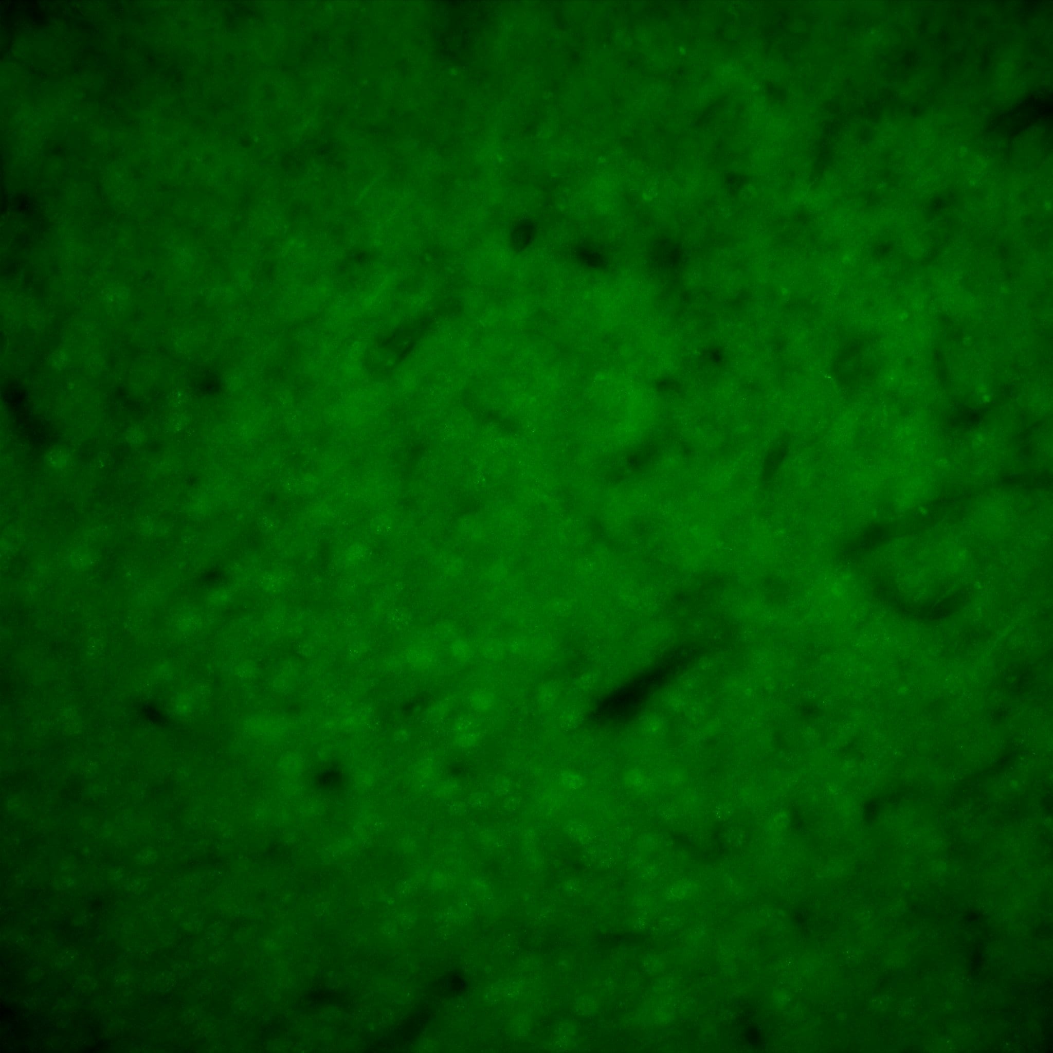
Immunohistochemistry-Paraffin: P2Y12/P2RY12 Antibody [NBP2-33870] - Staining of human cerebral cortex shows strong cytoplasmic positivity in microglia.![Immunohistochemistry-Paraffin: P2Y12/P2RY12 Antibody [NBP2-33870]](https://resources.rndsystems.com/images/products/P2Y12-P2RY12-Antibody-Immunohistochemistry-Paraffin-NBP2-33870-img0013.jpg "Immunohistochemistry-Paraffin: P2Y12/P2RY12 Antibody [NBP2-33870]")
Immunohistochemistry-Paraffin: P2Y12/P2RY12 Antibody [NBP2-33870]
Immunohistochemistry-Paraffin: P2Y12/P2RY12 Antibody [NBP2-33870] - Staining of human liver shows no positivity as expected.![Immunohistochemistry-Paraffin: P2Y12/P2RY12 Antibody [NBP2-33870]](https://resources.rndsystems.com/images/products/P2Y12-P2RY12-Antibody-Immunohistochemistry-Paraffin-NBP2-33870-img0016.jpg "Immunohistochemistry-Paraffin: P2Y12/P2RY12 Antibody [NBP2-33870]")
Immunohistochemistry-Paraffin: P2Y12/P2RY12 Antibody [NBP2-33870]
Immunohistochemistry-Paraffin: P2Y12/P2RY12 Antibody [NBP2-33870] - Staining of human testis shows no positivity in cells in seminiferous ducts as expected.![Immunohistochemistry-Paraffin: P2Y12/P2RY12 Antibody [NBP2-33870]](https://resources.rndsystems.com/images/products/P2Y12-P2RY12-Antibody-Immunohistochemistry-Paraffin-NBP2-33870-img0017.jpg "Immunohistochemistry-Paraffin: P2Y12/P2RY12 Antibody [NBP2-33870]")
Immunohistochemistry-Paraffin: P2Y12/P2RY12 Antibody [NBP2-33870]
Immunohistochemistry-Paraffin: P2Y12/P2RY12 Antibody [NBP2-33870] - Staining of human cerebellum shows strong cytoplasmic positivity in microglia.
Immunohistochemistry: P2Y12/P2RY12 Antibody [NBP2-33870] -
Immunohistochemistry: P2Y12/P2RY12 Antibody [NBP2-33870] - Images demonstrating colocalization of CD105/MAB1097 with microglial markers (a) panels A, B Low magnification images of high-plaque (HP) case (panel A) & AD case (panel B) showing staining with CD105/MAB1097 (purple arrows) & IBA-1 (brown arrows) in MTG sections. Both images at same magnification: scale bars represent 100 μm. (b) (panel A) CD105 microglia (purple arrows) with ramified morphology stained with antibody to P2RY12 (brown arrow) in low-plaque (LP) case. (panel B) CD105 microglia (purple arrows) & HLA-DR (brown arrow) in AD case. (panel C) CD105 microglia (purple arrow) & CD45 (brown arrow) in AD case. All sections were from MTG. Images at same magnification: scale bars represent 50 μm. (c) (panel A–E). Patterns of staining of CD105 (purple) & IBA-1 (brown) in MTG sections of cases with progressively increasing pathology from LP to AD. (panel F). CD105-positive microglia in hippocampus (HPC) of AD case. Images at same magnification: scale bars represent 50 μm. Image collected & cropped by CiteAb from the following publication (https://pubmed.ncbi.nlm.nih.gov/31340569), licensed under a CC-BY license. Not internally tested by Novus Biologicals.Applications for P2Y12/P2RY12 Antibody - BSA Free
Application
Recommended Usage
Immunohistochemistry
1:500 - 1:1000
Immunohistochemistry-Paraffin
1:500 - 1:1000
Multiplex Immunofluorescence
1:100
Application Notes
IHC-Paraffin, HIER pH 6 retrieval is recommended.
Reviewed Applications
Read 3 reviews rated 3.3 using NBP2-33870 in the following applications:
Formulation, Preparation, and Storage
Purification
Affinity purified
Formulation
PBS (pH 7.2) and 40% Glycerol
Format
BSA Free
Preservative
0.02% Sodium Azide
Concentration
Concentrations vary lot to lot. See vial label for concentration. If unlisted please contact technical services.
Shipping
The product is shipped with polar packs. Upon receipt, store it immediately at the temperature recommended below.
Stability & Storage
Store at 4C short term. Aliquot and store at -20C long term. Avoid freeze-thaw cycles.
Background: P2Y12/P2RY12
Long Name
P2Y Purinergic Receptor 12
Alternate Names
ADPG-R, HORK3, P2RY12, SP1999
Gene Symbol
P2RY12
Additional P2Y12/P2RY12 Products
Product Documents for P2Y12/P2RY12 Antibody - BSA Free
Certificate of Analysis
To download a Certificate of Analysis, please enter a lot or batch number in the search box below.
Product Specific Notices for P2Y12/P2RY12 Antibody - BSA Free
This product is for research use only and is not approved for use in humans or in clinical diagnosis. Primary Antibodies are guaranteed for 1 year from date of receipt.
Related Research Areas
Citations for P2Y12/P2RY12 Antibody - BSA Free
 Powered by Bioz
Customer Reviews for P2Y12/P2RY12 Antibody - BSA Free (3)
3.3 out of 5
3 Customer Ratings
Have you used P2Y12/P2RY12 Antibody - BSA Free?
Submit a review and receive an Amazon gift card!
$25/€18/£15/$25CAN/¥2500 Yen for a review with an image
$10/€7/£6/$10CAN/¥1110 Yen for a review without an image
Submit a review
Customer Images

Showing
1
-
3 of
3 reviews
Showing All
Filter By:
-
Application: Immunohistochemistry-Fixed; cryoprotectedSample Tested: Mouse brainSpecies: MouseVerified Customer | Posted 02/07/2020Mouse cortex 40X Leica DM6000Free floating coronal sections 50um. 72 hr primary @ RT with 4hr secondary @ RT.
 Bio-Techne ResponseThis review was submitted through the legacy Novus Innovators Program, reflecting a new species or application tested on a primary antibody.
 -
Application: Immunohistochemistry-FrozenSample Tested: Optic nerveSpecies: CanineVerified Customer | Posted 08/07/2018NBP2-33870 immunofluorescent signal was detected using Alexa Fluor 488 conjugated secondary antibody (green).
-
Application: ImmunocytochemistrySample Tested: optic nerveSpecies: FelineVerified Customer | Posted 05/11/2017Green-P2Y12/P2RY12, Blue-DAPI1:200 dilution for fixed tissue sections.
There are no reviews that match your criteria.
Protocols
Find general support by application which include: protocols, troubleshooting, illustrated assays, videos and webinars.
- Antigen Retrieval Protocol (PIER)
- Antigen Retrieval for Frozen Sections Protocol
- Appropriate Fixation of IHC/ICC Samples
- Cellular Response to Hypoxia Protocols
- Chromogenic IHC Staining of Formalin-Fixed Paraffin-Embedded (FFPE) Tissue Protocol
- Chromogenic Immunohistochemistry Staining of Frozen Tissue
- ClariTSA™ Fluorophore Kits
- Detection & Visualization of Antibody Binding
- Fluorescent IHC Staining of Frozen Tissue Protocol
- Graphic Protocol for Heat-induced Epitope Retrieval
- Graphic Protocol for the Preparation and Fluorescent IHC Staining of Frozen Tissue Sections
- Graphic Protocol for the Preparation and Fluorescent IHC Staining of Paraffin-embedded Tissue Sections
- Graphic Protocol for the Preparation of Gelatin-coated Slides for Histological Tissue Sections
- IHC Sample Preparation (Frozen sections vs Paraffin)
- Immunofluorescent IHC Staining of Formalin-Fixed Paraffin-Embedded (FFPE) Tissue Protocol
- Immunohistochemistry (IHC) and Immunocytochemistry (ICC) Protocols
- Immunohistochemistry Frozen Troubleshooting
- Immunohistochemistry Paraffin Troubleshooting
- Preparing Samples for IHC/ICC Experiments
- Preventing Non-Specific Staining (Non-Specific Binding)
- Primary Antibody Selection & Optimization
- Protocol for Heat-Induced Epitope Retrieval (HIER)
- Protocol for Making a 4% Formaldehyde Solution in PBS
- Protocol for VisUCyte™ HRP Polymer Detection Reagent
- Protocol for the Preparation & Fixation of Cells on Coverslips
- Protocol for the Preparation and Chromogenic IHC Staining of Frozen Tissue Sections
- Protocol for the Preparation and Chromogenic IHC Staining of Frozen Tissue Sections - Graphic
- Protocol for the Preparation and Chromogenic IHC Staining of Paraffin-embedded Tissue Sections
- Protocol for the Preparation and Chromogenic IHC Staining of Paraffin-embedded Tissue Sections - Graphic
- Protocol for the Preparation and Fluorescent IHC Staining of Frozen Tissue Sections
- Protocol for the Preparation and Fluorescent IHC Staining of Paraffin-embedded Tissue Sections
- Protocol for the Preparation of Gelatin-coated Slides for Histological Tissue Sections
- TUNEL and Active Caspase-3 Detection by IHC/ICC Protocol
- The Importance of IHC/ICC Controls
- Troubleshooting Guide: Immunohistochemistry
- View all Protocols, Troubleshooting, Illustrated assays and Webinars
Loading...