PD-L1 Antibody (2096A) - Azide and BSA Free
Novus Biologicals | Catalog # MAB90782
Recombinant Monoclonal Antibody
![Immunohistochemistry: PD-L1 Antibody (2096A) - Azide and BSA Free [MAB90782]](https://resources.rndsystems.com/images/products/B7-H1-PD-L1-CD274-Antibody-2096A-Immunohistochemistry-MAB90782-img0002.jpg "Immunohistochemistry: PD-L1 Antibody (2096A) - Azide and BSA Free [MAB90782]")
Key Product Details
Species Reactivity
Mouse
Applications
Immunohistochemistry, Immunohistochemistry-Frozen, Flow Cytometry, CyTOF-ready
Label
Unconjugated
Antibody Source
Monoclonal Rabbit IgG Clone # 2096A
Format
Azide and BSA Free
Loading...
Product Specifications
Immunogen
Mouse myeloma cell line NS0-derived recombinant mouse B7-H1/PD-L1
Met1-Thr239
Accession # Q9EP73
Met1-Thr239
Accession # Q9EP73
Clonality
Monoclonal
Host
Rabbit
Isotype
IgG
Scientific Data Images for PD-L1 Antibody (2096A) - Azide and BSA Free
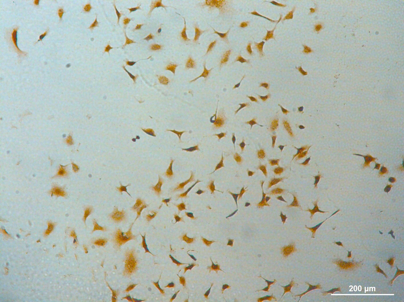
Immunohistochemistry: PD-L1 Antibody (2096A) - Azide and BSA Free [MAB90782]
Immunohistochemistry: B7-H1/PD-L1/CD274 Antibody (2096A) [MAB90782] - B7-H1 was detected in perfusion fixed frozen sections of mouse thymus using B7-H1 Monoclonal Antibody at 1.7 ug/ml. Tissue was stained using Rabbit IgG VisUCyte HRP Polymer Antibody (brown) and counterstained with hematoxylin (blue). Specific staining was localized to the cytoplasm and plasma membrane.Applications for PD-L1 Antibody (2096A) - Azide and BSA Free
Application
Recommended Usage
Immunohistochemistry
1 - 2 ug/ml
Immunohistochemistry-Frozen
1 - 2 ug/ml
Application Notes
This antibody is Cytof ready.
Reviewed Applications
Read 1 review rated 5 using MAB90782 in the following applications:
Flow Cytometry Panel Builder
Bio-Techne Knows Flow Cytometry
Save time and reduce costly mistakes by quickly finding compatible reagents using the Panel Builder Tool.
Advanced Features
- Spectra Viewer - Custom analysis of spectra from multiple fluorochromes
- Spillover Popups - Visualize the spectra of individual fluorochromes
- Antigen Density Selector - Match fluorochrome brightness with antigen density

Formulation, Preparation, and Storage
Purification
Protein A or G purified
Formulation
PBS
Format
Azide and BSA Free
Preservative
No Preservative
Concentration
1.0 mg/ml
Shipping
The product is shipped with polar packs. Upon receipt, store it immediately at the temperature recommended below.
Stability & Storage
Store at -20C. Avoid freeze-thaw cycles.
Background: PD-L1/B7-H1
PD-L1 binding with receptor PD-1 results in phosphorylation of in the inhibitory tyrosine-based switch motif (ITSM) domain of PD-1, which leads to recruitment of Src homology 2 domain-containing protein tyrosine-phosphatase 2 (SHP-2) and eventual downstream phosphorylation of spleen tyrosine kinase (Syk) and phospholipid inositol-3-kinase (PI3K) (1,3). Under normal conditions, the PD-L1/PD-1 signaling axis helps maintain immune tolerance and prevent destructive immune responses by inhibiting T cell activity such as proliferation, survival, cytokine production, and cytotoxic T lymphocyte (CTL) cytotoxicity (1-3). In the tumor microenvironment (TME), however, the PD-L1/PD-1 signaling axis is hijacked to promote tumor cell survival and limit anti-tumor immune response (1,3). More precisely, tumor cells can escape killing and immune surveillance due to T cell exhaustion and apoptosis (1-3).
Given the role the PD-L1/PD-1 signaling axis plays in tumor cells' ability to evade immune surveillance, it has become a target of several immunotherapeutic agents in recent years (3,5). Antibody immunotherapies that target these inhibitory checkpoint molecules has shown great promise for cancer treatment (3,5). PD-L1 and PD-1 blocking agents have been approved for treatment in a number of cancers including melanoma, non-small cell lung cancer (NSCLC), urothelial carcinoma, and Merkel-cell carcinoma (3,5). In many cancers the expression of PD-L1 in the TME has predictive value for response to blocking agents (3). Pembrolizumab, for example, is a PD-1 inhibitor that has been approved by the FDA as a second-line therapy for treatment of metastatic NSCLC in patients whose tumors express PD-L1 with a Tumor Proportion Score (TPS) greater than 1%, but also for first-line treatment in cases where patients' tumors expression PD-L1 with a TPS greater than 50%) (5). The most promising cancer immunotherapy treatments seem to point to combination therapy with both anti-cancer drugs (e.g. Gefitibin, Metformin, Etoposide) with PD-L1/PD-1 antibody blockade inhibitors (e.g. Atezolizumab, Nivolumab) (6).
References
1. Han, Y., Liu, D., & Li, L. (2020). PD-1/PD-L1 pathway: current researches in cancer. American journal of cancer research, 10(3), 727-742.
2. Jiang, Y., Chen, M., Nie, H., & Yuan, Y. (2019). PD-1 and PD-L1 in cancer immunotherapy: clinical implications and future considerations. Human vaccines & immunotherapeutics, 15(5), 1111-1122. https://doi.org/10.1080/21645515.2019.1571892
3. Sun, C., Mezzadra, R., & Schumacher, T. N. (2018). Regulation and Function of the PD-L1 Checkpoint. Immunity, 48(3), 434-452. https://doi.org/10.1016/j.immuni.2018.03.014
4. Cha, J. H., Chan, L. C., Li, C. W., Hsu, J. L., & Hung, M. C. (2019). Mechanisms Controlling PD-L1 Expression in Cancer. Molecular cell, 76(3), 359-370. https://doi.org/10.1016/j.molcel.2019.09.030
5. Tsoukalas, N., Kiakou, M., Tsapakidis, K., Tolia, M., Aravantinou-Fatorou, E., Baxevanos, P., Kyrgias, G., & Theocharis, S. (2019). PD-1 and PD-L1 as immunotherapy targets and biomarkers in non-small cell lung cancer. Journal of B.U.ON. : official journal of the Balkan Union of Oncology, 24(3), 883-888.
6. Gou, Q., Dong, C., Xu, H., Khan, B., Jin, J., Liu, Q., Shi, J., & Hou, Y. (2020). PD-L1 degradation pathway and immunotherapy for cancer. Cell death & disease, 11(11), 955. https://doi.org/10.1038/s41419-020-03140-2
Long Name
Programmed Death Ligand 1
Alternate Names
B7-H1, B7H1, CD274, PDCD1L1, PDCD1LG1, PDL1
Gene Symbol
CD274
Additional PD-L1/B7-H1 Products
Product Documents for PD-L1 Antibody (2096A) - Azide and BSA Free
Certificate of Analysis
To download a Certificate of Analysis, please enter a lot or batch number in the search box below.
Product Specific Notices for PD-L1 Antibody (2096A) - Azide and BSA Free
This product is for research use only and is not approved for use in humans or in clinical diagnosis. Primary Antibodies are guaranteed for 1 year from date of receipt.
Customer Reviews for PD-L1 Antibody (2096A) - Azide and BSA Free (1)
5 out of 5
1 Customer Rating
Have you used PD-L1 Antibody (2096A) - Azide and BSA Free?
Submit a review and receive an Amazon gift card!
$25/€18/£15/$25CAN/¥2500 Yen for a review with an image
$10/€7/£6/$10CAN/¥1110 Yen for a review without an image
Submit a review
Customer Images
Showing
1
-
1 of
1 review
Showing All
Filter By:
-
Application: ImmunocytochemistrySample Tested: A549 human lung carcinoma cell lineSpecies: HumanVerified Customer | Posted 01/22/202010x DIC2 ug/mL MAB90782 antibody diluted in 3%BSA/PBS for 3 h, at room temperature Stained with DAB from Leica Novocastra #RE7150-K

There are no reviews that match your criteria.
Protocols
Find general support by application which include: protocols, troubleshooting, illustrated assays, videos and webinars.
- 7-Amino Actinomycin D (7-AAD) Cell Viability Flow Cytometry Protocol
- Antigen Retrieval Protocol (PIER)
- Antigen Retrieval for Frozen Sections Protocol
- Appropriate Fixation of IHC/ICC Samples
- Cellular Response to Hypoxia Protocols
- Chromogenic IHC Staining of Formalin-Fixed Paraffin-Embedded (FFPE) Tissue Protocol
- Chromogenic Immunohistochemistry Staining of Frozen Tissue
- ClariTSA™ Fluorophore Kits
- Detection & Visualization of Antibody Binding
- Extracellular Membrane Flow Cytometry Protocol
- Flow Cytometry Protocol for Cell Surface Markers
- Flow Cytometry Protocol for Staining Membrane Associated Proteins
- Flow Cytometry Staining Protocols
- Flow Cytometry Troubleshooting Guide
- Fluorescent IHC Staining of Frozen Tissue Protocol
- Graphic Protocol for Heat-induced Epitope Retrieval
- Graphic Protocol for the Preparation and Fluorescent IHC Staining of Frozen Tissue Sections
- Graphic Protocol for the Preparation and Fluorescent IHC Staining of Paraffin-embedded Tissue Sections
- Graphic Protocol for the Preparation of Gelatin-coated Slides for Histological Tissue Sections
- IHC Sample Preparation (Frozen sections vs Paraffin)
- Immunofluorescent IHC Staining of Formalin-Fixed Paraffin-Embedded (FFPE) Tissue Protocol
- Immunohistochemistry (IHC) and Immunocytochemistry (ICC) Protocols
- Immunohistochemistry Frozen Troubleshooting
- Immunohistochemistry Paraffin Troubleshooting
- Intracellular Flow Cytometry Protocol Using Alcohol (Methanol)
- Intracellular Flow Cytometry Protocol Using Detergents
- Intracellular Nuclear Staining Flow Cytometry Protocol Using Detergents
- Intracellular Staining Flow Cytometry Protocol Using Alcohol Permeabilization
- Intracellular Staining Flow Cytometry Protocol Using Detergents to Permeabilize Cells
- Preparing Samples for IHC/ICC Experiments
- Preventing Non-Specific Staining (Non-Specific Binding)
- Primary Antibody Selection & Optimization
- Propidium Iodide Cell Viability Flow Cytometry Protocol
- Protocol for Heat-Induced Epitope Retrieval (HIER)
- Protocol for Liperfluo
- Protocol for Making a 4% Formaldehyde Solution in PBS
- Protocol for VisUCyte™ HRP Polymer Detection Reagent
- Protocol for the Characterization of Human Th22 Cells
- Protocol for the Characterization of Human Th9 Cells
- Protocol for the Preparation & Fixation of Cells on Coverslips
- Protocol for the Preparation and Chromogenic IHC Staining of Frozen Tissue Sections
- Protocol for the Preparation and Chromogenic IHC Staining of Frozen Tissue Sections - Graphic
- Protocol for the Preparation and Chromogenic IHC Staining of Paraffin-embedded Tissue Sections
- Protocol for the Preparation and Chromogenic IHC Staining of Paraffin-embedded Tissue Sections - Graphic
- Protocol for the Preparation and Fluorescent IHC Staining of Frozen Tissue Sections
- Protocol for the Preparation and Fluorescent IHC Staining of Paraffin-embedded Tissue Sections
- Protocol for the Preparation of Gelatin-coated Slides for Histological Tissue Sections
- Protocol: Annexin V and PI Staining by Flow Cytometry
- Protocol: Annexin V and PI Staining for Apoptosis by Flow Cytometry
- TUNEL and Active Caspase-3 Detection by IHC/ICC Protocol
- The Importance of IHC/ICC Controls
- Troubleshooting Guide: Fluorokine Flow Cytometry Kits
- Troubleshooting Guide: Immunohistochemistry
- View all Protocols, Troubleshooting, Illustrated assays and Webinars
Loading...