
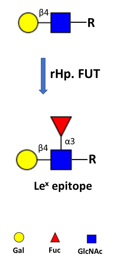
Lewis X (LeX), a fucosylated trisaccharide glycan epitope (Gal beta 1,4 [Fuc alpha 1,3]GlcNAc beta ) also known as CD15, and sialylated Lewis X (sLeX) are distributed throughout eukaryotes and are determinants of many functional glycoconjugates that play central roles in numerous physiological and pathological processes (1, 2). Lex-bearing glycans are also found in the infectious bacterium Helicobacter pylori to mask the bacterium from the host immune surveillance (3). H. pylori is the pathogen that causes peptic ulcers that can further lead to stomach cancer and gastritis. H. pylori fucosyltransferase is responsible for generating the LeX glycans, therefore is potentially a drug target for curing peptic ulcers, gastritis and stomach cancer. In molecular terms, H. pylori fucosyltransferase is an alpha 1,3 fucosyltransferase that shares function with human FUT4, FUT5, FUT6, and FUT9 (4). Remarkably, H. pylori fucosyltransferase can transfer IgG antibody to the glycocalyx on the surfaces of live cells when the antibody is conjugated to the enzyme's natural donor substrate GDP-Fucose (5). The activity of this enzyme has been measured with a phosphatase coupled method (6).
Recombinant H. pylori Fucosyltransferase Protein, CF
R&D Systems | Catalog # 11072-GT

Loading...
Key Product Details
- R&D Systems E. coli-derived Recombinant H. pylori Fucosyltransferase Protein (11072-GT)
- Quality control testing to verify active proteins with lot specific assays by in-house scientists
- All R&D Systems proteins are covered with a 100% guarantee
Source
E. coli
Accession Number
Applications
Enzyme Activity
Loading...
Product Specifications
Source
E. coli-derived h. pylori Fucosyltransferase protein
Met1-Tyr392, with a C-terminal 6-His tag
Met1-Tyr392, with a C-terminal 6-His tag
Purity
>95%, by SDS-PAGE visualized with Silver Staining and quantitative densitometry by Coomassie® Blue Staining.
Endotoxin Level
<1.0 EU per 1 μg of the protein by the LAL method.
N-terminal Sequence Analysis
Met1
Predicted Molecular Mass
46 kDa
SDS-PAGE
39 kDa, under reducing conditions.
Activity
Measured by its ability to transfer fucose from GDP-fucose to N-Acetyllactosamine
The specific activity is >1500 pmol/min/μg, as measured under the described conditions.
The specific activity is >1500 pmol/min/μg, as measured under the described conditions.
Scientific Data Images for Recombinant H. pylori Fucosyltransferase Protein, CF
Recombinant H. pylori Fucosyltransferase Protein Enzyme Activity Diagram.
Recombinant H. pylori Fucosyltransferase His-tag Protein (Catalog # 11072-GT) Enzyme Activity Diagram.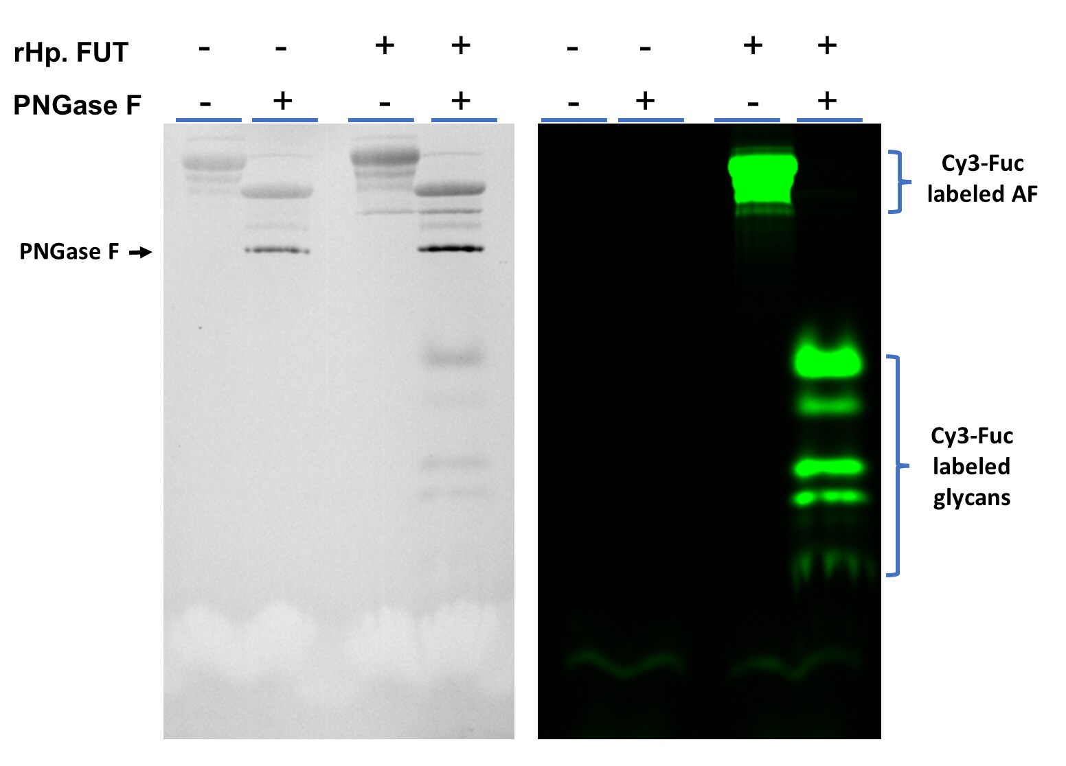
Fluorescent glycan labeling of asialofetuin (AF) with Cy3-Fuc using rHp. FUT.
Asialofetuin (4 μg) was incubated with 0.2 nmol of GDP-Cy3-Fucose (ES401) with or without rHp. FUT (Catalog # 11072) (1.0 μg) in 25 μL of 25 mM Tris pH 7.5, 10 mM MnCl2 at 37 °C for 30 minutes. Following labeling, half of the AF was deglycosylated with PNGase F N-glycan Releasing Kit (EA006) and resolved in 15% SDS-PAGE. Left side is the TCE image and right side is the Fluorescent image of the same gel.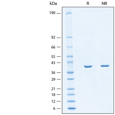
Recombinant H. pylori Fucosyltransferase Protein SDS-PAGE.
2 μg/lane of Recombinant H. pylori Fucosyltransferase Protein (Catalog # 11072-GT) was resolved with SDS-PAGE under reducing (R) and non-reducing (NR) conditions and visualized by Coomassie® Blue staining, showing bands at 39 kDa.Formulation, Preparation, and Storage
11072-GT
| Formulation | Supplied as a 0.2 μm filtered solution in Tris and NaCl. |
| Shipping | The product is shipped with polar packs. Upon receipt, store it immediately at the temperature recommended below. |
| Stability & Storage | Use a manual defrost freezer and avoid repeated freeze-thaw cycles.
|
Background: Fucosyltransferase
References
- Gooi, H.C. et al. (1981) Nature 292:156.
- Phillips, M.L. et al. (1990) Science 250:1130.
- Moran, A.P. et al. (1996) FEMS Immunol Med Microbiol 16:105.
- de Vries, T. et al. (2001). Glycobiology 11:119R.
- Li, J. et al. (2018) ACS Cent. Sci. 4:1633.
- Wu, Z.L. et al. (2011) Glycobiology 21:727.
Long Name
alpha-(1,3) Fucosyltransferase FucT
Alternate Names
FucT
Gene Symbol
fucT
UniProt
Additional Fucosyltransferase Products
Product Documents for Recombinant H. pylori Fucosyltransferase Protein, CF
Certificate of Analysis
To download a Certificate of Analysis, please enter a lot or batch number in the search box below.
Note: Certificate of Analysis not available for kit components.
Product Specific Notices for Recombinant H. pylori Fucosyltransferase Protein, CF
For research use only
Related Research Areas
Citations for Recombinant H. pylori Fucosyltransferase Protein, CF
 Powered by Bioz
Customer Reviews for Recombinant H. pylori Fucosyltransferase Protein, CF
There are currently no reviews for this product. Be the first to review Recombinant H. pylori Fucosyltransferase Protein, CF and earn rewards!
Have you used Recombinant H. pylori Fucosyltransferase Protein, CF?
Submit a review and receive an Amazon gift card!
$25/€18/£15/$25CAN/¥2500 for a review with an image
$10/€7/£6/$10CAN/¥1110 for a review without an image
Submit a review
Protocols
View specific protocols for Recombinant H. pylori Fucosyltransferase Protein, CF (11072-GT):
Materials
- Glycosyltransferase Activity Kit (Catalog # EA001)
- Assay Buffer: 25 mM Tris, 10 mM CaCl2, pH 7.0
- Recombinant H. pylori Fucosyltransferase (rHp FUT) (Catalog # 11072-GT)
- Lactosamine (Dextra Laboratories, Catalog # GN204), 50 mM stock in deionized water
- GDP-Fucose (Sigma, Catalog # G4401), 1.6 mM stock in deionized water
- 96-well Clear Plate (Catalog # DY990)
- Plate Reader (Model: SpectraMax Plus by Molecular Devices) or equivalent
- Dilute 1 mM Phosphate Standard provided by the Glycosyltransferase Activity Kit by adding 40 μL of the 1 mM Phosphate Standard to 360 μL of Assay Buffer for a 100 μM stock. This is the first point of the standard curve.
- Complete the standard curve by performing six one-half serial dilutions of the 100 μM Phosphate stock in Assay Buffer. The standard curve has a range of 0.078 to 5 nmol per well.
- Prepare Reaction Mixture containing 2.4 mM Lactosamine, 0.240 mM GDP-Fucose, and 4 μg/mL Coupling Phosphatase 1 (provided in kit) in Assay Buffer.
- Dilute rHp FUT to 1 μg/mL in Assay Buffer.
- Load 50 μL of each dilution of the standard curve into a plate. Include a curve blank containing 50 μL of Assay Buffer.
- Load 25 μL of 1 μg/mL of rHp FUT into the plate. Include a Control containing 25 μL of Assay Buffer.
- Start the reactions by adding 25 μL of Reaction Mixture to the wells, excluding the standard curve.
- Incubate sealed plate at 37 °C for 20 minutes.
- Add 30 μL of the Malachite Green Reagent A to all wells. Mix briefly.
- Add 100 μL of deionized water to all wells.
- Add 30 μL of the Malachite Green Reagent B to all wells. Mix and incubate for 20 minutes at room temperature.
- Read plate at 620 nm (absorbance) in endpoint mode.
- Calculate specific activity:
Specific Activity (pmol/min/µg) = | Phosphate released* (nmol) x (1000 pmol/nmol) |
| Incubation time (min) x amount of enzyme (µg) |
*Derived from the phosphate standard curve using linear or 4-parameter fitting and adjusted for Control.
Per Reaction:
- rHp FUT: 0.025 µg
- Coupling Phosphatase 1: 0.1 μg
- Lactosamine: 1.2 mM
- GDP-Fucose: 0.12 mM
Loading...